Mass and thrombus formation are known complications of ventriculoatrial shunts.Reference Yavuzgil, Ozerkan, Erturk, Islekel, Atay and Buket 1 , Reference Chaw, Buxton and Wong 2 Large thrombi do not resolve spontaneously and need more careful management depending on the clinical scenario.Reference Wilkinson, Sood, Ham, Gilmer, Flemming and Rajpurkar 3 , Reference Mukharji and Peterson 4 We describe a case of percutaneous removal of a large mass/thrombus with the aid of trans-oesophegeal echocardiogram and fluoroscopy.
Case report
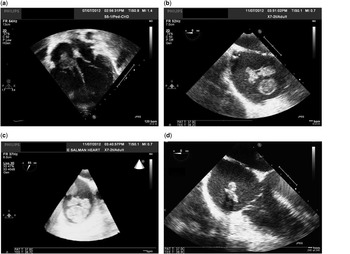
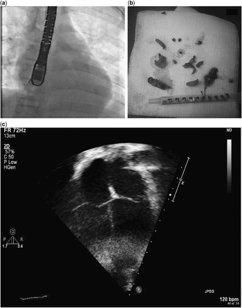
We present the case of a 12-year-old boy weighing 24 kg with traumatic hydrocephalus secondary to car accident 4 years back. Owing to progressive increase in hydrocephalus, a ventriculoatrial shunt was inserted after multiple failing venticuloperitoneal shunts. He had persistent fever, and blood culture showed multi-drug-resistant staphylococcus aureus. Therefore, the ventriculoatrial shunt was removed and was replaced by an external ventricular drain and he receieved various antibiotics. Echocardiogram showed a large mobile thrombus in the right atrium measuring 10×10 cm, protruding through the tricuspid valve (Fig 1a). The thrombus did not resolve for 4 weeks. Although the patient was clinically stable but had thrombocytopenia and a deranged coagulation profile, so was not thrombolysed. Owing to the possibility of embolisation and persistent bacteraemia, it was decided to remove it by cardiac catheterisation. Trans-oesophageal echocardiography confirmed the above findings (Fig 1b and c). The giant thrombus in the right atrium was removed by suction technique. A 6 Fr sheath was introduced into the right femoral vein, which was later upgraded gradually to 12 Fr. A Mullins sheath was positioned near the thrombus with the help of trans-oesopheageal echocardiography and fluoroscopy (Fig 1d). The thrombus was suctioned into the tip of the sheath with negative pressure applied through a 20 cc syringe (Fig 2a). It was removed in small pieces suctioned through the negative pressure (Fig 2b). A total of 500 ml of blood was drawn during this manoeuvre. The withdrawn blood was warmed and filtered by using transfusion microfilters (Pall LipiGuard; Terumo Cardiovascular Systems, Ann Arbor, Minnesota, United States of America) and reinfused into the patient. The above-mentioned filters remove particles as small as 40 μm, including microthrombi, fat globules, and leukocytes. Therefore, reinfusion of blood minimised blood loss. At the end of the procedure, the mass was no longer visible by trans-oesophegeal echocardiogram. The microscopy and culture of mass did not yield any organism. The patient was started on subcutaneous heparin 3 days before being discharged home. The patient remained haemodynamically stable and was discharged home. The serial follow-up echocardiogram done at 1, 3, and 6 months showed normal right atrium with no recurrence of mass (Fig 2c). The patient remained stable and is doing fine now. The subcutaneous heparin was discontinued after 3 months of thrombus removal.
( a ) Apical four-chamber view showing thrombus in the right atrium. ( b ) Trans-oesophegeal echocardiogram (TEE) two-dimensional image showing thrombus in the right atrium. ( c ) Trans-oesophegeal echocardiogram three-dimensional image showing thrombus in the right atrium. ( d ) Trans-oesophegeal echocardiogram (TEE) still image showing thrombus suctioned into the tip of the sheath.

( a ) Frontal view still frame showing the sheath positioned in the right atrium near the thrombus with the help of TEE. ( b ) The photo of the thrombus after the removal. ( c ) Apical four-chamber view 6 months after thrombus removal showing a normal right atrium with no thrombus.

Discussion
Ventriculoatrial shunts are known to be complicated by thrombus formation at the tip in the right atrium and bacteraemia.Reference Yavuzgil, Ozerkan, Erturk, Islekel, Atay and Buket 1 , Reference Chaw, Buxton and Wong 2 Large thrombi do not resolve spontaneously and require removal by intervention as they may lead to pulmonary embolism.Reference Wilkinson, Sood, Ham, Gilmer, Flemming and Rajpurkar 3 , Reference Mukharji and Peterson 4 There is high incidence of mortality with right atrial thrombi reported as high as 40%.Reference Starkey and de Bono 5 The choice of therapy for intracardiac thrombi is still a matter of debate.
They may be treated by thrombolytic therapy, surgical embolectomy, or percutaneously by cardiac catheterisation.Reference Wilkinson, Sood, Ham, Gilmer, Flemming and Rajpurkar 3 – Reference Engelman, Ransohoff, Cortes and Spencer 7 The conservative management with thrombolysis is a successful and better choice, but carries the risk of fragmentation and embolisation in cases of large, mobile thrombi.Reference Starkey and de Bono 5 , Reference Duidy, Kronzon, Cohen and Ruiz 8 , Reference Ferrari, Vagnarelli, Gargano, Roversi, Biagioni and Ranzi 9 Moreover, there is high risk of major bleeding including intracranial haemorrhage with thrombolysis. According to the current literature, medical management options include use of heparin, warfarin, streptokinase, urokinase, and recombinant tissue-type plasminogen activator.Reference Starkey and de Bono 5 , Reference Duidy, Kronzon, Cohen and Ruiz 8 , Reference Ferrari, Vagnarelli, Gargano, Roversi, Biagioni and Ranzi 9 The use of recombinant tissue-type plasminogen activator has proven successful in dissolving intracardiac thrombi in children and adults.Reference Starkey and de Bono 5 , Reference Duidy, Kronzon, Cohen and Ruiz 8 , Reference Ferrari, Vagnarelli, Gargano, Roversi, Biagioni and Ranzi 9 Timely diagnosis by echocardiogram before a thrombus gets bigger or organised leads to effective, safe, and rapid thrombolysis by use of recombinant tissue-type plasminogen activator.Reference Starkey and de Bono 5 , Reference Duidy, Kronzon, Cohen and Ruiz 8 , Reference Ferrari, Vagnarelli, Gargano, Roversi, Biagioni and Ranzi 9
The chances of haemorrhage are more in newborn babies. There is current evidence that recombinant tissue-type plasminogen activator is safe and effective in dissolving intracardiac thrombi even in preterm infants.Reference Ferrari, Vagnarelli, Gargano, Roversi, Biagioni and Ranzi 9
The operative management is complicated and has higher morbidity and mortality.Reference Engelman, Ransohoff, Cortes and Spencer 7 The percutaneous technique avoids surgical trauma and is safe and effective.Reference Mukharji and Peterson 4 Although the patient was clinically stable, thrombolysis was not considered because of the deranged coagulation profile and thrombocytopaenia. We removed the thrombus by percutaneous method to avoid embolisation and persistent infection.
In conclusion, the percutaneous removal of an intracardiac thrombus is a novel technique that is safe, feasible, effective, and avoids surgical trauma.