Exercise testing is frequently selected as a diagnostic modality in the assessment of functional status in the child with CHD. Formal exercise testing has also proven to be useful in the risk assessment of patients at risk for sudden cardiac death. This population includes patients with known or suspected cardiac electrical abnormalities and patients with congenital or acquired structural heart disease. Formal exercise testing can help confirm a suspected diagnosis and help rule out a tentative diagnosis. It is also useful for assessing the response of the patient to medical or device therapy. Given that the risk for sudden cardiac arrest is over twice as likely for athletes during sports participation compared with that for non-athletes,Reference Corrado, Basso, Rizzoli, Schiavon and Thiene 1 exercise testing is a reasonably good stressor to unmask disease. No testing modality is perfect. Therefore, a normal exercise test does not absolutely rule out risk. Notwithstanding, the formal exercise test is usually capable of distinguishing benign cardiac conditions from potentially dangerous and life-threatening entities. The goal of this article is to familiarise the reader with the value of exercise testing in patient risk assessment using hypothetical historical case examples. This article is not intended to be comprehensive but, rather, is intended to highlight potentially helpful tips and pitfalls for clinicians who have interest in paediatric exercise testing with regard to the management of patients who carry a risk for sudden death. A more comprehensive review of sudden death in athletes can be found elsewhere.Reference Noakes 2 , Reference Maron 3
Key components of the formal exercise test
Although the measurement of metabolic data using volumetric assessment of inspired oxygen and excreted carbon dioxide is useful in the assessment of some patients at risk for cardiac death, most investigators would agree that exercise testing in the absence of these data is still worthwhile. Therefore, assessment of the patient’s rhythm alone during exercise can be useful to the clinician assessing the patient. The author considers the essential components of exercise testing to include an experienced exercise technologist/physiologist, a blood pressure cuff, a pulse oximeter, an electrocardiographic recorder/writer, and an exercise bike or treadmill. Having a metabolic cart to measure inspired and expired gases, measure the anaerobic threshold, and compute ventilatory equivalents can provide additional valuable data in the determination of prognosis for certain CHD and acquired cardiac defect.Reference Giardini, Hager and Lammers 4
Preparation for testing day
Patient preparation
On occasion, patients arrive at the exercise laboratory ill prepared to engage in maximally strenuous exercise. Teenagers are more likely than younger patients to opine that the test, particularly an unplanned test, is egregious. Explaining that at-risk patients are in a potentially safer environment in a hospital or medical centre exercise testing facility than on the playing field in the event of a dangerous cardiac event usually helps assuage the patient and their family. Patient should refrain from eating two hours prior to the test, should wear comfortable clothing – preferably “gym” clothes – and should have available sneakers or athletic shoes. Many busy exercise laboratories find it worthwhile to maintain an inventory of athletic shoes for those patients who are not prepared to exercise.
Provider preparation
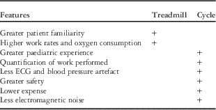
The exercise laboratory team should always be equipped with clinical information that will allow the providers to optimise information yield. Choosing the correct protocol helps to optimise the yield. For example, if exercise-provoked chest pain is present in a patient with a known coronary arterial abnormality, then bicycle ergometry is preferred because of less electrocardiographic motion artefact. Or, if long QT syndrome is suspected, then careful measurement of the QT interval during early recovery may help arrive at a definitive diagnosis. Table 1 highlights the advantages and disadvantages of choosing a treadmill protocol over cycle ergometry.
Benefits of treadmill versus cycle ergometer.

ECG=electrocardiogram.
Case illustration 1
An 8-year-old female patient is scheduled to undergo tonsillectomy and adenoidectomy at a community hospital. Upon anaesthesia induction, a wide QRS tachycardia is noted. Her past medical history includes two previous episodes of chest pain, near syncope, abdominal pain, and nausea, during which she appeared pale. These episodes always resolved after a bowel movement. Family history was unremarkable.
A formal treadmill exercise test was undertaken because of the patient’s small size. During exercise an arrhythmia occurred. Fig 1 shows a wide QRS tachycardia with a right bundle branch block pattern. The patient was haemodynamically stable throughout the arrhythmia. Findings are typical of fascicular ventricular tachycardia, which is usually responsive to treatment with a calcium channel agent.Reference German, Packer and Bardy 5 This arrhythmia, although disconcerting, is not typically associated with sudden cardiac death. Contrast this test result with the following case illustration.
Exercise-provoked sustained wide QRS tachyarrhythmia with right bundle branch block consistent with fascicular ventricular tachycardia. Illustrated are the precordial leads V1–V6.

Case illustration 2
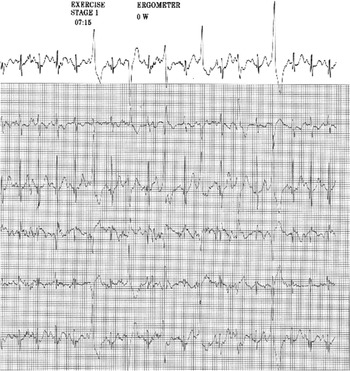
An 11-year-old faints 1 minute into a basketball playoff game. The patient appears to have a generalised tonic–clonic seizure. The patient recovers and is able to finish the basketball game. Family history includes seizures in first- and second-degree relatives. In one, sudden death occurred during sleep. In another, seizures were brought on by anxiety. The bicycle ergometry exercise test in the patient showed the onset of multiform premature ventricular depolarisations during middle to late exercise (Fig 2).
Multiform premature ventricular complexes in an 11-year-old during bicycle exercise stress testing (see text). Illustrated are the limb leads, I, II, III, aVR, aVL, and aVF.

Although the test result in case illustration 2 seems less dangerous than the finding of fascicular ventricular tachycardia shown in the previous case illustration, the finding of polymorphic premature ventricular complexes in a symptomatic patient is quite disturbing and should prompt treatment. The onset of premature ventricular complexes that increase as exercise progresses is a common finding in patients with catecholaminergic polymorphic ventricular tachycardia, a known cause of sudden cardiac death in athletes.Reference Coumel, Fidelle and Lucet 6 The patient’s family history is also a clue that the patient’s premature ventricular complexes during exercise can be a potentially ominous sign.
Case illustrations 3
A 14-year-old male patient is referred to the exercise laboratory with a history of palpitations. A baseline electrocardiogram reveals ventricular pre-excitation (Fig 3a). Exercise testing demonstrates abrupt loss of pre-excitation during the latter stages of exercise with no exercise-induced arrhythmias (Fig 3b). Loss of pre-excitation, particularly abrupt loss, is thought to be associated with a lower risk for sudden cardiac death in patients with Wolff–Parkinson–White syndrome (WPW).Reference Cohen, Triedman and Cannon 7 Careful observation of the rhythm in patients with known Wolff–Parkinson–White syndrome is essential throughout the exercise and recovery phases of the stress test. Assessing for the loss of pre-excitation during exercise is not always straightforward. The recovery phase of exercise offers clinicians the opportunity to determine whether and when pre-excitation returns during recovery and to assess for post-exercise ectopy. On the basis of recent guidelines, the unequivocal sudden loss of pre-excitation during exercise has been used by clinicians to permit young athletes to return to sports participation.Reference Cohen, Triedman and Cannon 7 Tragic exceptions to this “rule” have been anecdotally described, however, and the Wolff–Parkinson–White syndrome pattern, especially in the asymptomatic youngster, remains a conundrum for the electrophysiologist. It is anticipated that the aforementioned guidelines may be further revised as new data become available.
A 12-lead electrocardiogram in a 14-year-old with Wolff–Parkinson–White syndrome at rest ( a ) and during exercise ( b ) illustrating abrupt loss of pre-excitation.

Case illustration 4
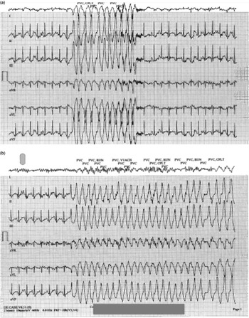
An 18-year-old rower is referred to the Exercise Laboratory, having fainted during rowing practice, fallen out of the scull, and then regained consciousness in the water, not requiring team-mate assistance to get back into the boat. The patient’s electrocardiographic findings during exercise and recovery are shown in Fig 4a and b, respectively. The patient was found to have subtle MRI findings consistent with arrhythmogenic right ventricular dysplasia. Despite multiple medications and catheterisation-based treatments, the patient continued to develop new arrhythmogenic foci. Recent data suggest that frequent vigorous exercise is more likely to result in clinical recognition of this disorder of the right – and to a lesser extent the left – ventricle at younger ages and to accelerate its pathogenesis.Reference James, Bhonsale and Tichnell 8
Limb lead electrocardiographic recordings during exercise ( a ) and during recovery ( b ) from an 18-year-old competitive rower, demonstrating non-sustained ventricular tachycardia related to arrhythmogenic right ventricular cardiomyopathy.

Coronary abnormalities and sudden cardiac death
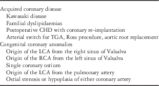
Coronary artery abnormalities comprise the second most common cause of sudden death in athletes, following only hypertrophic cardiomyopathy. An abbreviated list of congenital and acquired coronary arterial abnormalities is shown in Table 2. There are special considerations when performing exercise testing in children with these conditions.
An abridged list of coronary artery abnormalities that may affect the young.

TGA=transposition of the great arteries; LCA=left coronary artery; RCA=right coronary artery.
Case illustration 5
An 8-year-old male patient was admitted and discharged from an inpatient cardiology unit after a diagnostic work-up for syncope during exercise yielded no positive findings. Stress test results were normal, with no exercise-induced arrhythmias or ST segment changes suggestive of ischaemia. Dehydration was thought to have caused the patient’s symptoms. Two years later, the patient had a seizure during exercise. A repeat echocardiogram showed the left coronary artery arising immediately to the right of the intercoronary commissure, consistent with left coronary origin from the opposite-facing sinus. The echocardiographic findings were confirmed with MRI, and coronary unroofing was performed. The patient remains symptom free one decade after the surgical intervention and competes at a collegiate level.
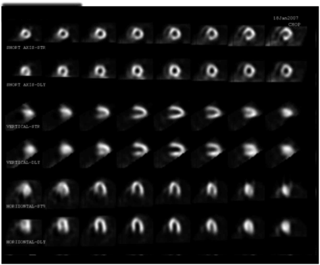
This case illustration demonstrates that false-negative results occur with exercise testing. Although ST segment changes during exercise can be seen in patients with anomalous coronary arteries,Reference Brothers, Carter and McBride 9 an exercise stress test that yields no positive findings does not mean the patient is not at risk. Patients with long QT syndrome, anomalous coronary arteries, and hypertrophic cardiomyopathy may have normal exercise stress test results. ST segment changes that occur during exercise are usually a late finding and are usually preceded by abnormalities that can be detected with myocardial perfusion imaging (Fig 5), positron emission tomography, or stress echocardiography.Reference Kimball 10
Myocardial perfusion imaging demonstrating a medium-sized reversible filling defect in the inferior basal septal region in an 8-year-old with right coronary artery from the left-facing sinus.

Case illustration 6
A 15-year-old male patient is referred because of syncope during a sporting event. The echocardiogram demonstrated mild concentric left ventricular hypertrophy, mild right ventricular hypertrophy, and a left ventricular end-diastolic internal dimension of 4.6 cm. Exercise stress testing demonstrated no evidence of exercise-induced arrhythmias and no ischaemic changes. His oxygen consumption was >45 ml/kg/minute. Family history was non-contributory. A tentative diagnosis of hypertrophic cardiomyopathy was made, and exercise restriction was recommended. However, sudden cardiovascular collapse occurred during a casual sporting event. Marked left ventricular hypertrophy was noted on a follow-up echocardiogram. The patient did not survive the event.
The above case highlights the challenges in the diagnosis and treatment of hypertrophic cardiomyopathy. Despite the classic teachings, not all patients with the malignant form of this disease will have a reduced left ventricular internal dimension (<4.5 cm), reduced oxygen consumption (<45 ml/kg/minute), or a positive family history.Reference Pelliccia, Maron and Maron 11 Exercise stress testing can be normal in these patients and is not synonymous with the absence of risk.
Stress echocardiographic measurement of outflow tract gradients has been used in this patient population to guide therapy.Reference Maron, Rowin and Olivotto 12 Elevated outflow tract gradients from intra-cavitary obstruction has also been demonstrated in normal adult and children.Reference Wittlieb-Weber, Cohen and McBride 13 Using outflow tract gradients alone to make a diagnosis of hypertrophic cardiomyopathy could potentially lead to a misdiagnosis of normal patients.
Predictive value of metabolic data
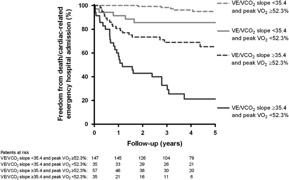
Diller, Dimopoulos,Reference Diller, Dimopoulos and Okonko 14 , Reference Dimopoulos, Okonko and Diller 15 and others have shown the value of using metabolic data obtained during exercise in patients with CHD. Giardini et alReference Giardini, Hager and Lammers 4 found that oxygen consumption (VO2) combined with a high slope of the ventilatory equivalent for carbon dioxide (VE/VCO2), the latter a measurement of ventilatory inefficiency, was shown to be a predictor of poor outcomes in this patient population (Fig 6). Patients with CHD referred to the exercise laboratory should undergo gas analysis, if the laboratory is equipped.
Exercise-derived metabolic data and survival curves in patients with transposition of the great arteries after atrial switch operation.

Conclusions
Exercise laboratory testing is an integral part of the assessment of children at risk for sudden cardiac death. The use of electrocardiography and metabolic data, as well as the occasional use of stress echocardiography and myocardial perfusion imaging, allows medical care providers a unique opportunity to assess the patient in a safe environment that stresses the cardiopulmonary system.
Acknowledgements
The author wishes to acknowledge the Children’s Hospital of Philadelphia Exercise Laboratory physiologists, Shannon O’Malley, Elizabeth Dougherty Ford, and Sarah DiMaio and Dr Michael McBride, for their invaluable assistance in the preparation of this manuscript.
Financial Support
This research or review received no specific grant from any funding agency or from commercial or not-for-profit sectors.
Conflicts of Interest
None.
Ethical Standards
The authors assert that all referenced work contributing to this review complies with the ethical standards of biomedical or medicolegal investigation.