

%20--%3e%3c!DOCTYPE%20svg%20PUBLIC%20'-//W3C//DTD%20SVG%201.0//EN'%20'http://www.w3.org/TR/2001/REC-SVG-20010904/DTD/svg10.dtd'%3e%3csvg%20version='1.0'%20id='Layer_1'%20xmlns='http://www.w3.org/2000/svg'%20xmlns:xlink='http://www.w3.org/1999/xlink'%20x='0px'%20y='0px'%20width='64px'%20height='64px'%20viewBox='5.5%20-3.5%2064%2064'%20enable-background='new%205.5%20-3.5%2064%2064'%20xml:space='preserve'%3e%3cg%3e%3ccircle%20fill='%23FFFFFF'%20fill-opacity='0'%20cx='37.785'%20cy='28.501'%20r='28.836'/%3e%3cpath%20fill='%23595959'%20d='M37.441-3.5c8.951,0,16.572,3.125,22.857,9.372c3.008,3.009,5.295,6.448,6.857,10.314%20c1.561,3.867,2.344,7.971,2.344,12.314c0,4.381-0.773,8.486-2.314,12.313c-1.543,3.828-3.82,7.21-6.828,10.143%20c-3.123,3.085-6.666,5.448-10.629,7.086c-3.961,1.638-8.057,2.457-12.285,2.457s-8.276-0.808-12.143-2.429%20c-3.866-1.618-7.333-3.961-10.4-7.027c-3.067-3.066-5.4-6.524-7-10.372S5.5,32.767,5.5,28.5c0-4.229,0.809-8.295,2.428-12.2%20c1.619-3.905,3.972-7.4,7.057-10.486C21.08-0.394,28.565-3.5,37.441-3.5z%20M37.557,2.272c-7.314,0-13.467,2.553-18.458,7.657%20c-2.515,2.553-4.448,5.419-5.8,8.6c-1.354,3.181-2.029,6.505-2.029,9.972c0,3.429,0.675,6.734,2.029,9.913%20c1.353,3.183,3.285,6.021,5.8,8.516c2.514,2.496,5.351,4.399,8.515,5.715c3.161,1.314,6.476,1.971,9.943,1.971%20c3.428,0,6.75-0.665,9.973-1.999c3.219-1.335,6.121-3.257,8.713-5.771c4.99-4.876,7.484-10.99,7.484-18.344%20c0-3.543-0.648-6.895-1.943-10.057c-1.293-3.162-3.18-5.98-5.654-8.458C50.984,4.844,44.795,2.272,37.557,2.272z%20M37.156,23.187%20l-4.287,2.229c-0.458-0.951-1.019-1.619-1.685-2c-0.667-0.38-1.286-0.571-1.858-0.571c-2.856,0-4.286,1.885-4.286,5.657%20c0,1.714,0.362,3.084,1.085,4.113c0.724,1.029,1.791,1.544,3.201,1.544c1.867,0,3.181-0.915,3.944-2.743l3.942,2%20c-0.838,1.563-2,2.791-3.486,3.686c-1.484,0.896-3.123,1.343-4.914,1.343c-2.857,0-5.163-0.875-6.915-2.629%20c-1.752-1.752-2.628-4.19-2.628-7.313c0-3.048,0.886-5.466,2.657-7.257c1.771-1.79,4.009-2.686,6.715-2.686%20C32.604,18.558,35.441,20.101,37.156,23.187z%20M55.613,23.187l-4.229,2.229c-0.457-0.951-1.02-1.619-1.686-2%20c-0.668-0.38-1.307-0.571-1.914-0.571c-2.857,0-4.287,1.885-4.287,5.657c0,1.714,0.363,3.084,1.086,4.113%20c0.723,1.029,1.789,1.544,3.201,1.544c1.865,0,3.18-0.915,3.941-2.743l4,2c-0.875,1.563-2.057,2.791-3.541,3.686%20c-1.486,0.896-3.105,1.343-4.857,1.343c-2.896,0-5.209-0.875-6.941-2.629c-1.736-1.752-2.602-4.19-2.602-7.313%20c0-3.048,0.885-5.466,2.658-7.257c1.77-1.79,4.008-2.686,6.713-2.686C51.117,18.558,53.938,20.101,55.613,23.187z'/%3e%3c/g%3e%3c/svg%3e)
%20--%3e%3c!DOCTYPE%20svg%20PUBLIC%20'-//W3C//DTD%20SVG%201.0//EN'%20'http://www.w3.org/TR/2001/REC-SVG-20010904/DTD/svg10.dtd'%3e%3csvg%20version='1.0'%20id='Layer_1'%20xmlns='http://www.w3.org/2000/svg'%20xmlns:xlink='http://www.w3.org/1999/xlink'%20x='0px'%20y='0px'%20width='64px'%20height='64px'%20viewBox='5.5%20-3.5%2064%2064'%20enable-background='new%205.5%20-3.5%2064%2064'%20xml:space='preserve'%3e%3cg%3e%3ccircle%20fill='%23FFFFFF'%20fill-opacity='0'%20cx='37.637'%20cy='28.806'%20r='28.276'/%3e%3cg%3e%3cpath%20fill='%23595959'%20d='M37.443-3.5c8.988,0,16.57,3.085,22.742,9.257C66.393,11.967,69.5,19.548,69.5,28.5c0,8.991-3.049,16.476-9.145,22.456%20C53.879,57.319,46.242,60.5,37.443,60.5c-8.649,0-16.153-3.144-22.514-9.43C8.644,44.784,5.5,37.262,5.5,28.5%20c0-8.761,3.144-16.342,9.429-22.742C21.101-0.415,28.604-3.5,37.443-3.5z%20M37.557,2.272c-7.276,0-13.428,2.553-18.457,7.657%20c-5.22,5.334-7.829,11.525-7.829,18.572c0,7.086,2.59,13.22,7.77,18.398c5.181,5.182,11.352,7.771,18.514,7.771%20c7.123,0,13.334-2.607,18.629-7.828c5.029-4.838,7.543-10.952,7.543-18.343c0-7.276-2.553-13.465-7.656-18.571%20C50.967,4.824,44.795,2.272,37.557,2.272z%20M46.129,20.557v13.085h-3.656v15.542h-9.944V33.643h-3.656V20.557%20c0-0.572,0.2-1.057,0.599-1.457c0.401-0.399,0.887-0.6,1.457-0.6h13.144c0.533,0,1.01,0.2,1.428,0.6%20C45.918,19.5,46.129,19.986,46.129,20.557z%20M33.042,12.329c0-3.008,1.485-4.514,4.458-4.514s4.457,1.504,4.457,4.514%20c0,2.971-1.486,4.457-4.457,4.457S33.042,15.3,33.042,12.329z'/%3e%3c/g%3e%3c/g%3e%3c/svg%3e)
Introduction
Candidozyma auris is an emerging multidrug-resistant fungal pathogen associated with healthcare-associated infections and high mortality rates. Reference Sears and Schwartz1,2 Since its identification in 2009, its global spread and increasing incidence represent a significant concern for infection prevention. 3,4 Its ability to persist on surfaces and colonize multiple anatomical sites, including those involved in endocavitary ultrasound procedures (eg, rectum, vagina), poses a risk of transmission via inadequately disinfected reusable medical devices. Reference Keighley, Garnham and Harch5,Reference Fu, Le and Liu6
High-level disinfection (HLD) of endocavitary ultrasound probes is therefore essential to prevent cross-contamination. However, uncertainties remain regarding the effectiveness of some chemical disinfectants against C. auris, especially under realistic use conditions. Reference Rutala, Kanamori, Gergen, Sickbert-Bennett and Weber7 UV-C disinfection represents a rapid, residue-free alternative based on a physical mechanism of action. However, data on its performance against C. auris remain limited.
This study evaluated the efficacy of UV-C disinfection using the Chronos® (Germitec, France) device against C. auris.
Methods
An experimental study was conducted to assess the fungicidal efficacy of a UV-C disinfection device (Chronos®, Germitec, France) against C. auris. Testing was performed by an independent accredited laboratory (Eurofins Biotech-Germande) in accordance with AOAC Official Method 955.17, at an ambient temperature (20°C to 25°C).
Silicone carriers (20 × 60 mm) were inoculated with a high concentration (>106 CFU per carrier) of C. auris (CDC B11903 strain) in the presence of organic (5% serum) and inorganic soil. After drying, carriers were exposed to UV-C irradiation within the UV-C chamber from 15 to 90 seconds under conditions simulating clinical probe positioning with the carrier positioned in the center of the disinfection chamber, approximately 2.5 cm from the UV-C sources. A total of 30 inoculated carriers were used across 5 exposure time points, comprising 25 UV-C-treated carriers (5 replicates per time point) and 5 untreated control carriers.
Following exposure, viable microorganisms were quantified using standard microbiological methods. Results were expressed as log10 reduction compared with untreated controls. A reduction ≥4.7-log10 was required to meet AOAC acceptance criteria. The adapted testing protocol is summarized in Table 1.
Carrier testing plan, including comparison between the standards methods and the adapted methods. (AOAC 955.17-1955, fungicidal activity of disinfectants)

Table 1. Long description
A table comparing different methods for carrier testing of microorganisms. The table has 1 row and 7 columns. Column headers are Microorganisms, Carrier type, Inoculation, Initial conc., Interferents, Contact time, No of assays, and Criteria. Row 1: Microorganisms, Candidozyma auris CDC B11903; Carrier type, Silicone flat carrier; Inoculation, 1,056 droplets of 0.05uL/5cm2; Initial conc., 10^6 CFU/carrier; Interferents, 5% Calf serum Hard water; Contact time, Chronos UV-C set cycle [15 to 90s]; No of assays, 25; Criteria, 4.7-log10 reduction.
Gray shading indicates no adaptation from AOAC Official Method 955.17.
* Minimum log reduction estimated to pass the growth/no growth AOAC Official Method 955.17 test.
Results
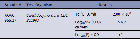
UV-C disinfection using the device achieved a reduction ≥6.7-log10 of C. auris per carrier, exceeding the AOAC 955.17 acceptance criterion of ≥4.7-log10. All test replicates showed a recovered microbial load below the limit of detection (log10(E) < 1) in 100% of cycles (0/25 with detectable recovery; SD = 0). Specifically, even the shortest UV-C cycle demonstrated complete inactivation of a high initial challenge (test concentration: 2.00 × 10⁸ CFU/mL), with a recovered microbial load below the limit of detection (log10(E) < 1), corresponding to a reduction exceeding 6.7 log10 per carrier. The results are presented in Table 2.
Determination of the activity of the cycle of the disinfection process Chronos® against C. auris. Results at the shortest UV-C cycle are presented

Table 2. Long description
A table with three columns and four rows, including a header row. The columns are labeled Standard, Test Organism, and Results. The first row under the header shows the standard AOAC 955.17, the test organism Candidozyma auris CDC B11903, and the results including Tc (CFU/ml) with a value of 2.00 x 10^8, Log10Nw (CFU/carrier) with a value greater than 6.7, and Log10(E) ± SD with a value of less than 1.
TC = Test concentration. Log10Nw = Log10 microbial recovery from untreated control carriers, Log10 (E) = Log10 microbial recovery after UV-C exposure.
These findings demonstrate substantial fungicidal activity under stringent test conditions.
In contrast, existing evidence on chemical high-level disinfectants remains limited, as most published studies, including Rutala et al. (2019), evaluated performance using substantially lower inoculum levels (<104 CFU). Consequently, the ability of chemical disinfectants to consistently achieve >4-log10 reductions under high contamination conditions remains uncertain. Reference Rutala, Kanamori, Gergen, Sickbert-Bennett and Weber7
Discussion
This study demonstrated that UV-C disinfection using the UV-C device achieves HLD efficacy against C. auris, exceeding standardized AOAC requirements. These findings demonstrate that UV-C may represent an effective alternative to conventional chemical disinfection methods for endocavitary ultrasound probes.
Previous studies have reported that the efficacy of UV-C against C. auris may vary according to exposure time, distance from the UV source, and strain characteristics. De Groot et al demonstrated that increasing exposure duration and reducing distance improved fungal inactivation. Our results extend these observations by demonstrating complete inactivation under AOAC-standardized high inoculum conditions. Reference de Groot, Chowdhary, Meis and Voss8
Beyond efficacy, UV-C offers practical advantages, including rapid processing time, absence of chemical residues, and reduced risk of user-related variability. These characteristics could improve workflow efficiency and limit device degradation associated with repeated exposure to chemical agents. Reference Koibuchi, Fujii and Kotani9 In addition, previous studies have shown that UV-C disinfection demonstrates strong sporicidal activity compared with chemical sterilants. Reference Pineau, Radix and Weber10
In contrast, published evidence suggests that the efficacy of chemical disinfectants against C. auris appears to be limited, particularly under high contamination conditions. Reference Rutala, Kanamori, Gergen, Sickbert-Bennett and Weber7 However, direct comparisons with chemical HLD approaches under standardized high-load conditions remain limited, highlighting a need for further investigation.
This study has several limitations, including the use of controlled laboratory conditions, flat carriers instead of complete ultrasound probes, a single C. auris strain (CDC B11903, clade IV), and the absence of clinical outcome data. Consequently, the efficacy of UV-C disinfection against isolates from other clades requires further confirmation.
Further studies are required to confirm these findings in real-world clinical settings and across multiple strains.
Overall, these findings support the potential of UV-C disinfection as an effective approach for limiting transmission of C. auris associated with the reuse of ultrasound probes in clinical practice.
In addition to its demonstrated antimicrobial efficacy, UV-C disinfection offers several operational advantages, including rapid processing, the absence of chemical residues, and no requirement for postdisinfection rinsing. Together, these features highlight its value as an efficient and environmentally responsible option for HLD.
Data availability statement
The data supporting the findings of this study are available from the corresponding author upon reasonable request.
Acknowledgements
The authors thank Eurofins Biotech Germande (Aix-en-Provence, France) for conducting the independent microbiological analyses included in this study.
Germitec was involved in study design, funding, and manuscript preparation.
Financial support
This study, including microbiological testing, data generation, and manuscript preparation, was funded by Germitec (Bordeaux, France).
Competing interests
C.C and Prof. D.J.W serve as consultants for Germitec and receive consulting fees from the company. Dr F.P is an employee of Germitec. The authors report no other conflicts of interest relevant to this article.
Eurofins Biotech Germande (Aix-en-Provence, France), the independent laboratory that performed the microbiological analyses, received funding from Germitec for conducting these analyses. The authors report no other conflicts of interest relevant to this article.


 Open access

