Introduction
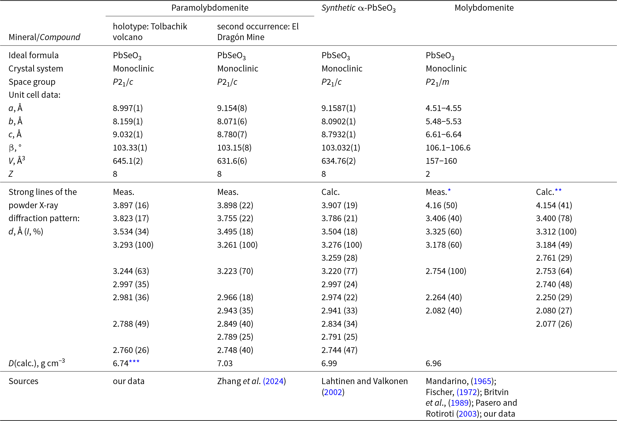
A simple lead(II) selenite has been known in nature for almost one and a half centuries. In 1882 it was described as the new mineral molybdomenite from the oxidation zone of selenide-bearing ores at Cerro de Cacheuta, Cacheuta District, Mendoza Province, Argentina (Bertrand, Reference Bertrand1882). However, this mineral species was only clearly defined as PbSe4+O3 significantly later (Mandarino, Reference Mandarino1965). The crystal structure of molybdomenite was studied by Fischer (Reference Fischer1972) and Pasero and Rotiroti (Reference Pasero and Rotiroti2003) who found the monoclinic space group P21/m for the mineral and unit-cell parameters as reported in Table 1. The synthetic analogue of molybdomenite has also been known for a while. Its crystal structure was first solved by Popovkin et al. (Reference Popovkin, Cheremisinov and Simanov1963) and later refined by Koskellina and Valkonen (Reference Koskellina and Valkonen1977). In the beginning of 21st Century, another monoclinic, with space group P21/c, modification of PbSeO3 was synthesized, structurally characterized and designated as α-PbSeO3 which meant the ‘old’, molybdomenite-type modification was named β-PbSeO3 (Lahtinen and Valkonen, Reference Lahtinen and Valkonen2002).
Comparative data for paramolybdomenite, its synthetic analogue α-PbSeO3 and molybdomenite

Table 1 Long description
Comparative crystallographic and powder X-ray diffraction data are given for paramolybdomenite from two localities, a synthetic alpha lead selenite analogue, and molybdomenite. All four entries list the same ideal chemical formula and a monoclinic crystal system. Paramolybdomenite from both localities and the synthetic analogue share space group P21/c, with closely matching unit-cell edges near nine, eight, and nine angstroms and beta angles near one hundred three degrees; their unit-cell volumes cluster around about six hundred thirty to six hundred forty five cubic angstroms, with eight formula units per cell. Molybdomenite is monoclinic but uses space group P21/m, has much smaller unit-cell edges around four and a half, five and a half, and six and a half angstroms, a beta angle around one hundred six degrees, a unit-cell volume around one hundred fifty seven to one hundred sixty cubic angstroms, and two formula units per cell. Strong powder diffraction spacings for the P21/c materials include prominent lines near three point nine, three point eight, three point five, and a strongest line near three point two nine angstroms, whereas molybdomenite shows its strongest measured line near two point seven five angstroms and also strong lines near four point one six and three point three three angstroms. Calculated densities are similar across entries, roughly about six point seven to seven point zero grams per cubic centimeter, with one paramolybdomenite value noted as lower due to compositional admixture.
* after Britvin et al. (Reference Britvin, Burakov, Nikitin and Bogdanova1989); **calculated based on structure data by Fischer (Reference Fischer1972): JCPDS-ICDD, card #71-0831; ***density is relatively low due to significant S admixture (see composition table below).
A natural analogue of the α-PbSeO3 synthesized by Lahtinen and Valkonen (Reference Lahtinen and Valkonen2002) was discovered by us in the weathering zone of paleo-fumarolic deposits at the Tolbachik volcano, Kamchatka, Russia. In June 2023, the Commission on New Minerals, Nomenclature and Classification of the International Mineralogical Association (IMA-CNMNC) approved it, based on our proposal (IMA2023-025), as a new mineral species. It was named paramolybdomenite (Cyrillic: парамолибдоменит) due to the dimorphism with molybdomenite, for which the name originates from the Greek μόλυβδος for ‘lead’ and μήυη for ‘moon’, because the chemical element selenium is named for the moon. The IMA-accepted symbol of paramolybdomenite is Pmdm (Pekov et al., Reference Pekov, Britvin, Agakhanov, Vigasina, Turchkova and Zhegunov2023). Two fragments of its holotype specimen are deposited in the systematic collection of the Fersman Mineralogical Museum, Moscow with the catalogue numbers 98314 (polished sample from which chemical and reflectance data were obtained) and 98315 (free crystal).
In 2024, the same mineral was characterized as ‘molybdomenite-P21/c, a natural counterpart of synthetic α-PbSeO3’ from the oxidation zone of a selenide ore at the El Dragón Mine, Potosí, Bolivia. Its electron-microprobe analysis (EMPA), Raman spectrum, single-crystal and powder X-ray diffraction (XRD) data and crystal structure were reported (Zhang et al., Reference Zhang, Yang and Gibbs2024). It was surprising that this paper, received by The Canadian Journal of Mineralogy and Petrology 26 January 2024 and accepted for publication after revision 29 March 2024, contains neither any mention of paramolybdomenite nor a citation for the IMA CNMNC Newsletter No. 74 announcing this new mineral (Pekov et al., Reference Pekov, Britvin, Agakhanov, Vigasina, Turchkova and Zhegunov2023), which was published in Mineralogical Magazine in autumn 2023 (published online 29 July 2023).
In the present article, we describe paramolybdomenite from the type locality.
Occurrence and general appearance
The Tolbachik volcano is a well-known mineral locality which became famous originally due to the discoveries of more than 150 new mineral species found in deposits of active, hot oxidizing-type fumaroles (Pekov et al., Reference Pekov, Agakhanov, Zubkova, Koshlyakova, Shchipalkina, Sandalov, Yapaskurt, Turchkova and Sidorov2020 and references therein). Paramolybdomenite is different in origin: it is a supergene mineral formed in the weathering zone of the deposits of volcanic fumaroles extinct for a long time.
Paramolybdomenite was found at the Western paleo-fumarole field located at the western slope of Mountain 1004 (Tolbachik volcano, Kamchatka peninsula, Far-Eastern Region, Russia). Mountain 1004 is one of the numerous ancient monogenetic volcanoes belonging to the huge Tolbachik volcanic complex. This scoria cone is situated 20 km SSW of the active volcano Ploskiy Tolbachik and 2 km SSW of the Second scoria cone of the Northern Breakthrough of the Great Tolbachik Fissure Eruption 1975–1976, a famous locality of the majority of Tolbachik new minerals. Like scoria cones of the Northern Breakthrough of the Great Tolbachik Fissure Eruption, Mountain 1004 was formed as a result of a basalt eruption of the Plinian type, however, not recently but approximately two thousand years ago (Fedotov and Markhinin, Reference Fedotov and Markhinin1983). All fumaroles at Mountain 1004 are extinct, and the weathering zone was formed on the paleo-fumarolic fields to date. Like the well-studied active fumaroles at young scoria cones born by the Great Tolbachik Fissure Eruption (Vergasova and Filatov, Reference Vergasova and Filatov2016; Pekov et al., Reference Pekov, Agakhanov, Zubkova, Koshlyakova, Shchipalkina, Sandalov, Yapaskurt, Turchkova and Sidorov2020), ancient fumaroles at Mountain 1004 belonged to the oxidizing type. By analogy with these contemporary Tolbachik fumaroles, we believe that they produced a rich ‘ore’ sublimate mineralization including oxysalts (sulfates, selenites, vanadates, arsenates, molybdates, etc.), halides (chlorides and fluorides) and oxides with Cu and Pb as the major ‘ore’ components. These sublimate minerals are mainly unstable under atmospheric conditions and easily decompose with the release of acidic (H2SO4, HCl, HF) solutions which reacted with host rocks and mobilized their constituents, initially, Si, Al and Ca. As a result, during a period as short (in geological scales) as two thousand years, a rich weathering zoneFootnote 1, with an outstanding chemical and species diversity of supergene minerals, was formed here.
The first data on the mineralogy of paleo-fumarole fields of Mountain 1004 were published by Naboko and Glavatskikh (Reference Naboko and Glavatskikh1992) and Serafimova et al. (Reference Serafimova, Semenova and Sulimova1994), however, systematic studies of the supergene mineralization at these localities have been started by us since 2016. The Western paleo-fumarole field became the co-type locality of fluorpyromorphite (Kasatkin et al., Reference Kasatkin, Pekov, Škoda, Chukanov, Nestola, Agakhanov, Kuznetsov, Koshlyakova, Plášil and Britvin2023), and we found have found numerous supergene minerals here, including paramolybdomenite. The latter is associated with chrysocolla, allophane, atacamite, opal, gypsum, anglesite, linarite, caledonite, brochantite, spangolite, connellite, munakataite, cerussite, hydrocerussite, wulfenite, vanadinite, mottramite, volborthite, pyromorphite, fluorpyromorphite, fluorphosphohedyphane, phoenicochroite, vauquelinite, murdochite, fluorite, gearksutite, and iron hydroxides (limonite). Relict fumarolic (sublimate) minerals in this assemblage are tenorite, hematite, magnesioferrite, sanidine, diopside and fluorophlogopite.
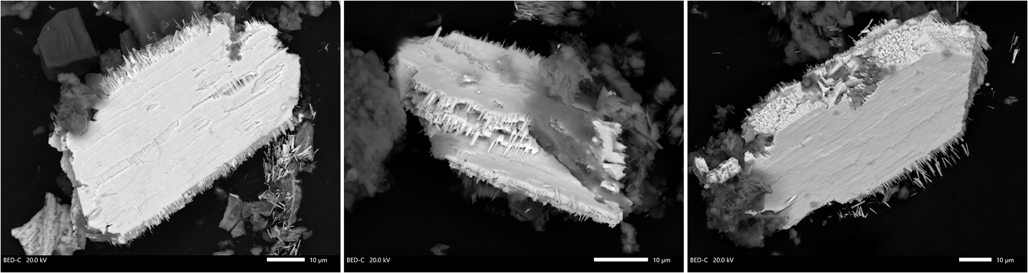
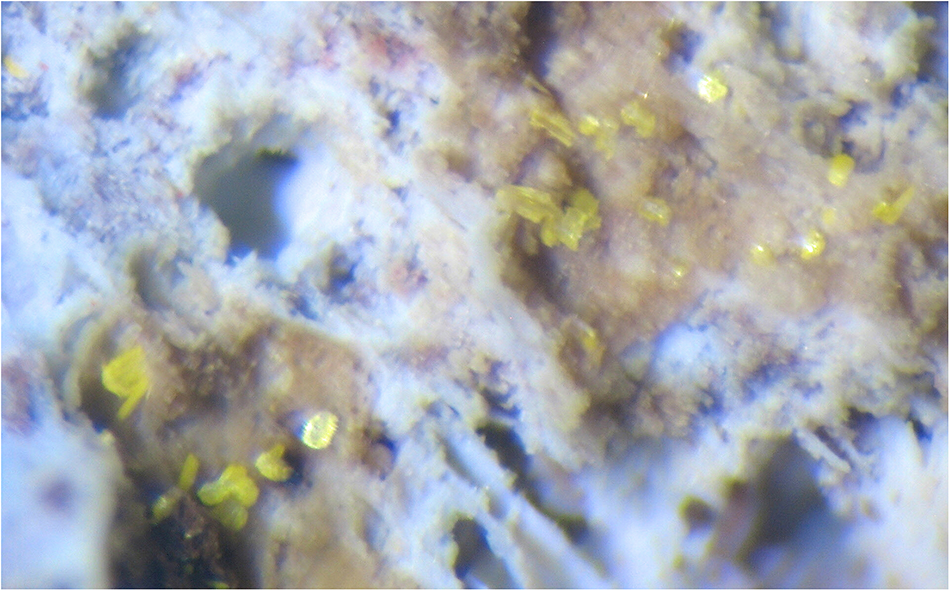
Paramolybdomenite occurs in cavities as tabular to lamellar, usually crude, crystals up to 0.1 × 0.05 × 0.01 mm. They are flattened on (100) and elongated on [010]. The major crystal form is {100}, the lateral faces are not indexed. Paramolybdomenite crystals are isolated or, rarely, combined in open-work clusters up to 0.15 mm across (Figs 1 and 2). The crystals demonstrate different signs of the skeletal growth: they are surrounded by fringed aggregates of acicular or columnar tiny subindividuals grown on lateral faces and are typically cavernous and ‘waffle-like’ (Fig. 1).
Three scanning electron images in back-scattered electron mode of paramolybdomenite crystals (white) with overgrowing allophane particles (grey to dark grey). These crystals were extracted from the holotype specimen.

Bright yellow crystals (up to 0.08 mm in size) of paramolybdomenite on white to brownish crust consisting mainly of allophane and opal. The holotype specimen. FOV width: 1.75 mm. Photo: I.V. Pekov and A.V. Kasatkin.

In another part of the Western paleo-fumarole field an ‘ordinary’ molybdomenite was found. It was first reported here by Serafimova et al. (Reference Serafimova, Semenova and Sulimova1994) and additionally studied by us. Molybdomenite also occurs in the weathering zone of fumarole deposits. It forms transparent lemon-yellow flattened prismatic, board-like, sometimes curved crystals up to 1 mm long and their clusters. Molybdomenite overgrows tenorite (relict of fumarolic, sublimate mineral association) and is associated with opal, chrysocolla, linarite, anglesite and pyromorphite.
Physical properties and optical data
Paramolybdomenite is transparent, bright yellow, with yellowish streak and adamantine lustre. It is brittle. Cleavage or parting was not observed. The fracture is uneven (observed under the scanning electron microscope). The density calculated using the empirical formula and unit-cell volume found from the single-crystal XRD data is 6.743 g cm–3.
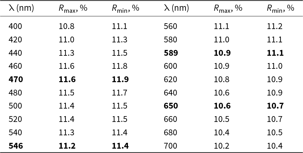
The porosity of paramolybdomenite crystals (Fig. 1) and high values of refractive indices prevented its optical study in transmitted light. Under the microscope in reflected light, paramolybdomenite is grey, pleochroism was not observed. Bireflectance is very weak. The mineral is strongly anisotropic, with colour change from brownish-yellow to dark brown. Internal reflections are bright yellow. The reflectance values measured in air by means of the Opton UMSP-50 microspectrophotometer using the No. 545 Zeiss SiC standard are reported in Table 2.
The reflectance data of paramolybdomenite

Table 2 Long description
Reflectance measurements for paramolybdomenite are listed by wavelength from 400 to 700 nanometers, giving maximum and minimum reflectance percentages at each wavelength. Values are tightly clustered near 11 percent overall. The highest reflectance occurs around 460 to 470 nanometers, with maximum about 11.6 percent and minimum about 11.8 to 11.9 percent. From about 500 nanometers onward, both maximum and minimum reflectance slowly decrease, reaching about 10.2 percent maximum and 10.4 percent minimum at 700 nanometers. The spread between maximum and minimum at each wavelength is small, generally a few tenths of a percent.
The values for wavelengths (λ) recommended by the IMA Commission on Ore Mineralogy are marked in boldtype.
Raman spectroscopy
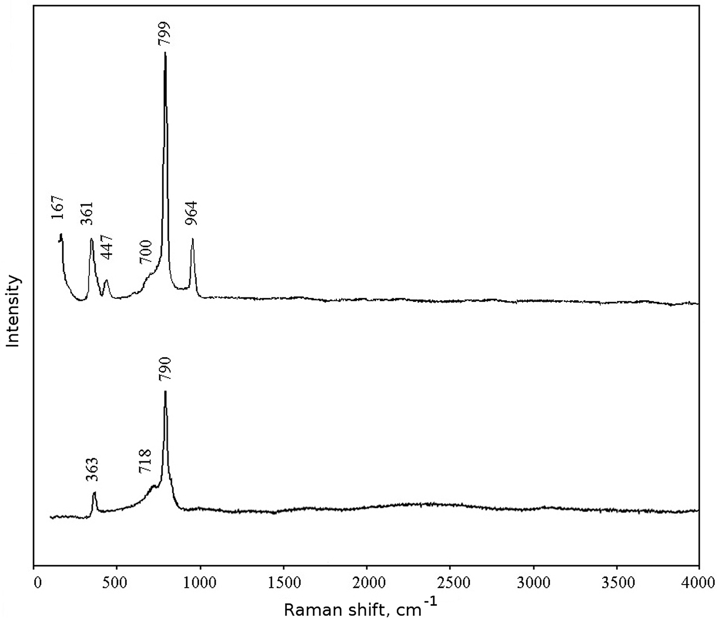
The Raman spectra of paramolybdomenite and its dimorph molybdomenite are given in Fig. 3. The spectrum of paramolybdomenite was obtained for a randomly orientated crystal. The spectrum of molybdomenite was obtained for a fine-grained (powdery) sample; this sample, from the Svetloe selenide occurrence, South Karelia, Russia, chemically corresponding to the end-member PbSeO3, was described by Britvin et al. (Reference Britvin, Burakov, Nikitin and Bogdanova1989). Both spectra were recorded using an EnSpectr R532 spectrometer (Dept. of Mineralogy, Lomonosov Moscow State University, Moscow, Russia) with a green laser (532 nm) at room temperature. The spectrometer is equipped with a CCD detector. The calibration of the device was carried out along a 520 cm−1 crystalline silicon line. The output power of the laser beam was ∼6 mW. The diameter of the laser spot on the sample was ∼7 μm. The back-scattered Raman signal was collected with 40× objective. Signal acquisition time for a single scan was 2 s for sample of paramolybdomenite and 1 s for molybdomenite. For molybdomenite the signal was averaged over 200 scans and 100 scans, respectively. The spectrum was processed using the EnSpectr expert mode program in the range from 150 to 4000 cm–1 with the use of a holographic diffraction grating with 1800 lines per millimetre and a resolution of 6 cm–1.
The Raman spectra of paramolybdomenite (upper) from this work and molybdomenite (lower) with composition corresponding to the end-member PbSeO3 from the Svetloe selenide occurrence, South Karelia, Russia (the specimen described by Britvin et al., Reference Britvin, Burakov, Nikitin and Bogdanova1989).

The spectral bands were assigned according to the general data provided in the reference book by Nakamoto (Reference Nakamoto1986).
In the Raman spectrum of paramolybdomenite, bands at 964 and 447 cm–1 correspond to symmetric stretching and antisymmetric bending vibrations, respectively, of (SO4)2– groups which partially substitute (SeO3)2– groups (see below). The most intense band at 799 cm–1 corresponds to symmetric stretching vibrations and the shoulder near 700 cm–1 corresponds to antisymmetric stretching vibrations of the (SeO3)2– anion. Antisymmetric bending vibrations of (SeO3)2– groups give the band at 361 cm–1.
The Raman spectrum of molybdomenite contains bands in the region 800–200 cm–1 corresponding to symmetric (790 cm–1) and antisymmetric (718 cm–1) stretching and antisymmetric bending (363 cm–1) vibrations of (SeO3)2– groups.
The absence of bands with frequencies higher than 1000 cm–1 indicates the absence of groups with O–H, C–H, C–O, N–H, N–O and B–O bonds in both minerals.
Chemical data
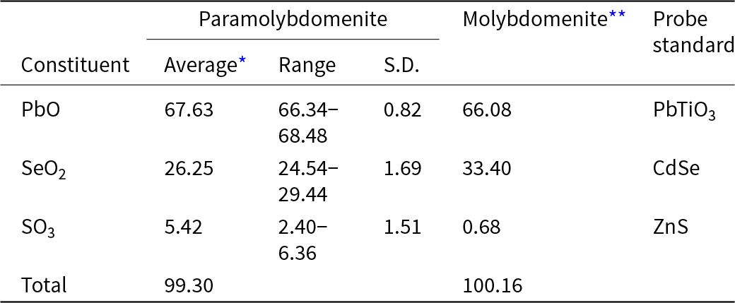
The chemical composition of paramolybdomenite and molybdomenite from Mountain 1004 was studied by electron microprobe using a Jeol IT-500 scanning electron microscope equipped with an X-Max-50 energy-dispersive spectrometer in the Laboratory of Analytical Techniques of High Spatial Resolution, Dept. of Petrology, Lomonosov Moscow State University. The operating conditions were: an acceleration voltage 20 kV, a beam current 2 nA, and the electron beam diameter 3 μm. We used EDS mode because paramolybdomenite is damaged under the electron beam with the current needed for WDS; the correctness of the determination of S content due to the overlap of analytical lines of S and Pb in EDS has been controlled by WDS data which showed the same atomic proportions Pb:Se:S in the samples analysed. The chemical data for both minerals are given in Table 3. The contents of other elements with atomic numbers higher than C are below detection limits.
Chemical composition (wt.%) of paramolybdomenite and molybdomenite from the Western paleo-fumarole field of Mountain 1004, Tolbachik volcano

Table 3 Long description
Chemical composition in weight percent is reported for paramolybdomenite and for a representative molybdomenite analysis, listing PbO, SeO2, SO3, and totals. Paramolybdomenite averages 67.63 wt percent PbO with a range from 66.34 to 68.48 and a standard deviation of 0.82. Its SeO2 averages 26.25 wt percent, ranging from 24.54 to 29.44, with a standard deviation of 1.69. SO3 in paramolybdomenite averages 5.42 wt percent, ranges from 2.40 to 6.36, and has a standard deviation of 1.51. Molybdomenite has similar PbO at 66.08 wt percent but higher SeO2 at 33.40 wt percent and much lower SO3 at 0.68 wt percent. Totals are near complete for both minerals, at 99.30 wt percent for paramolybdomenite and 100.16 wt percent for molybdomenite. Probe standards used were PbTiO3 for PbO, CdSe for SeO2, and ZnS for SO3, and only paramolybdomenite includes variability statistics across multiple spots.
* For six spot analyses. **Typical, representative analysis. S.D. – standard deviation
The empirical formula of paramolybdomenite was calculated on the basis of 2 Pb+Se+S atoms per formula unit. We avoid the anionic basis of formula calculation because of the partial substitution (SO4)2– → (SeO3)2–: see the crystal structure data below. The empirical formula is Pb1.00Se4+0.78S6+0.22O3.22 and the simplified formula can be written as Pb[(SeO3),(SO4)]. The idealised, end-member formula PbSeO3 requires PbO 66.79, SeO2 33.21, total 100 wt.%.
The character of Se and S distribution in crystals of paramolybdomenite, observed under the scanning electron microscope in back-scattered electron mode, is spotty, with no sign of growth zonation or other regularities.
The empirical formula of molybdomenite from the Western paleo-fumarole field of Mountain 1004, calculated on the same basis, is Pb0.98Se4+0.99S6+0.03O3.05.
X-ray crystallography and crystal structure determination
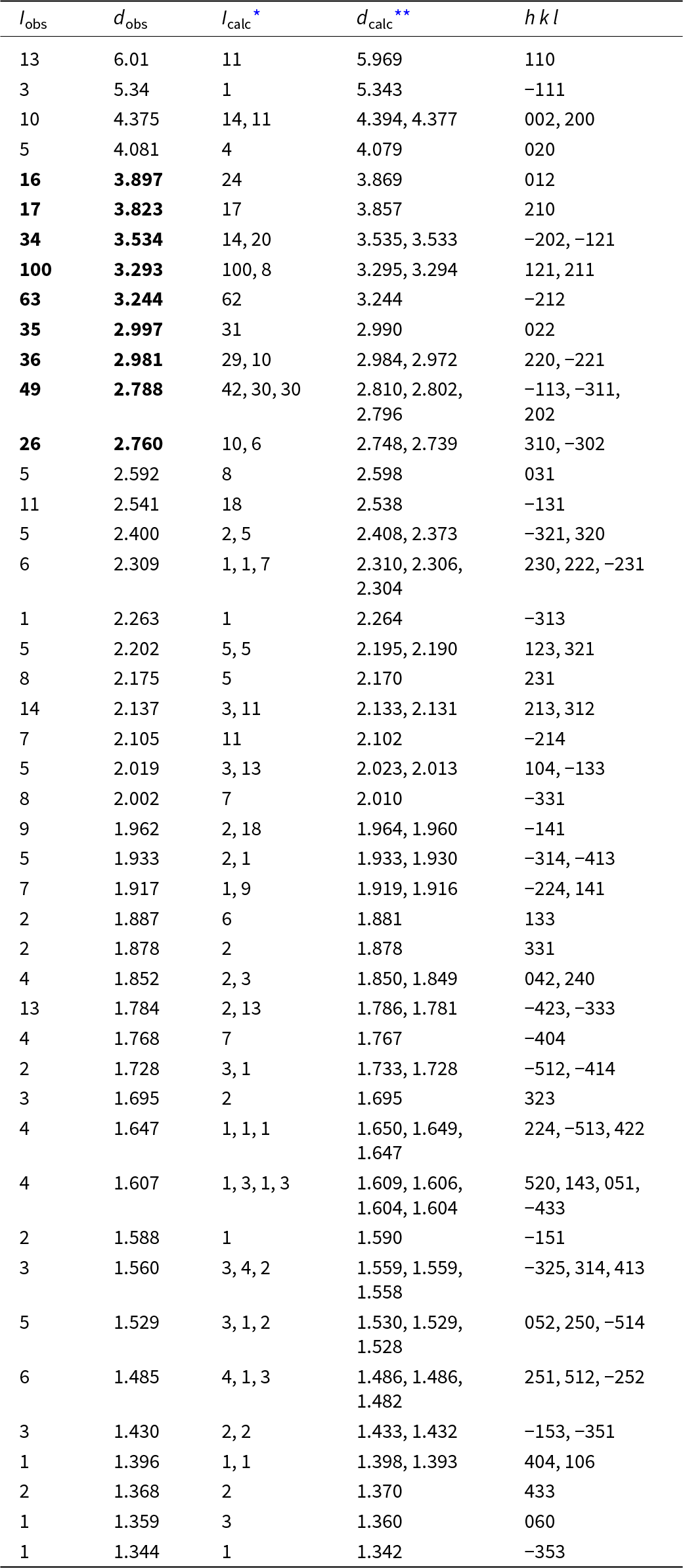
Powder XRD data for paramolybdomenite (Table 4) were collected in Debye-Scherrer geometry by means of a Rigaku RAXIS Rapid II diffractometer equipped with a curved (semi-cylindrical) imaging plate detector (r = 127.4 mm), using CoKα radiation (λ = 1.79021 Å) generated by a rotating anode (40 kV, 15 μA) with microfocus optics; exposure time was 15 min. The imaging plate was calibrated against a NIST Si standard. The image-to-profile data processing was performed using osc2xrd software (Britvin et al., Reference Britvin, Dolivo-Dobrovolsky and Krzhizhanovskaya2017). Parameters of the monoclinic unit cell refined from the powder data are: a = 8.983(5), b = 8.161(2), c = 9.055(5) Å, β = 103.42(3)° and V = 645.7(8) Å3.
Powder X-ray diffraction data (d in Å) of paramolybdomenite

Table 4 Long description
Powder X-ray diffraction peak list for paramolybdomenite, giving observed intensity and d spacing alongside calculated intensity and d spacing, with Miller indices for each reflection. The strongest observed reflection is at dobs 3.293 Å with Iobs 100, matching calculated lines at about 3.295 and 3.294 Å and indexed as 121 and 211. Other prominent observed peaks include 3.244 Å (Iobs 63, indexed −212), 2.788 Å (Iobs 49, indexed −113, −311, and 202), 2.981 Å (Iobs 36, indexed 220 and −221), 2.997 Å (Iobs 35, indexed 022), and 3.534 Å (Iobs 34, indexed −202 and −121). Several rows show multiple calculated reflections close to one observed d value, indicating overlapping peaks in the powder pattern (for example near 4.375 Å, 3.534 Å, 3.293 Å, and 2.309 Å). Across the list, observed and calculated d spacings generally agree closely, supporting the indexing, while many low-intensity lines occur from about 2.6 Å down to about 1.34 Å.
* For the calculated pattern (calculated using STOE WinXPOW, version 2.22), only reflections with intensities ≥1 are given; **for the unit-cell parameters obtained from single-crystal data.
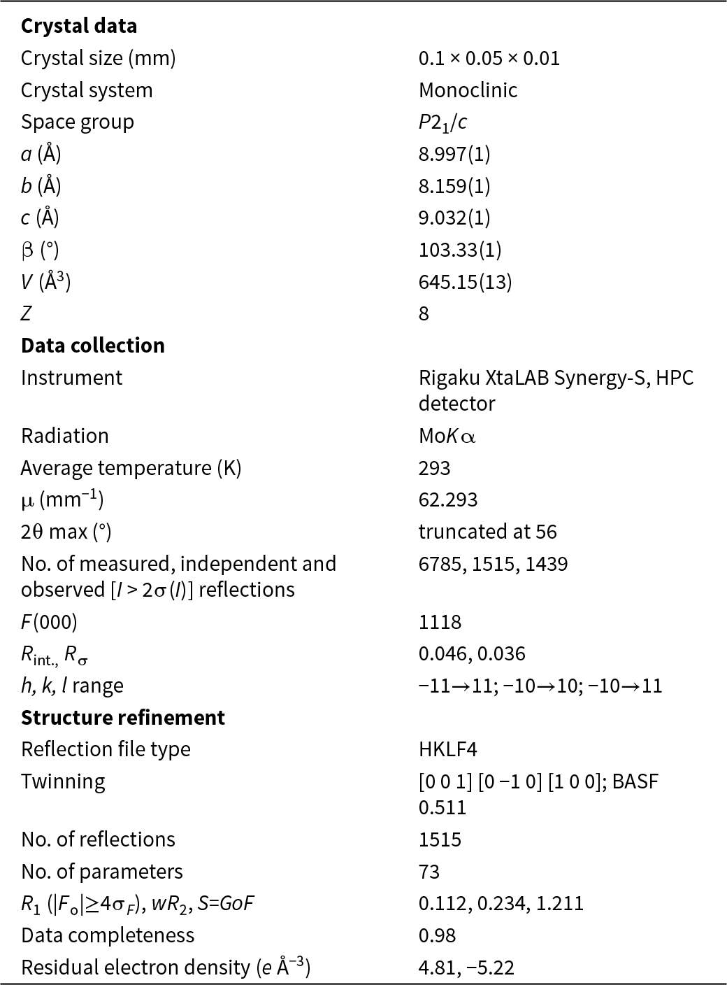
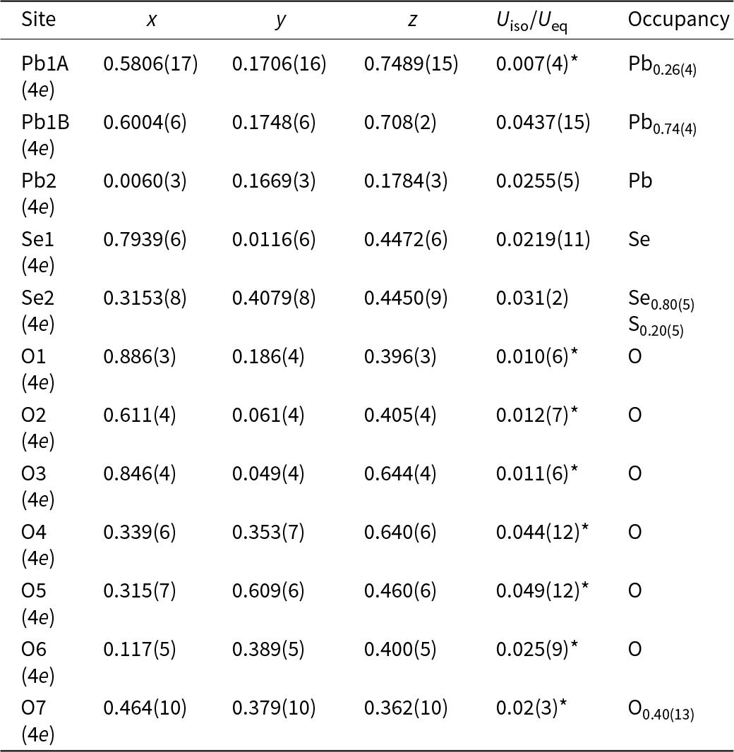
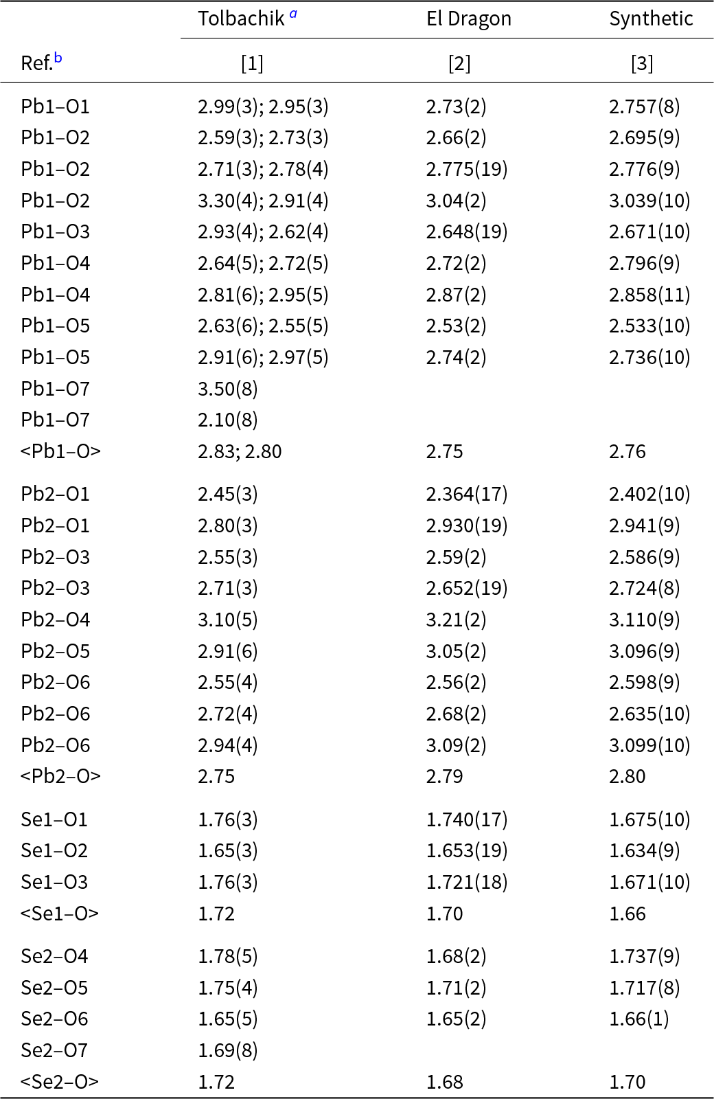
Single-crystal XRD studies of paramolybdomenite were carried out at room temperature using a Rigaku-Oxford diffraction XtaLAB Synergy-S diffractometer equipped with a microfocus X-ray tube and HyPix-6000 hybrid photon counting detector (MoKα radiation). The dataset was collected and processed by means of CrysAlis Pro software (Rigaku Oxford Diffraction, 2022). The structure was solved using SHELXT program (Sheldrick, Reference Sheldrick2015a) on the basis of 1439 independent reflections with I > 2σ(I), followed by refinement with the SHELXL software (Sheldrick, Reference Sheldrick2015b) operated under the Olex2 v.1.4 graphical user environment (Dolomanov et al., Reference Dolomanov, Bourhis, Gildea, Howard and Puschmann2009). The crystal data and the experimental details are presented in Table 5, atom coordinates and displacement parameters in Tables 6 and 7, and selected interatomic bond lengths in Table 8. The crystallographic information file has been deposited and is available as Supplementary material (see below). The data presented in the tables were collected on the best crystal out of four suitable for the single-crystal study. The poor quality of even that best crystal imposed limitations on the quality of the collected single-crystal XRD dataset and consequently, on the quality of the structure refinement. In particular, oxygen atom sites, as well as the Pb1A site (Table 6) could only be refined with isotropic displacement parameters (Table 6), with significant phantoms of residual electron density (4.8 e Å–3) nearby the Pb2 site (Table 5). Due to the relatively low quality of the data, the results of the structure refinement should be treated as a structural model. However, it can be seen (Table 8) that they are, in general, very consistent with the data on paramolybdomenite and synthetic α-PbSeO3. The occupancy of the mixed Se2/S2 site was freely refined based on the scattering atomic factors of Se and S. The occupancy of the partially vacant O7 site was also freely refined based on the scattering factor of neutral oxygen atom.
Crystal parameters, data collection information and structure refinement details for paramolybdomenite

Table 5 Long description
Crystal, data-collection, and refinement parameters are reported for paramolybdomenite from single-crystal X-ray diffraction. The crystal is monoclinic in space group P21/c with unit-cell lengths a 8.997 angstroms, b 8.159 angstroms, c 9.032 angstroms, beta 103.33 degrees, volume 645.15 cubic angstroms, and Z 8. Data were collected on a Rigaku XtaLAB Synergy-S with Mo K alpha radiation at an average temperature of 293 kelvin, with a absorption coefficient of 62.293 per millimeter and a two-theta limit of 56 degrees. A total of 6785 reflections were measured, yielding 1515 independent and 1439 observed reflections, with Rint 0.046 and Rsigma 0.036; index ranges span h minus 11 to 11, k minus 10 to 10, and l minus 10 to 11. Refinement used an HKLF4 reflection file and included twinning with a BASF value of 0.511, refining 73 parameters against 1515 reflections. Fit indicators are R1 0.112 for strong reflections, weighted R2 0.234, and goodness of fit 1.211, with data completeness 0.98. Residual electron density ranges from 4.81 to minus 5.22 electrons per cubic angstrom.
Weighting scheme: w=1/[σ2(F o2) + (330.8148P)2] where P = (F o2 + 2F c2)/3
Fractional atomic coordinates and isotropic or equivalent isotropic displacement parameters (Å2) for paramolybdomenite

Table 6 Long description
Fractional atomic coordinates are given for Pb, Se, S, and O sites in paramolybdomenite, along with isotropic or equivalent isotropic displacement values and site occupancies. The Pb1 position is split into two components: Pb1A at x 0.5806, y 0.1706, z 0.7489 with occupancy Pb 0.26, and Pb1B at x 0.6004, y 0.1748, z 0.708 with occupancy Pb 0.74. Pb2 is fully occupied Pb at x 0.0060, y 0.1669, z 0.1784. Se1 is fully occupied Se at x 0.7939, y 0.0116, z 0.4472, while Se2 shows mixed occupancy with Se 0.80 and S 0.20 at x 0.3153, y 0.4079, z 0.4450. Oxygen sites O1 through O6 are fully occupied, and O7 is partially occupied at 0.40. Displacement values vary notably, with Pb1B higher than Pb1A, and several oxygen sites (especially O4 and O5) showing comparatively larger displacement values than O1 to O3.
*U iso
Anisotropic displacement parameters (Å2) for Pb and Se sites in the structure of paramolybdomenite

Table 7 Long description
Anisotropic displacement parameters for four atomic sites are listed as U11, U22, U33 and U12, U13, and U23, with values reported with uncertainties in parentheses. Pb1B shows the greatest directional motion, especially along U33 at 0.075(4), compared with U11 0.037(2) and U22 0.0272(17). Pb2 is lower overall, with U11 0.0351(12), U22 0.0183(8), and U33 0.0255(11). Se1 is relatively small and nearly isotropic, with U11 and U22 both 0.019 and U33 0.023(3). Se2/S2 is larger than Se1 and more anisotropic, with U33 0.039(4) and U22 0.029(3) exceeding U11 0.016(3). Cross terms are generally modest; Se1 and Se2/S2 have negative U12 values (−0.008(2) and −0.014(3)), while Pb sites have small positive U12 values.
Selected interatomic bond lengths (Å) in the structure of paramolybdomenite and its synthetic counterpart

Table 8 Long description
Interatomic bond lengths in angstroms are listed for Pb1, Pb2, Se1, and Se2 to oxygen atoms, comparing Tolbachik, El Dragon, and a synthetic sample. Pb–O distances span roughly 2.10 to 3.50 Å, whereas Se–O distances are much shorter, about 1.63 to 1.78 Å. Mean Pb1–O is about 2.83 and 2.80 Å for the two Tolbachik Pb1 subpositions, compared with 2.75 Å at El Dragon and 2.76 Å in the synthetic sample. Mean Pb2–O is similar across samples, about 2.75 Å in Tolbachik, 2.79 Å in El Dragon, and 2.80 Å in the synthetic sample. Se1–O means decrease slightly from 1.72 Å in Tolbachik to 1.70 Å in El Dragon and 1.66 Å in the synthetic sample. Se2–O means are 1.72 Å in Tolbachik, 1.68 Å in El Dragon, and 1.70 Å in the synthetic sample, with some Se2–O7 and Pb1–O7 entries missing outside Tolbachik.
a The Pb1 site in the holotype mineral from Tolbachik is split into Pb1A and Pb1B; the data for subpositions are separated by semicolon
b References: [1] This work; [2] Zhang et al. (Reference Zhang, Yang and Gibbs2024); [3] Lahtinen and Valkonen (Reference Lahtinen and Valkonen2002).
A single-crystal XRD study of molybdomenite from the Western paleo-fumarole field of Mountain 1004 was carried out using an Xcalibur S diffractometer equipped with a CCD detector (MoKα radiation) and gave the following parameters of a monoclinic unit cell: a = 4.53(3), b = 5.53(3), c = 6.614(16) Å, β = 106.1(4)° and V = 159(1) Å3.
Discussion
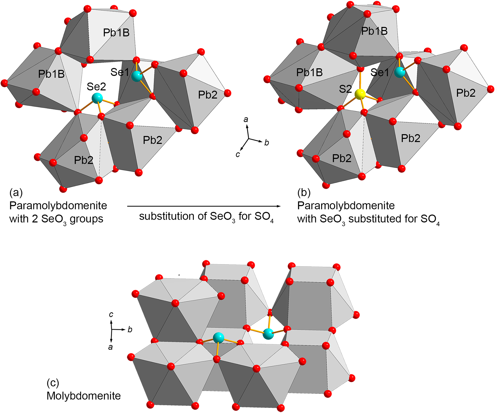
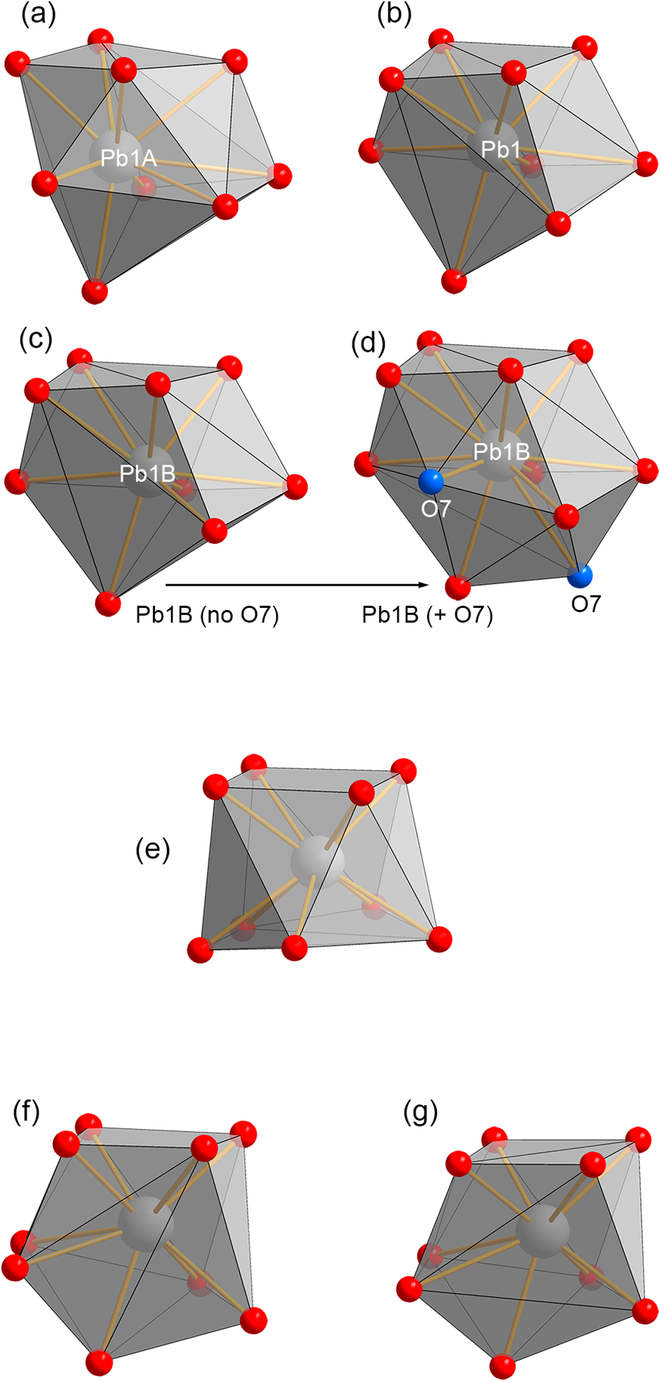
Paramolybdomenite is a natural analogue of α-PbSeO3 (Lahtinen and Valkonen, Reference Lahtinen and Valkonen2002; see Table 1). The structures of paramolybdomenite (Fig. 4a,b) and molybdomenite (Fig. 4c), or α- and β-PbSeO3, respectively (Lahtinen and Valkonen, Reference Lahtinen and Valkonen2002), are essentially different, including the topological aspect. Both modifications contain Pb in nine-fold coordination, which can be described as distorted monocapped square antiprisms [Pb2+O9] (Lahtinen and Valkonen, Reference Lahtinen and Valkonen2002). The [Pb2+O9] polyhedra are cross-connected via the common edges with [Se4+O3] groups. However, the arrangement of the lead-centred and selenite polyhedra in the structure of paramolybdomenite forms a quasi-framework (Fig. 4a), while in molybdomenite, the polyhedra are aligned in a quasi-layered structure (Fig. 4c). Selenium atoms in paramolybdomenite occupy two crystallographically independent sites: the apexes of trigonal-pyramidal [SeO3]2– anions (Table 6). The arrangement of [SeO3] groups relative to [PbO9] polyhedra is principally different in paramolybdomenite (Fig. 4a) and molybdomenite (Fig. 4c). In addition, the geometry of the lead–oxygen polyhedron is substantially different in paramolybdomenite and molybdomenite (Fig. 5). Therefore, these modifications of PbSeO3 cannot be considered as polytypes, polytypoids or topologically-similar polymorphs.
Crystal structures of holotype paramolybdomenite from Tolbachik and molybdomenite. (a) The quasi-framework of paramolybdomenite with Pb1B site devoid of an additional O7 oxygen atom: the case of a pure PbSeO3 structure containing two symmetrically independent [SeO3] groups. (b) The same fragment showing the incorporation of an O7 atom and simultaneous introduction of a sulfur atom, with the appearance of a [SO4] tetrahedron substituting for one of the two [SeO3] groups. (c) The quasi-layered structure of molybdomenite. Note the substantially different arrangement of [SeO3] groups in paramolybdomenite and molybdomenite. Drawn based on our data (a, b) and Pasero and Rotiroti (Reference Pasero and Rotiroti2003) (c). Legend: (a) Se – turquoise balls; S – yellow ones; oxygen – red, Pb-centred polyhedra – grey. Drawn using Diamond v.3 software (Brandenburg, Reference Brandenburg1996).

Figure 4 Long description
The image consists of three images illustrating the crystal structures of paramolybdomenite and molybdomenite. The first image shows paramolybdomenite with two [SeO] groups, featuring a quasi-framework structure. The Pb1B site is highlighted and the arrangement includes Se2 and Se1 atoms. The second image depicts paramolybdomenite with a substitution of [SeO] for [SO], showing the incorporation of a sulfur atom and the appearance of a [SO] tetrahedron. The Pb1B site is again highlighted, with Se1 and sulfur atoms visible. The third image illustrates the quasi-layered structure of molybdomenite, showing a different arrangement of [SeO] groups compared to paramolybdomenite. Each image includes red spheres representing oxygen atoms, grey polyhedra centered on lead atoms, turquoise spheres for selenium and yellow for sulfur. Axes are labeled with ′a′, ′b′ and ′c′ to indicate orientation.
Geometry of the lead-oxygen polyhedra in the crystal structure of holotype paramolybdomenite, synthetic α-PbSeO3 and molybdomenite. (a–d) The oxygen shell around the Pb1 site. (a) Configuration of the Pb1A polyhedron in paramolybdomenite. (b) The Pb1 polyhedron in synthetic α-PbSeO3. (c) The Pb1B oxygen shell in paramolybdomenite with the omitted additional O7 atom. (d) The full coordination shell of Pb1B in paramolybdomenite, including additional O7 oxygen sites that are highlighted by blue. (e) Coordination polyhedron [PbO9] in molybdomenite. (f, g) The Pb1B polyhedra in paramolybdomenite (f) and in synthetic α-PbSeO3 (g). The source of structural data: paramolybdomenite – this work; molybdomenite – Pasero and Rotiroti (Reference Pasero and Rotiroti2003); synthetic α-PbSeO3 – Lahtinen and Valkonen (Reference Lahtinen and Valkonen2002). Drawn using Diamond v.3 software (Brandenburg, Reference Brandenburg1996).

Figure 5 Long description
The diagram consists of seven images labeled a to g, illustrating lead-oxygen polyhedra configurations. Image a shows the Pb1A polyhedron in paramolybdomenite, with red spheres as oxygen atoms and gray faces representing the polyhedral structure. Image b depicts the Pb1 polyhedron in synthetic α-PbSeO, maintaining a similar coordination. Sub-image c presents the Pb1B oxygen shell in paramolybdomenite without the O7 atom, while sub-image d includes the O7 atom, highlighted in blue, indicating a change in coordination geometry. Image e illustrates the [PbO] polyhedron in molybdomenite, showing a distinct arrangement. Images f and g display paired Pb1B polyhedra in paramolybdomenite and synthetic α-PbSeO, respectively, highlighting differences in coordination environments. The diagram uses a ball-and-stick style with central lead atoms and bonds connecting to oxygen atoms.
The structural differences cause the principal difference in the unit-cell metrics and consequently, in powder XRD patterns of paramolybdomenite and molybdomenite (Table 1).
Unlike the synthetic counterpart and the mineral from the El Dragón Mine in Bolivia (so-called ‘molybdomenite-P21/c’, Zhang et al., Reference Zhang, Yang and Gibbs2024: the second occurrence of paramolybdomenite), both corresponding to the ideal, end-member formula, the holotype paramolybdomenite from Tolbachik volcano contains a significant amount of the sulfate anion (SO4)2–, which partially substitutes for selenite (SeO3)2– in one of the two structural positions, Se2/S2 (Table 6, Fig. 4, 5). This substitution, although subordinate, results in the appearance of two distinctive structural features of holotype paramolybdomenite relative to that of ideal α-PbSeO3 (Lahtinen and Valkonen, Reference Lahtinen and Valkonen2002). First, it is a splitting of the Pb1 site of α-PbSeO3 (Lahtinen and Valkonen, Reference Lahtinen and Valkonen2002) into Pb1A and Pb1B subsites (Table 6) separated by 0.45 Å. The Pb1B site corresponds to Pb1 in the ideal, undisturbed α-PbSeO3 structure (Lahtinen and Valkonen, Reference Lahtinen and Valkonen2002). The Pb1A site is shifted away relative to the ideal Pb1B position. The next distinguishing feature is the appearance of an ‘additional’, incompletely populated O7 site (Table 6, Fig. 4b, 5c,d) which is absent in pure α-PbSeO3. The incorporation of the O7 oxygen atom increases the coordination number of Pb1B from 9 to 11 (Fig. 5d). It is even more important that the introduction of the O7 site, which is located closely to the sulfur position, allows the appearance of a new, additional S2–O7 bond, thereby completing the tetrahedral oxygen shell around the sulfur atom: the emergence of the (SO4)2– anion (Fig. 4b). In the case of the pair Se2–Pb1B (with no O7 site), the absence of the Se2–O7 bond is compensated for by the presence of a lone electron pair of (SeO3)2– (Fig. 4a), being identical to that in the ideal α-PbSeO3 (Lahtinen and Valkonen, Reference Lahtinen and Valkonen2002). Therefore, in the holotype paramolybdomenite from Tolbachik, one can observe a rare type of substitution of Se4+ for S6+ [homovalent substitution of trigonal pyramidal selenite ion (SeO3)2– for the tetrahedral sulfate ion (SO4)2–], accompanied by the emergence of a new, low-occupied oxygen site (O7, freely refined site occupation factor = 0.2) which substitutes the lone electron pair of Se4+ according to the scheme [::SeO3] (selenite) → [OSO3] (sulfate). The presence of the partially occupied O7 site with the S2–O7 distance equal to 1.69 Å (Table 8) clearly confirms, together with the Raman spectrum (Fig. 3), the presence of sulfur in the form of sulfate but not sulfite anion in paramolybdomenite. The same type of partial substitution of (SeO3)2– for (SO4)2– was reported in munakataite, ideally Pb2Cu2(Se4+O3)(SO4)(OH)4, an ordered selenite–sulfate analogue of linarite PbCu(SO4)(OH)2 (Matsubara et al., Reference Matsubara, Mouri, Miyawaki, Yokoyama and Nakahara2008; Kampf et al., Reference Kampf, Mills and Housley2010).
Paramolybdomenite is represented in Nature, as it is known now, by two chemical varieties: an S-enriched one, containing up to 6.4 wt.% SO3 (the holotype: Tolbachik, Table 3), and S-free, corresponding to the end-member (the second occurrence: El Dragón Mine, Zhang et al., Reference Zhang, Yang and Gibbs2024). They are also structural varieties of the mineral species paramolybdomenite: the sample from the El Dragón Mine is completely identical in terms of structure to synthetic α-PbSeO3 (Lahtinen and Valkonen, Reference Lahtinen and Valkonen2002) whereas the structure of the holotype contains additional partially occupied positions. These varieties of paramolybdomenite can be distinguished easily using Raman spectroscopy: the Raman spectrum of the S-bearing variety contains a distinct band of symmetric stretching vibrations of (SO4)2– groups (at 964 cm–1, Fig. 3) which is, of course, absent in the spectrum of the S-free variety reported by Zhang et al. (Reference Zhang, Yang and Gibbs2024). In the general pattern, the Raman spectra of paramolybdomenite are similar to the spectrum of molybdomenite (except for bands corresponding to sulfate groups), however, positions of bands in the spectra of these dimorphous lead(II) selenites are not identical. Both spectra of molybdomenite, one obtained by us (Fig. 3) and that given in Zhang et al. (Reference Zhang, Yang and Gibbs2024), demonstrate the same position of the strongest band corresponding to symmetric stretching vibrations of the (SeO3)2– anion: 790 cm–1. In the spectrum of S-free paramolybdomenite from the El Dragón Mine the band of symmetric stretching vibrations of (SeO3)2– is split to form a doublet (804 and 788 cm–1, Zhang et al., Reference Zhang, Yang and Gibbs2024) whilst S-enriched paramolybdomenite from Tolbachik gives a singlet with maximum at 799 cm–1 (Fig. 3).
It looks likely that α-PbSeO3 has a stronger affinity for S6+ incorporation (which selectively substitutes Se4+ at the Se2 site) compared to β-PbSeO3. It is indirectly evidenced by the comparison of Se:S ratios in paramolybdomenite and molybdomenite from Tolbachik (Table 3), and should be taken into account in the syntheses and studies of lead(II) selenite in technological applications. In the finite case, the substitution of (SeO3)2– for (SO4)2– could inspire the possibility of the existence of a hypothetical double selenite–sulfate Pb2(SeO3)(SO4) isotypic to paramolybdomenite, Pb2(SeO3)(SeO3), like in the case of the above-mentioned pair munakataite Pb2Cu2(Se4+O3)(SO4)(OH)4 – linarite Pb2Cu2(SO4)(SO4)(OH)4. Also of note is adanite Pb2(Te4+O3)(SO4) (Kampf et al., Reference Kampf, Housley, Rossman, Yang and Downs2020) as the tellurite–sulfate formula-analogue of the above-mentioned hypothetical selenite–sulfate Pb2(Se4+O3)(SO4).
Supplementary material
The supplementary material for this article can be found at https://doi.org/10.1180/mgm.2025.10166.
Acknowledgements
We are grateful to three anonymous reviewers and Associate Editor Irina O. Galuskina for valuable comments. This work was supported by the Russian Science Foundation, grant no. 25-17-00005. The XRD study was carried out at the Center for X-Ray Diffraction Research of the Science Park of St. Petersburg State University within the framework of project 125021702335-5.
Competing interests
The authors declare none.