Introduction
The genus Stirtonia A. L. Sm. comprises inconspicuous, little-studied elements of the lichen flora in the tropics. Only 18 species have been validly described, 12 of which are accepted in the monograph of the genus (Makhija & Patwardhan Reference Makhija and Patwardhan1998). Since then, two additional species have been described from Thailand and Indonesia by Wolseley & Aptroot (Reference Wolseley and Aptroot2009). Fewer than a dozen publications exist that record species of Stirtonia, and these include precursors to the aforementioned monograph (e.g. Makhija & Patwardhan Reference Makhija and Patwardhan1987, Reference Makhija and Patwardhan1994). Furthermore, most of the publications refer only to the foliicolous species that are now classified in the genera, Amazonomyces Bat. andEremothecella Syd. (see Lücking Reference Lücking2008).
Stirtonia is characterized by the absence of a true hamathecium between the round asci, which produce thick-walled trans-septate ascospores. The absence of a hymenium can be best observed at the upper level where the densely anastomosing hyphae peter out and do not form an epihymenium of a different texture or colour. The interascal hyphae are not glued together and are never held in a hymenial gel. The ascigerous areas usually do contain crystals, and sometimes also algal cells. The asci are generally round to ovoid and contain mostly 8 ascospores that are only transversely septate and, at least initially, hyaline. The asci are thus formed in ascigerous areas, not in apothecia in the strict sense. These ascigerous areas can be very similar to the thallus, but can also differ markedly and become convex and/or pruinose or lirelline, and can even be surrounded by a margin of a different colour, for example, when the ascigerous areas break through the thallus. These structures do resemble apothecia and these species are distinguished from Arthonia by the absence of a gelatinous hymenium. The genus Stirtonia was lectotypified by Makhija & Patwardhan (Reference Makhija and Patwardhan1998) with S. obvallata (Stirt.) A. L. Sm.
The genus is close to Cryptothecia Stirt. in its key characters except for the muriform ascospores in that genus. There are no intermediate taxa known and it seems unlikely that the two genera are different developmental stages of the same lineage. However, in the absence of a phylogenetic study, it is not known whether both genera, as currently circumscribed, do or do not present monophyletic groups.
The different species of Stirtonia are mainly characterized by the thallus structure, crystals, colour and iodine reaction, and the colour, crystals, algae, iodine reaction and shape of the ascigerous areas, and ascospore size and septation and the absence or presence of a distinctly enlarged upper cell.
The present paper provides a key to and a short description of all the accepted species. Type specimens have been studied of nearly all species and special attention paid to the following characters: chemistry, organization of the ascigerous areas, presence of calcium oxalate or other crystals in the thallus and ascigerous areas, and iodine reactions of the tissues. Illustrations already given in Makhija & Patwardhan (Reference Makhija and Patwardhan1998). Ascospore drawings and habitus pictures of most species, are not repeated.
Material and Methods
Identification and descriptive work was carried out at the Adviesbureau voor Bryologie en Lichenologie in the Netherlands and at the Natural History Museum, London, using an Olympus SZX7 stereomicroscope and an Olympus BX50 compound microscope with interference contrast, connected to a Nikon Coolpix digital camera. The materials are preserved in ABL, AMH, B, BM, L, NY and hb. Schumm. TLC was performed using the solvent TDA, and in some instances also HEF.
Key to the species of Stirtonia
1 Ascigerous zones mostly rounded, often raised above thallus level; ascospores always with thickened septa and wall, never with an enlarged end cell ... 2
Ascigerous zones linear, branched or anastomosing, usually not raised above thallus level; ascospores not always with thickened septa and wall, sometimes with enlarged end cell ... 7
2(1) Thallus Pd+ yellow, with psoromic acid ... 3
Thallus Pd−, without psoromic acid ... 4
3(2) Ascospores 50–60 μm long; ascigerous zones >1 mm wide ... S. indica
Ascospores 75–87 μm long; ascigerous zones <1 mm wide ... S. psoromica
4(2) Ascospores 36–45 μm long ... S. schummii
Ascospores 45–100 μm long ... 5
5(4) Thallus UV+ white, with 2′-O-methylsuperphyllinic acid; ascospores 85–110 μm long ... S. alboverruca
Thallus UV−, with 2′-O-methylperlatolic acid, without substances or with terpenoids; ascospores 45–100 μm long ... 6
6(5) Thallus whitish, with 2′-O-methylperlatolic acid; ascigerous zones raised ... ... S. macrocarpa
Thallus brownish, without substances or with terpenoids; ascigerous zones not raised ... S. obvallata
7(1) Thallus felty; ascospores 2–3-septate, septa and wall not thickened ... S. biseptata
Thallus smooth; ascospores 4–15-septate, septa and wall thickened ... 8
8(7) Ascospores with enlarged upper cell ... 9
Ascospores without enlarged upper cell (except in immature ascospores) ... 10
9(8) Thallus whitish, with perlatolic acid ... S. alba
Thallus brownish, without substances ... S. santessonii
10(8) Ascospores 15–30 μm long; ascigerous zones inconspicuous ... S. dubia
Ascospores 35–65 μm long; ascigerous zones usually conspicuous ... 11
11(10) Ascospores 4–6-septate; ascigerous zones pale anastomosing lines ... S. ramosa
Ascospores 7–11-septate; ascigerous zones different ... 12
12(11) Thallus and ascigerous zones C−, with calcium oxalate crystals; ascigerous area IKI+ violet above, IKI+ blue at the base ... S. curvata
Thallus and ascigerous zones partly C+ red, without calcium oxalate crystals; ascigerous area IKI− ... S. neotropica
The Species
Stirtonia alba Makhija & Patw
Mycotaxon 67: 296 (1998); type: Indonesia, Java, Bay of Serang, S. of Blitar, on tree, January 1959, Groenhart 691 (L—holotype!).
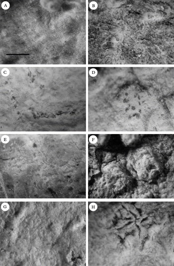
Stirtonia species, habitus. A, S. biseptata (holotype); B, S. curvata (Groenhart 9555a, L); C, S. neotropica (isotype, B). D, S. macrocarpa (isotype, ABL); E, S. obvallata (holotype of S. mellea); F, S. psoromica (holotype) G, S. ramosa (isotype, ABL); H, S. santessonii (isotype, ABL). Scale (all to same scale) = 0·5 mm.

(Fig. 2A)
Thallus spreading, covering an area of up to 10 cm diam., contiguous, smooth, dirty whitish, less than 0·1 mm thick, lightly shiny, with calcium oxalate crystals, IKI+ blue.
Ascigerous zones delimited, lirelliform, irregularly stellately branched, c. 0·1–0·2 mm wide, up to 1·5 mm long, not raised, pale brown, not pruinose, not dotted, with calcium oxalate crystals, without algal cells, IKI+ blue. Asci invisible in surface view, globose to ovoid, with 8 ascospores. Ascospores 4–5-septate, ellipsoid, 38–60 × 14–22 μm, upper cell largest, walls and septa >1 μm thick.
Chemistry. Perlatolic acid present (TLC). Thallus and ascigerous zones C−, Pd−, KOH−, UV−.
Stirtonia alboverruca Makhija & Patw
Biovigyanam 13: 48 (1987); type: India, South Andaman, Chidya Tapu, 15 February 1985, Patwardhan & Nagarkar (AMH 85.476—holotype).
Thallus spreading, covering an area of up to 5 cm diam., contiguous, smooth, dirty whitish, less than 0·1 mm thick, lightly shiny.
Ascigerous zones delimited, round to slightly elongated in outline, raised, always higher than the thallus, white, pruinose, inconspicuously dotted when abraded, with large crystals, without algal cells. Asci visible as pale brown dots in surface view, globose to ovoid, with 8 ascospores. Ascospores 11–15-septate, ellipsoid, 85–110 × 40–45 μm, cells generally equal, walls and septa >1 μm thick.
Chemistry. 2′-O-methylsuperphyllinic acid present (TLC). Thallus and ascigerous zones C−, Pd−, KOH−, UV+ white.
Stirtonia biseptata Aptroot & Wolseley
In Wolseley & Aptroot, Bibliotheca Lichenologica 99: 418 (2009); type: Thailand, Uthai Thani Prov., Khao Nang Rum, Viewpoint track, 12 January 1992, Wolseley & Aguirre-Hudson 4249 (BM—holotype!).
(Fig. 1A)
Thallus spreading, covering an area of up to 5 cm diam., contiguous, felty, pale olivaceous green, 0·1–0·2 mm thick, flaking off in one vellum, dull, without calcium oxalate crystals, but densely encrusted with tiny crystals, presumably of psoromic acid, IKI−.
Ascigerous zones delimited, linear to branched in outline, anastomosing to form a net of several cm, not raised, white, not pruinose, conspicuously pink dotted, without calcium oxalate crystals, but densely encrusted with tiny crystals, presumably of psoromic acid, without algal cells, IKI+ violet. Asci in surface view pink, ovoid, with 8 ascospores. Ascospores 2–3-septate, fusiform, 15–17 × 5–6 μm, cells generally equal, walls and septa <1 μm thick.
Chemistry. Psoromic and conpsoromic acids present (TLC). Thallus and ascigerous zones C−, Pd+ yellow, KOH−, UV−.
Note. This species is the most deviant currently accepted in the genus, characterized by the felty thallus and thin ascospore wall and septa.
Additional specimens examined. Marshall Islands: Ujae Atoll, Bock islet, on dead coconut tree, 1952, Fosberg 34370 (L!).—Indonesia: Java, Bay of Nglijep, S of Donomuljo, 1937, Groenhart 1454 & 7275 (L!); Cibodas, Mt. Gede, Pantjuran Mas., 22 iv 1050, Nurta & Madrodji (L!).
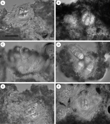
Stirtonia species, sections through ascigerous areas. A, S. alba showing ascigerous area with calcium oxalate crystals and ascospores with enlarged end cells (holotype); B, S. curvata in IKI, showing 4-spored ascus and blue coloration in lower parts and around the asci and a violet reaction in upper part (bottom: bark cells) (holotype); C, S. neotropica showing dark tiny crystals above the asci (isotype, B); D, S. obvallata showing large ascospores and algal cells in ascigerous area (holotype of S. mellea); E, S. ramosa (isotype, ABL); F, S. santessonii showing ascospores with enlarged end cells (isotype, ABL). All in tap water except B which is in IKI. Scale = 50 μm (all to same scale).

Stirtonia curvata Aptroot sp. nov
Stirtonia areis ascigeris lirelliformis thallo acidis perlatolicis continens.
Typus: Indonesia, Irian Jaya, Manokwari, c. 2 m alt., February 1959, W. Vink s.n. (L—holotypus!).
Thallus spreading, covering an area of up to 5 cm diam., contiguous, smooth, whitish yellow to brownish, less than 0·1 mm thick, shiny, with calcium oxalate crystals, IKI−.
Ascigerous zones delimited, angular to linear, branched or not, c. 0·1–0·2 mm wide, up to 1 mm long, erumpent, slightly raised, white, somewhat byssoid, not pruinose, erumpent, conspicuously red-brown or black dotted when abraded, with calcium oxalate crystals, without algal cells, IKI+ blue at the base and around the asci, upper layer IKI+ violet. Asci in surface view red-brown to black, single or in lines, ovoid, with 48 ascospores. Ascospores 7–11-septate, ellipsoid, 35–55 × 12–19 μm, often curved, cells generally equal, walls and septa > 1 μm thick.
Chemistry. Perlatolic acid present (TLC). Thallus and ascigerous zones C−, Pd−, KOH−, UV−.
Note. This new species differs from all other known species except Stirtonia schummii by its chemistry; it differs from S. schummii by its yellowish to brownish thallus.
Additional specimen examined. Indonesia: Java, West Bantam, track along the South brink of the Tjitadjur ravine, on tree, 1959, Groenhart 9555a (L!).
Stirtonia dubia A. L. Sm
Trans. Brit. Mycol. Soc. 11: 195 (1926); type: India, Bengal, Chinsurah, on tree, s.d., Watt (BM—isolectotype!).
Thallus spreading, covering an area of up to 5 cm diam., contiguous, smooth, grey whitish, less than 0·1 mm thick, slightly shiny.
Ascigerous zones seemingly not delimited but in fact lirelline and branched, c. 0·1 mm wide, not raised, whitish, pruinose, inconspicuously dotted when abraded, with small crystals, without algae. Asci visible as pale brown dots in surface view, globose to ovoid, with 8 ascospores. Ascospores 6–7-septate, ellipsoid, 15–30 × 6–8 μm, cells generally equal, walls and septa >1 μm thick.
Chemistry. No lichen substances detected (TLC). Thallus and ascigerous zones C−, Pd−, KOH−, UV−.
Stirtonia indica Makhija & Patw
Mycotaxon 67: 300 (1998); type: India, South India, Eastern Ghats, Tamil Nadu, Chittery, 21 January 1983, Sethy (AMH 83.47—holotype).
Thallus spreading, covering an area of up to 5 cm diam., contiguous, smooth, dirty whitish, 0·2–0·3 mm thick, slightly shiny.
Ascigerous zones delimited, round to slightly elongated in outline, 1–5 mm diam., raised, always higher than the thallus, white, pruinose, inconspicuously dotted when abraded, with calcium oxalate crystals, without algal cells. Asci visible as pale brown dots in surface view, globose to ovoid, with 8 ascospores. Ascospores 6–9-septate, ellipsoid, 50–60 × 15–22 μm, cells generally equal, walls and septa >1 μm thick.
Chemistry. Psoromic and conpsoromic acids present (TLC). Thallus and ascigerous zones C−, Pd+ yellow, KOH−, UV−.
Stirtonia macrocarpa Makhija & Patw
Biovigyanam 13: 48 (1987); type: India, North Andaman, Diglipur Range, Milangram, 3 January 1986, Patwardhan & Sethy (AMH 86.276—holotype; ABL—isotype!); (AMH 86.347—ABL, topotype!);
(Fig. 1D)
Thallus spreading, covering an area of up to 5 cm diam., contiguous, smooth, grey whitish, < 0·1 mm thick, lightly shiny, with calcium oxalate crystals, IKI+ blue.
Ascigerous zones delimited, round to slightly elongated in outline, 0·2–0·4 mm diam., raised, always higher than the thallus, white, pruinose, conspicuously brown to black dotted when abraded, with calcium oxalate crystals, without algal cells, IKI+ blue. Asci visible as brown to black dots in surface view, globose to ovoid, with 8 ascospores. Ascospores 11–16-septate, ellipsoid, 50–110 × 20–35 μm, cells generally equal, walls and septa >1 μm thick.
Chemistry. 2′-O-methylperlatolic acid present (TLC). Thallus and ascigerous zones C−, Pd−, KOH−, UV−.
Additional specimens examined. Bangla Desh: Bhawal National Park, 60 km N of Dhaka city, 9 March 2002, Hopman (ABL!).—Seychelles: Praslin, National Park Sentier Glacier Noire, 30 September 2008, Schumm 14455 & Frahm (hb. Schumm!).
Stirtonia neotropica Aptroot sp. nov
Stirtonia ascis catenulatis rubigineis.
Typus: Netherlands Antilles, Sint Eustatius, Quill National Park, Quill Trail, c. 200–300 m alt., 29 January 2008, W. R. Buck 53004 (NY—holotype!, ABL—isotypus!, B—isotypus!).
Thallus spreading, covering an area of up to 10 cm diam., contiguous, smooth, pale olivaceous, < 0·1 mm thick, slightly shiny, without calcium oxalate crystals, IKI−.
Ascigerous zones delimited, linear, branched and anastomosing, not raised, white, pruinose, conspicuously red-brown dotted when abraded, without calcium oxalate crystals but with tiny crystals above the asci causing the upper surface to be dark dotted, without algal cells. Asci in surface view red-brown, in lines, ovoid, with 8 ascospores, IKI−. Ascospores (7–)10–11-septate, ellipsoid, 35–38 × 10–12 μm, cells equal, walls and septa >1 μm thick.
Chemistry. Gyrophoric acid present (TLC). Thallus and ascigerous zones patchily C+ red, furthermore C−, Pd−, KOH−, UV−.
Note. This new species is the first of the genus to be described from the Neotropics. Its chemistry differs from all other known species.
Additional specimen examined. Costa Rica: Limón, 65 km SE of Limón, Manzanilla, along beach to Punta Vargas, 11 March 2004, Sipman 51694 (B!).
Stirtonia obvallata (Stirt.) A. L. Sm
Trans. Brit. Mycol. Soc. 11: 195 (1926); type: India, Bengal, Chinsurah, on tree, s.d., Watt (BM—isolectotype!).
Stirtonia aggregata Makhija & Patw., Mycotaxon 67: 295 (1998); type: Indonesia, Java, S. coast near Kampong Nglijep, S. of Malang, on tree, January 1959, Groenhart 1452 (L—holotype!); Groenhart 4691 (L—isotype!).
New synonyms: Stirtonia gibberulosa Makhija & Patw., Mycotaxon 67: 299 (1998); type: Indonesia, Java, Bay of Nglijep, S. of Donomuljo, on tree, January 1959, Groenhart 1455 (L—holotype!); Groenhart 557 (L—isotype!).
Stirtonia marginata Makhija & Patw., Mycotaxon 67: 302 (1998); type: Indonesia, Java, Bay of Nglijep, S. of Donomuljo, on tree, September 1959, Groenhart 578 (L—holotype!); Groenhart 4697 (L—isotype!).
Stirtonia mellea Makhija & Patw., Mycotaxon 67: 304 (1998); type: Indonesia, Java, Bay of Nglijep, S. of Donomuljo, on tree, September 1959, Groenhart 1453 (L—holotype!).
Thallus spreading, covering an area of up to 10 cm diam., contiguous, smooth, brownish to whitish, under 0·1 mm thick, mostly endophoeodal, lightly shiny, with calcium oxalate crystals, IKI+ blue.
Ascigerous zones delimited, round to elongated and often branched and anastomosing in outline, c. 0·2–0·3 mm wide, erumpent through the thallus and bark, higher than most of the surrounding thallus, white, pruinose, inconspicuously dotted when abraded, with calcium oxalate crystals, often with algal cells, IKI+ blue. Asci not visible in surface view, globose to ovoid, with 8 ascospores. Ascospores 7–13-septate, ellipsoid, 45–90 × 20–39 μm, cells generally equal, walls and septa >1 μm thick.
Chemistry. No diagnostic substances detected, but various terpenoids variably present which are originating from the bark (TLC). Thallus and ascigerous zones C−, Pd−, KOH−, UV−.
Notes. The differences reported between the taxa synonymized here are not taxonomically significant and, at most, reflect age difference and some bark properties. All Indonesian specimens originate from the same area and are virtually identical. The ascospore measurements reported forStirtonia gibberulosa by Makhija & Patwardhan (Reference Makhija and Patwardhan1998) must have been mostly based on immature material; the dimensions of the ascospore illustrated fits that species best. The Groenhart collecting numbers are not sequential but have been applied much later, and identical specimens bearing identical names can thus be regarded as isotypes; the typification is not problematical.
Additional specimens examined. Indonesia: Java, Bay of Tapen, Sout of Wlingi, on tree, Sept. 1959, Groenhart 4835, 4818, 7970, 4822, 4824, 4829 (L!, all essentially one specimen).—Thailand: Uthai Thani Prov.: Khao Nang Rum, Serrayut's plot, 1992, Aguirre-Hudson, James & Wolseley 2783 (BM!).
Stirtonia psoromica Aptroot & Wolseley
In Wolseley & Aptroot, Bibliotheca Lichenologica 99: 419 (2009); type: Thailand, Chiang Mai Prov., Doi Suthep, Wat Palad, 25 November 1991, Wolseley & Aguirre-Hudson 5930 (BM—holotype!).
(Fig. 1F)
Thallus spreading, covering an area of up to 5 cm diam., contiguous, smooth, dirty whitish, less than 0·1 mm thick, slightly shiny, with calcium oxalate crystals, IKI−.
Ascigerous zones delimited, round to slightly elongated in outline, 0·3–0·7 mm diam., always higher than the thallus, white, pruinose, inconspicuously black dotted when abraded, with calcium oxalate crystals, without algal cells, IKI+ blue. Asci in surface view black, ovoid, with 8 ascospores. Ascospores generally 7–9-septate, fusiform, 75–87 × 12–16 μm, cells generally equal, walls and septa >1 μm thick.
Chemistry: Psoromic and conpsoromic acids always present (TLC). Thallus C−, Pd+ yellow, KOH−, medulla UV−.
Additional specimen examined. Thailand: Uthai Thani Prov.: Khao Nang Rum, Serrayut's plot, 14 January 1992, Wolseley & Aguirre-Hudson 4273 (BM!).
Stirtonia ramosa Makhija & Patw
Biovigyanam 13: 49 (1987); type: India, South Andaman, Alexandria Island, 6 March 1983, Patwardhan, Nagarkar & Sethy (AMH 85.1122—holotype; ABL—isotype!).
Thallus spreading, covering an area of up to 5 cm diam., contiguous, smooth, dirty whitish to pale olivaceous, < 0·1 mm thick, slightly shiny, without calcium oxalate crystals, IKI−.
Ascigerous zones delimited, lirelliform, irregularly stellately branched, c. 0·1–0·3 mm wide, anastomosing to form a network of several cm, not or slightly raised, pale brown, not pruinose, not dotted, with calcium oxalate crystals (only near the upper surface with small crystals, probably of the secondary compounds mentioned below), without algal cells, IKI−. Asci invisible in surface view, globose to ovoid, with 8 ascospores. Ascospores (4–)5–6-septate, ellipsoid, 45–65 × 15–22 μm, all cells equal (upper cell or both end cells enlarged only in young ascospores), walls and septa >1 μm thick, postmature ascospores becoming brown.
Chemistry. 2′-O-methylnorsuperphyllinic and 4′-O-demethylsuperconfluentic acids present (TLC). Thallus and ascigerous zones C−, Pd−, KOH−, UV−.
Additional specimens examined. India: North Andaman, Diglipur Range, Milangram, 3 i 1986, Sethy & Patwardhan (AMH 86.272, ABL!); Little Andaman, Netaji Nagar, Krishna Nala, Nagarkar & Patwardhan (AMH 85.913, ABL!).
Stirtonia santessonii Makhija & Patw
Biovigyanam 13: 49 (1987); type: India, South Andaman, Redskin Island, 12 December 1985, Nagarkar & Patwardhan (AMH 85.1985—holotype; ABL—isotype!); (AMH 85.1986—topotype, ABL!).
Thallus spreading, covering an area of up to 5 cm diam., contiguous, smooth, pale brownish, < 0·1 mm thick, lightly shiny, with calcium oxalate crystals, IKI−.
Ascigerous zones delimited, whitish, round, c. 0·5–1·5 mm diam., raised, not pruinose, with pale to dark brownish asci in lirelliform, irregularly stellately branched, c. 0·1–0·3 mm wide lines, with calcium oxalate crystals, without algal cells, IKI+ blue. Asci visible as pale to dark brown lines in surface view, globose to ovoid, with 8 ascospores. Ascospores 7-septate, ellipsoid, 35–50 × 13–18 μm, upper cell largest, walls and septa >1 μm thick.
Chemistry. No diagnostic substances detected (TLC). Thallus and ascigerous zones C−, Pd−, KOH−, thallus UV−, ascigerous areas UV+ white.
Stirtonia schummii Aptroot sp. nov
Stirtonia ascosporis minimis thallo acidis perlatolicis continens.
Typus: Seychelles, Praslin, Anse Lazio, Hotel Bonbon Plume, 2 September 2008, F. Schumm 14438 & J. P. Frahm (hb. Schumm—holotypus!).
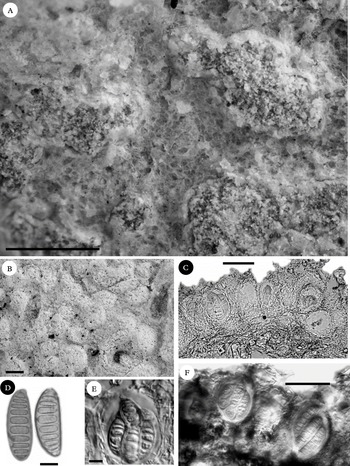
Stirtonia schummii (holotype). A & B, habitus; C, E & F, sections through ascigerous areas in IKI; D, ascospores. Scales: A & B = 0·5 mm; C & F = 50 μm; D & E, = 10 μm.

Thallus spreading, covering an area of up to 5 cm diam., contiguous, smooth, dirty whitish, less than 0·1 mm thick, slightly shiny, with calcium oxalate crystals, IKI−.
Ascigerous zones delimited, round to slightly elongated in outline, 0·3–0·8 mm diam., raised, always higher than the thallus, white, pruinose, inconspicuously dotted when abraded, with calcium oxalate crystals, without algae, IKI+ blue. Asci in surface view brown, ovoid, with 8 ascospores. Ascospores 7–9-septate, ellipsoid, 36–45 × 12–15 μm, cells generally equal, walls and septa >1 μm thick.
Chemistry. Perlatolic acid present (TLC). Thallus and ascigerous zones C−, Pd−, KOH−, UV−.
Note. This new species is the first of the genus to be described from Africa. It differs from all other known species except Stirtonia curvata by its chemistry, and differs from S. curvata by the white thallus. It is named in honour of the lichenologist Felix Schumm, who collected the type specimen and prepared the illustrations.
Additional specimen examined. Seychelles: Mahé, Dans Iles, on shrubs, 1973, Norkett 16379 (ABL!, BM!). Reported by Seaward & Aptroot (Reference Seaward and Aptroot2009) as S. gibberulosa.
Excluded species
Stirtonia amazonica Sambo
Annali di Botan. 22: 24 (1940).
Not studied, as it was already excluded by Makhija & Patwardhan (Reference Makhija and Patwardhan1998). = Arthonia sp.
Stirtonia macrocephala R. Sant
Symbol. Bot. Upsal. 12(1): 60 (1952).
Not studied, as it was already excluded by Makhija & Patwardhan (Reference Makhija and Patwardhan1998). = Eremothecella macrocephala (R. Sant.) G. Thor, Sérus., Lücking & Tat. Matsumoto.
Stirtonia monospora D. D. Awasthi & K.P. Singh
Geophytology 1: 97 (1971).
Not studied, as it was already excluded by Makhija & Patwardhan (Reference Makhija and Patwardhan1998) = ?Arthonia sp.
Stirtonia sprucei R. Sant
Symbol. Bot. Upsal. 12(1): 60 (1952).
Type and neotropical material not studied, as it was already excluded by Makhija & Patwardhan (Reference Makhija and Patwardhan1998). = Amazonomyces sprucei (R. Sant.) Lücking, Sérus. & G. Thor (Lücking et al. Reference Lücking, Sérusiaux, Maia and Pereira1998).
Discussion
After this revision, Stirtonia is still a small genus with only 13 accepted species. It is quite homogenous in some characters, such as the asci, ascospores and interascal filaments, but quite variable in other characters, such as iodine reactions and the gross morphology of the ascigerous areas. It is quite possible that in its present sense the genus is not monophyletic. The most aberrant element is S. biseptata with its byssoid thallus and thin septa.
After the previous exclusion of the foliicolous species assigned to this genus by Santesson (Reference Santesson1952), the remaining species in the genus were known only from tropical Asia. Here, one new species each is described from Africa and the neotropics, showing that it is a pantropical genus, but most diverse in the palaeotropics.
Stirtonia species are apparently rare or at least rarely collected and only locally more abundant. For example, among a set of about 100 Arthoniaceae specimens from Thailand in BM, only four specimens belonged to three species of Stirtonia; an even larger set of c. 200 Arthoniaceae specimens from Indonesia (mainly Java) in L contained 15 specimens (with 13 numbered duplicates) representing five species.
The author thanks Felix Schumm for sending a new species and preparing some illustrations, Urmila Makhija for kindly sending duplicates of types and other specimens upon request and Leo Spier for performing TLC on the specimens in ABL. Harrie Sipman is acknowledged for detecting the neotropical Stirtonia among a set of unidentified lichens and for comments on an earlier draft. The author also thanks Göran Thor for comments on an earlier draft and for various suggestions concerning the study of Cryptotheciaceae and Pat Wolseley (BM) and the staff at L for the loans.