Introduction
Phosphate minerals from the apatite supergroup possess a strong affinity for strontium, especially apatite, hedyphane and belovite-group minerals (Pasero et al., Reference Pasero, Kampf, Ferraris, Pekov, Rakovan and White2010). Although minerals with chemical formula close to Ca2Sr3(PO4)3F have been reported before in the space group P63 (Klevtsova, Reference Klevtsova1964; Pushcharovsky et al., Reference Pushcharovsky, Nadezhina and Khomyakov1987; Sghir et al., Reference Sghir, Hlil, Laghzizil, Boujrhal, Cherkaoui El Moursli and Fruchart2009), or as a second occurrence of fluorcaphite, ideally SrCaCa3(PO4)3F (Chakhmouradian et al., Reference Chakhmouradian, Hughes and Rakovan2005), unfortunately, none of these minerals has been approved by the Commission on New Minerals, Nomenclature and Classification of the International Mineralogical Association (IMA–CNMNC, Pasero et al., Reference Pasero2022).
The new mineral fluorsigaiite, ideally Ca2Sr3(PO4)3F, is the Sr analogue of fluorphosphohedyphane [Ca2Pb3(PO4)3F] and a new member of the apatite supergroup. It was discovered in lujavrite from the Saima alkaline complex, Liaoning Province, northeast China. Fluorsigaiite is named after the Chinese pronunciation, ‘si’ for strontium, and ‘gai’ for calcium with a prefix fluor, to indicate the composition of the mineral and the specific dominance of Ca at Ca1 site and Sr at Ca2 site in the crystal structure. Optical microscopy, electron probe microanalysis, X-ray diffraction and Raman spectroscopy were used to determine its petrographic features, chemical composition and crystal structure. The species and the name (symbol Fsig) have been approved by the IMA–CNMNC (IMA2021-87a, Wu et al., Reference Wu, Gu, Rao, Wang, Zhong and Wan2022). The type mineral is deposited at the Geological Museum of China, No.15 Yangrouhutong, Xisi, Beijing 100031, PR China, catalogue number M16130. This paper describes the physical, chemical properties, and crystal structure of fluorsigaiite, and then discusses its origin and relation to other similar species.
Occurrence
Fluorsigaiite was discovered in lujavrite from the Saima alkaline complex, (124°16′E, 40°58′N), ~50 km northeast of Fengcheng City, Liaoning Province, PR China. This alkaline complex is also the type locality for recently approved minerals gugiaite, hezuolinite and fengchengite (Peng et al., Reference Peng, Cao, Zou, Zhang, Yin and Ding1962; Yang et al., Reference Yang, Giester, Ding and Tillmanns2012; Shen et al., Reference Shen, Xu, Yao and Li2017), and it is primarily comprised of phonolite, nepheline syenite, lujavrite and alkaline pegmatite with significant mineralisation of U, rare earth elements, Nb and Zr (Wu et al., Reference Wu, Wang, Yang, Wu, Zhang, Gu and Zhang2016, Reference Wu, Wen, Bonnetti, Wang, Yang and Wu2019). The most recent geochemistry and chronological evidence from zircon, titanite and eudialyte show that all of the Saima rocks are commonly derived from the partial melting of a lithospheric mantle source with a contribution of recycled crust material, and they are almost coeval within an emplacement age range of 224–230 Ma (Wu et al., Reference Wu, Yang, Marks, Liu, Zhou, Ge, Yang, Zhao, Mitchell and Markl2010; Zhu et al., Reference Zhu, Yang, Sun, Zhang and Wu2016, Reference Zhu, Yang, Sun and Wang2017). Detailed petrography and mineral paragenesis of Saima rocks have been well described in previous studies (Wu et al., Reference Wu, Wang, Yang, Wu, Zhang, Gu and Zhang2015, 2016; Zhu et al., Reference Zhu, Yang, Sun, Zhang and Wu2016).
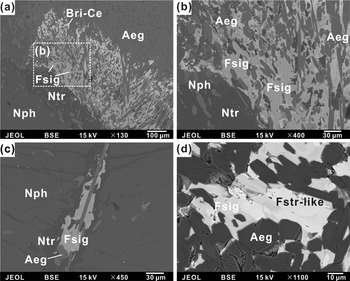
Fluorsigaiite commonly occurs as individual prismatic, columnar and platy crystals of 10 to 50 μm in the matrix of microcline and nepheline, associated with natrolite, eudialyte, fluorapatite, a fluorstrophite-like mineral, stronadelphite and calcite (Fig. 1). In some cases, fluorsigaiite crystals form prismatic or anhedral aggregates up to 100 μm across in interstices of aegirine and nepheline (Fig. 1a–c). Back-scattered electron (BSE) images show the crystals to be weakly heterogeneous (Fig. 1b), and that the brighter areas in fluorsigaiite are slightly richer in Sr. In addition, tiny grains of bastnäsite-(Ce), strontianite and britholite-(Ce) (<1 μm) are occasionally observed filling fractures or at grains boundaries between fluorsigaiite and nepheline.
Back-scattered electron images showing the occurrence and mineral association of fluorsigaiite from the Saima lujavrite. (a) Cluster of columnar fluorsigaiite crystals intergrown with aegirine, surrounded by natrolite and nepheline. (b) Enlarged frame area of (a) showing fluorsigaiite intergrown with aegirine in natrolite. (c) Prismatic fluorsigaiite with aegirine and natrolite cutting nepheline. (d) Fluorstrophite-like mineral associated with fluorsigaiite in interstices of aegirine aggregate. Mineral abbreviations: Aeg – aegirine, Bri-Ce – britholite-(Ce), Fsig – fluorsigaiite, Fstr-like – fluorstrophite-like mineral, Nph – nepheline and Ntr – natrolite.

Physical and optical properties
Individual prismatic, columnar or platy crystals of fluorsigaiite are translucent to transparent and colourless to yellowish white in transmitted light with a vitreous lustre. No fluorescence is shown in long- or short-wave ultraviolet light. The tenacity is brittle with uneven fracture. The Mohs hardness is ~5, and the calculated density is 3.842 g/cm3 based on the empirical formula and single-crystal unit-cell parameters. It is nonmagnetic as tested by a magnetic needle. Optically, fluorsigaiite is uniaxial (–), with ω = 1.64(1) and ɛ = 1.63(1), as determined in white light and without dispersion. According to the calculated density and the measured indices of refraction, the compatibility index [1 – (K P/K C)] is 0.021, and corresponds to the ‘excellent’ category (Mandarino, Reference Mandarino1981).
Raman spectroscopy
The Raman spectrum of fluorsigaiite was collected from a randomly oriented grain, using a Renishaw inVia RM2000 spectrometer equipped with a 50× Leica objective lens at the State Key Laboratory of Nuclear Resources and Environment, East China University of Technology. The excitation wavelength was 532 nm, and the output power of the laser was set at 20 mW. The spatial resolution was estimated to be 1 μm, and the spectral resolution was 1 cm–1. High-pure silicon with a 520 cm–1 Raman shift was chosen for calibration. The spectrum was collected from 100 to 4000 cm–1 Raman shift and the accumulation time for each spectrum was ~30 s with 2–3 accumulations.
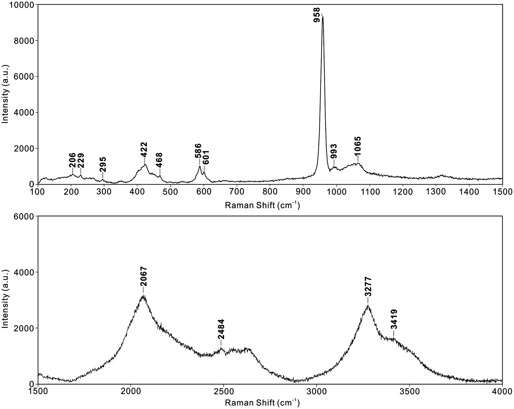
The Raman spectrum of fluorsigaiite (Fig. 2) contains strong sharp peaks at 958 cm–1; medium peaks at 1065, 601, 586, 468 and 422 cm–1; and weak peaks at 993, 295, 229 and 206 cm–1, respectively. In comparison with other apatite-supergroup minerals (Meegoda et al., Reference Meegoda, Bonner, Loutts, Stefanos and Miller1999; O'Donnell et al., Reference O'Donnell, Fredholm, de Rouffignac and Hill2008; Zhai et al., Reference Zhai, Shieh, Xue and Xie2015; Xue et al., Reference Xue, Zhai, Lin and Zhai2018), the Raman shifts of PO4 groups are observed at 958 (ν1), 422 (ν2), 1065 (ν3), 601 (ν4) and 586 cm–1 (ν4). The Ca–O vibration modes are most probably at 229 and 468 cm–1, and the Raman shifts at 206 and 295 cm–1 probably correspond to Sr–O vibrations. The bands of the OH stretching vibrations are present in the range of 3000–4000 cm–1.
The Raman spectrum for fluorsigaiite.

Chemical composition
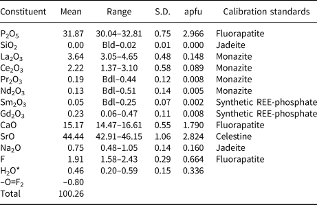
Quantitative elemental microanalyses of fluorsigaiite and a fluorstrophite-like mineral were conducted with a JEOL-JXA 8530F Plus electron microprobe in wavelength dispersive spectroscopy mode at 15 kV and 50 nA, with a defocused beam diameter of 2 μm, at the State Key Laboratory of Nuclear Resources and Environment, East China University of Technology. Measurement time for each element was 20 and 10 s on peaks and backgrounds, respectively. Standards for calibration were fluorapatite (CaKα, PKα and FKα), celestine (SrLα), jadeite (NaKα and SiKα), monazite (LaLα, CeLα, PrLβ and NdLα), and synthetic REE-phosphates (SmLα and GdLα). The Cl content was below detection limits of the electron microprobe (i.e. ≤200 ppm). The empirical formula, calculated on the basis of 13 total anions per formula unit and F+(OH) = 1 (apfu, Table 1), is: (Sr2.82Ca1.79Na0.16La0.15Ce0.09Pr0.01Nd0.01Gd0.01)Σ5.04P2.97O12[F0.66(OH)0.34]Σ1.00. The ideal formula of Ca2Sr3(PO4)3F requires P2O5 32.91, CaO 17.34, SrO 48.05, F 2.94, excessive O = 1.23 and total 100.00 (all in wt.%).
Chemical electron microprobe data (in wt.%) for fluorsigaiite.

*calculated on the basis of 13 total anions and F+(OH) = 1 apfu; Bdl = Below detection limits; S.D.= Standard deviation (2σ).
Powder X-ray diffraction determination
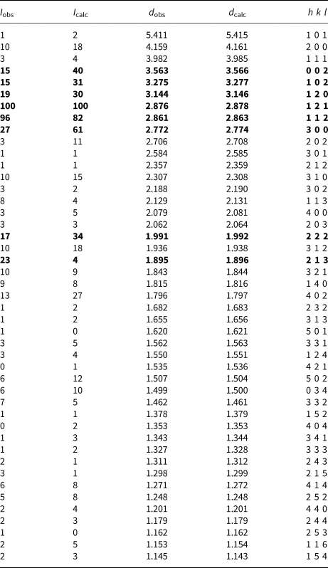
The powder X-ray diffraction (XRD) pattern of fluorsigaiite was collected on a Rigaku XtaLAB Synergy diffractometer (CuKa, λ = 1.54184 Å) in powder Gandolfi mode at 50 kV and 1 mA, at the School of Earth Sciences and Info-physics, Central South University, China. The total exposure time was ~4 h. The structural model of single crystal (see below) was used to index the powder XRD pattern of fluorsigaiite (Table 2). Unit-cell parameters refined from the powder data using the method of Holland and Redfern (Reference Holland and Redfern1997) show that fluorsigaiite belongs to hexagonal crystal system, with a = 9.6005(3) Å, c = 7.1119(4) Å, V = 567.67(4) Å3 and Z = 2.
Experimental and calculated* powder X-ray diffraction data (d in Å, I in %) for fluorsigaiite.

*The calculated values were obtained using VESTA 3 (Momma and Izumi, Reference Momma and Izumi2011); the strongest values are given in bold.
Crystal structure refinement
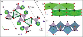
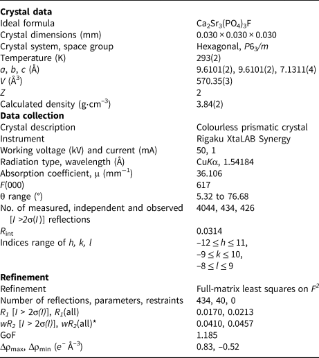
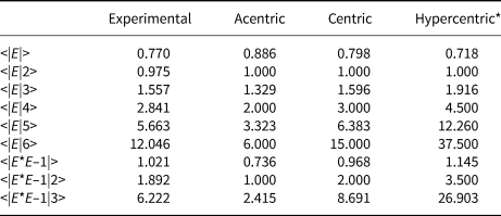
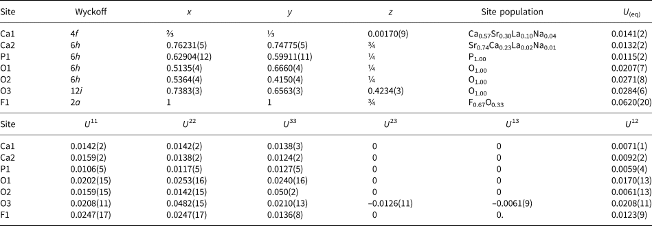
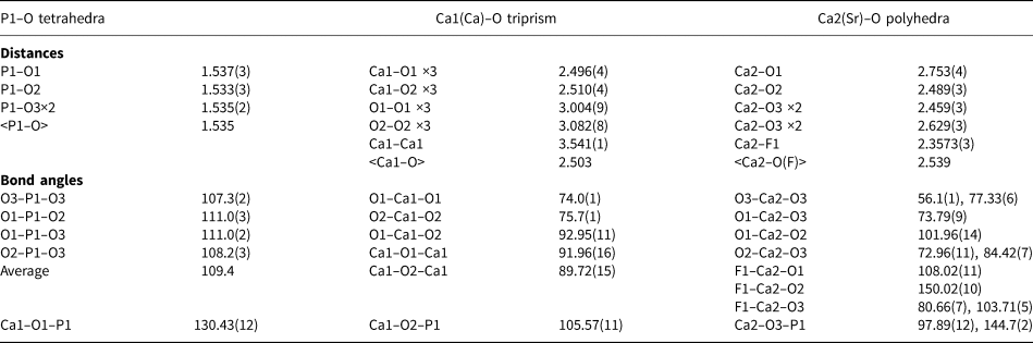
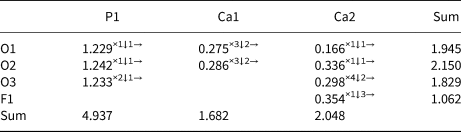
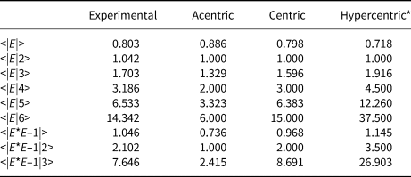
To characterise the structure of fluorsigaiite, single-crystal X-ray diffraction data on a crystal fragment were collected on a Rigaku XtaLAB Synergy diffractometer, at the School of Earth Sciences and Info-physics, Central South University, China, and refined using SHELX (Sheldrick, Reference Sheldrick2015a, Reference Sheldrick2015b). The crystallographic data and refinement statistics are given in Table 3. The diffraction data are treated with the program CrysAlisPro (Rigaku Oxford Diffraction, UK) and the space group is recognised as P63/m according to <E> parameters (Table 4). The structure was solved in the space group P63/m and refined with anisotropic vibrations for all sites. The occupancies of Ca and Sr are first freely refined and then manually adjusted, along with La, Na and F, towards minimum R 1, and in good agreement with the chemical compositions in Table 1. For simplicity, only La is considered in the refinement for rare earth elements as it is the dominant element according to microprobe analyses. The final atomic coordinates and displacement parameters are listed in Table 5, and selected bond lengths and angles in Table 6. The bond-valence sums of atoms, calculated using the parameters given by Brese and O'Keeffe (Reference Brese and O'Keeffe1991) are presented in Table 7. The structure is illustrated in Fig. 3. The crystallographic information file has been deposited with the Principal Editor of Mineralogical Magazine and is available as Supplementary material (see below).
Crystal structure of fluorsigaiite (unit cell outlined in black lines) plotted with VESTA 3 (Momma and Izumi, Reference Momma and Izumi2011). (a) Crystal structure viewed along the c-axis, showing that the chains of twisted Ca1–O triprism (green-white colour, with 22.5°-rotated top and base triangle faces) are linked through isolated PO4 tetrahedra (pink colour), forming open channels accommodating F and OH at the F1(O4) site, and Sr, Ca, La, Na atoms at Ca2 site sideway. (b) Face-sharing chains of Ca1–O triprism (green colour) parallel to the c-axis viewed along the b-axis. (c) Corner-sharing chains composed of Ca2–O polyhedra (blue colour) and PO4 tetrahedra (pink colour) are connected with each other by corner- and edge-sharing modes parallel to the c-axis.

Data collection and structure refinement details for fluorsigaiite.

*wR 2 = {∑[w(Fo2–Fc2)2]/ ∑[w(Fo2)2]}½.
The <E> parameters for determination of the space group of fluorsigaiite.

*Number of asymmetric units in one cell.
Wyckoff positions, fractional coordinates, inferred site population, equivalent isotropic and anisotropic displacement parameters (Å2) of atoms for fluorsigaiite.

Selected bond distances (Å) and angles (°) for fluorsigaiite.

Calculated bond-valence sums (in valence units) of atoms for fluorsigaiite, using the parameters of Brese and O'Keeffe (Reference Brese and O'Keeffe1991).

Fluorsigaiite is isostructural with minerals of the hedyphane group in the apatite supergroup (Pasero et al., Reference Pasero, Kampf, Ferraris, Pekov, Rakovan and White2010), in which exist three cation sites, Ca1(4f), Ca2(6h) and P1(6h) with a ratio of 2: 3: 3, and four anion sites, O1, O2, O3 and F1 (O4) (Table 5). The structure of fluorsigaiite is based on a framework consisting of [Ca1O6–Ca1O6] triprism chain and [Ca2O7–P1O4] chain, connected by corner-sharing mode (O1 and O2) along the c-axis (Fig. 3). The Ca-dominated Ca1 site (Ca0.57Sr0.30La0.10Na0.04) is bound to 6 O [O1 (×3) and O2 (×3)] with an average bonding length 2.503 Å (Table 6) to form a twisted triprism, and more weakly bound to further O3 (×3) with a distance of 2.873 Å. The top and base triangle faces of the triprism are 22.5°-rotated and composed by O1 (×3) and O2 (×3) atoms, respectively (Fig. 3a). The Ca1O6 triprisms share two triangle faces to form triprism chains parallel to the c-axis (Fig. 3b). P1 is bound to 4 O atoms at O1, O2 and O3 (×2) sites to form isolated PO4 tetrahedra with an average bonding length of 1.535 Å. The Ca2 site is dominated by the Sr atom (Sr0.74Ca0.23La0.02Na0.01) and bound to 6 O [O1, O2 and O3 (×4)] atoms and 1 F1(O4) atom with an average bonding length of 2.539 Å, forming a pentagonal bipyramid. The Ca2O7 polyhedra are bound to P1O4 tetrahedra by corner-sharing mode (O3) with the ratio of 1:1, to form chains parallel to [Ca1O6–Ca1O6] triprism chains along the c-axis. In addition, [Ca2O7–P1O4] chains are connected with each other by corner- and edge-sharing modes (Fig. 3c). From the view along the c-axis, the Ca1O6 triprism chains are joined by PO4 tetrahedra to form hexagonal channels, in which the Ca2 site is located hexagonally around the peripheral tunnels and F-dominated F1(O4) site located in the centre (Fig. 3a). The bond-valence sums for P1 (4.937) and Ca2 (2.048) are close to their theoretical values, whereas the slightly lower bond-valence sum for Ca1 (1.682) is probably attributed to higher light element Na in Ca1 site (Table 7).
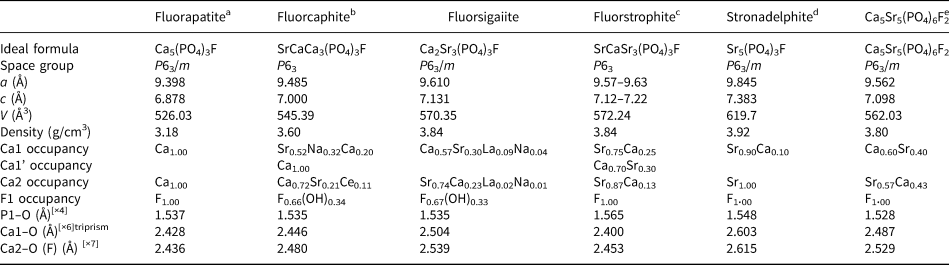
Fluorsigaiite is a new, and the only, natural intermediate member with significant contents of both Ca and Sr in the space group P63/m of the Ca5(PO4)3F–Sr5(PO4)3F solid-solution series. Comparative data for these solid-solution series members and synthetic Ca5Sr5(PO4)6F2 in Table 8 illustrate that the progressive replacement of Ca (effective ionic radius 1.06–1.18 Å, Shannon, Reference Shannon1976) in the structure of CaxSr5–x(PO4)3F by Sr (effective ionic radius 1.21–1.31 Å) leads to an increase in average Ca1–O[×6] and Ca2–O[7] bond length from 2.428 to 2.603 Å and from 2.463 to 2.615 Å, respectively, as well as increases in the unit-cell parameters and density.
Comparison among natural Ca5(PO4)3F–Sr5(PO4)3F solid-solution series and synthetic Ca5Sr5(PO4)6F2.

afrom Hughes et al. (Reference Hughes, Cameron and Crowley1989); bfrom Rastsvetaeva and Khomyakov (Reference Rastsvetaeva and Khomyakov1996) and Chakhmouradian et al. (Reference Chakhmouradian, Hughes and Rakovan2005); cfrom Klevtsova (Reference Klevtsova1964) and Pushcharovsky et al.(Reference Pushcharovsky, Nadezhina and Khomyakov1987); dfrom Pekov et al. (Reference Pekov, Britvin, Zubkova, Pushcharovsky, Pasero and Merlino2010); efrom Lim et al. (Reference Lim, Baikie, Pramana, Smith and White2011).
Relation to fluorstrophite
Although fluorsigaiite and fluorstrophite are similar in composition, fluorstrophite is in the non-centrosymmetric space group of P63, in which the Ca1 site is further split into two sites, namely the Ca1 site mainly occupied by Sr and the Ca1' site mostly occupied by Ca, corresponding to the ideal formula SrCaSr3(PO4)3F (Klevtsova, Reference Klevtsova1964; Pushcharovsky et al., Reference Pushcharovsky, Nadezhina and Khomyakov1987). Fluorsigaiite has the centrosymmetric space group P63/m according to <E> discriminating parameters of the diffraction data (Table 4). The refined atom numbers of fluorsigaiite Ca1.83Sr2.81 and F0.67 in P63/m is in good agreement with its actual composition of Ca1.79Sr2.82 and F0.66 with the Ca:Sr ratio of nearly 2:3, apparently larger than that of fluorstrophite according to its actual composition of Ca1.28Sr2.96 and F0.58, etc. (Efimov et al., Reference Efimov, Kravchenko and Vasileva1962). In contrast, we tried to refine the structure of fluorsigaiite in non-centrosymmetric space group P63 with R 1 = 0.0209 (Fsig-P63.cif in Supplementary material), but the refined atom numbers Ca1.56Sr3.12 and F1.16 are significantly different from the actual composition and have an unreasonable number of F atoms (1.16 F) and a Flack parameter of 0.43 suggesting missing symmetry (Fsig-P63_checkcif.pdf in Supplementary material).
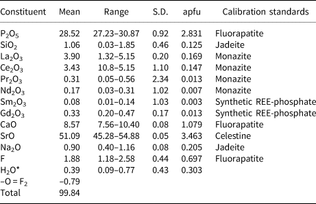
In addition, we also studied a fluorstrophite-like mineral coexisting with fluorsigaiite (Fig. 1d), whose empirical formula (Sr3.46Ca1.08Na0.21La0.17Ce0.15Pr0.01Nd0.01Gd0.01)Σ5.10(P2.83Si0.13)Σ2.96O12[F0.70(OH)0.30]Σ1.00, determined by electron microprobe (Table 9), is similar to the actual composition of fluorstrophite of Ca1.28Sr2.96 and F0.58. (Efimov et al., Reference Efimov, Kravchenko and Vasileva1962). Moreover, its cell parameters of a = 9.6517 Å, c = 7.1799 Å and V = 579.24 Å3 are also similar to those of fluorstrophite (Table 8). However, the <E> parameters of the diffraction data support the centrosymmetric space group P63/m (Table 10) and the structure refinement (R 1 = 0.0197) indicates that it belongs to stronadelphite with both Ca1 and Ca2 sites dominated by Sr (Fstr-P63_m.cif and Fstr-P63_m_checkcif.pdf in Supplementary material). Although it may also be refined in space group P63 in compliance with the definition of fluorstrophite (Fstr-P63.cif in Supplementary material, Klevtsova, Reference Klevtsova1964; Pasero et al., Reference Pasero, Kampf, Ferraris, Pekov, Rakovan and White2010), the unreasonable flack parameter of 0.51, along with other parameters (Fstr-P63_checkcif.pdf in Supplementary material), may favour the centrosymmetric structure of space group P63/m. Therefore, fluorsigaiite cannot be a variety of the fluorstrophite and it is unambiguously distinguished from the later both in composition and structure, however we could not confirm the validity of fluorstrophite due to the lack of type material available.
Chemical electron microprobe data (in wt.%) for a fluorstrophite-like mineral associated with fluorsigaiite.

*Calculated on the basis of 13 total anions and F + (OH) = 1 apfu; S.D.= Standard deviation (2σ).
The <E> parameters for determination of the space group of the fluorstrophite-like mineral associated with fluorsigaiite.

*Number of asymmetric units in one cell.
Origin and implications
Strontium is one of the most common substituents for Ca in apatite due to their similar crystal-chemistry characteristics. A complete solid-solution series exists between Ca and Sr in synthetic apatite (Khattech and Jemal, Reference Khattech and Jemal1997; Lim et al., Reference Lim, Baikie, Pramana, Smith and White2011), and their corresponding natural end-members (fluorapatite and stronadelphite) in the space group P63/m and intermediate members (fluorcaphite and fluorstrophite) in the space group of P63 have been already discovered (Klevtsova, Reference Klevtsova1964; Khomyakov et al., Reference Khomyakov, Kulikova and Rastsvetaeva1997; Pekov et al., Reference Pekov, Britvin, Zubkova, Pushcharovsky, Pasero and Merlino2010). Previous studies have demonstrated the affinity of Sr for the larger Ca2 site and its lesser influence on symmetry relative to Ca1 in the apatite structure (Hughes et al., Reference Hughes, Cameron and Crowley1991; Khattech and Jemal, Reference Khattech and Jemal1997; Lim et al., Reference Lim, Baikie, Pramana, Smith and White2011). Therefore, compared with fluorcaphite and fluorstrophite, the almost exclusive occupancy of Sr at the Ca2 site of ideal fluorsigaiite structure prevents the lowering of symmetry from P63/m to P63.
Fluorsigaiite was first discovered in lujavrite of the Saima alkaline complex, and compositionally similar Ca2Sr3(PO4)3F minerals, though in the space group of P63, were also reported in the Lovozero and Khibiny alkaline complexes, together with other belovite-group minerals (Klevtsova, Reference Klevtsova1964; Pushcharovsky et al., Reference Pushcharovsky, Nadezhina and Khomyakov1987; Khomyakov et al., Reference Khomyakov, Kulikova and Rastsvetaeva1997; Chakhmouradian et al., Reference Chakhmouradian, Hughes and Rakovan2005; Pekov et al., Reference Pekov, Britvin, Zubkova, Pushcharovsky, Pasero and Merlino2010). The type locality indicates that alkaline and peralkaline igneous rocks or related pegmatites with enrichments of large ion lithophile elements (LILE), rare earth elements (REE), high field strength elements (HFSE) and volatiles provide an essential prerequisite for the crystallisation of these minerals. In the Saima lujavrite, fluorsigaiite occurs in close association with bastnäsite-(Ce), britholite-(Ce), and strontianite in interstices of rock-forming minerals, thus suggesting that they may have crystallised in the late-magmatic or hydrothermal stage. The crystallisation evolution from Sr-poor fluorapatite, through Sr-rich fluorapatite, fluorcaphite, fluorsigaiite and a fluorstrophite-like mineral to ultimately stronadelphite with increasing Sr content has also been observed in our unpublished study. Consequently, the activity of Sr in late fluids seems to be an essential factor controlling the transition of apatite-group minerals and the crystallisation of fluorsigaiite. In fact, the high-Sr activity in late hydrothermal fluids affecting the Saima alkaline system has been documented in our previous work (Wu et al., Reference Wu, Wang, Yang, Wu, Zhang, Gu and Zhang2015, Reference Wu, Wen, Bonnetti, Wang, Yang and Wu2019). This high-Sr activity induces the intense replacement of primary wadeite and rinkite-(Ce) by a series of secondary hydrothermal minerals including Sr-bearing calcite and strontianite in the late stage of the Saima complex. However, the source of Sr in the hydrothermal fluids, responsible for the crystallisation of fluorsigaiite and these secondary Sr-rich minerals, remains to be determined in the future.
Acknowledgements
The authors thank Principal Editor Stuart Mills for handling the manuscript as well as the comments from reviewers Peter Leverett, Igor V. Pekov and another anonymous reviewer. This study is financially supported by the National Natural Science Foundation of China (Grant No. 42063006, 42072054 and 41702033), and the Natural Science Foundation of Jiangxi Province, China (Grant No. 20212BAB203003).
Supplementary material
To view supplementary material for this article, please visit https://doi.org/10.1180/mgm.2022.96
Competing interests
The authors declare none.