Case
Pulmonary atresia with intact ventricular septum was first described by Hunter in 1783.Reference Hunter 1 Congenital or acquired aneurysms of the interventricular septum have been previously reported in this group of patients and are associated with right-ventricular-dependent coronary circulation.Reference Nguyen, Srivastava, Ko and Lai 2 , Reference Patel, Farrell, Harris, Gaynor and Gillespie 3
We report a case of a 9-year-old girl with pulmonary atresia with intact ventricular septum and hypoplastic right ventricle who was referred for cardiac MRI as part of her long-term follow-up. Her past medical history included an initial neonatal palliation followed by bidirectional Glenn operation and completion of the total cavopulmonary connection. On initial operation, the tricuspid valve was hypoplastic and severely stenotic. The right ventricle was significantly underdeveloped with a thin-walled and dilated rudimentary right ventricular outflow tract. The pulmonary valve was found to be functionally atretic on previous echocardiogram, with only mild pulmonary regurgitation. Therefore, ligation of the main pulmonary artery was performed. In addition, a central shunt between the right pulmonary artery and the innominate artery was implanted.
A left modified Blalock–Taussig shunt was implanted a few days after the initial surgery because there was only minimal flow between the right and left pulmonary artery. The stenosis between the right and left pulmonary artery was felt to be related to ductal tissue.
The result of the initial palliation was good 6 months after surgery. Angiography showed that the left pulmonary artery was disconnected proximally, but the distal pulmonary arteries were of good size and both shunts were patent. After discussion in our multidisciplinary team meeting, a bidirectional cavopulmonary connection was performed at the age of 13 months, including anastomosis of the right and left pulmonary artery. Total cavopulmonary connection completion with an extracardiac fenestrated conduit was performed at the age of 5 years, followed by transcatheter fenestration closure a year later using a covered stent.Reference Marini, Boudjemline and Agnoletti 4 After surgery, the patient was commenced on oral anti-coagulation therapy, which was replaced by anti-platelet therapy as risk of paradoxical embolism through the fenestration was felt to be abolished.
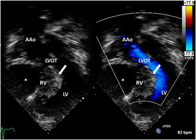
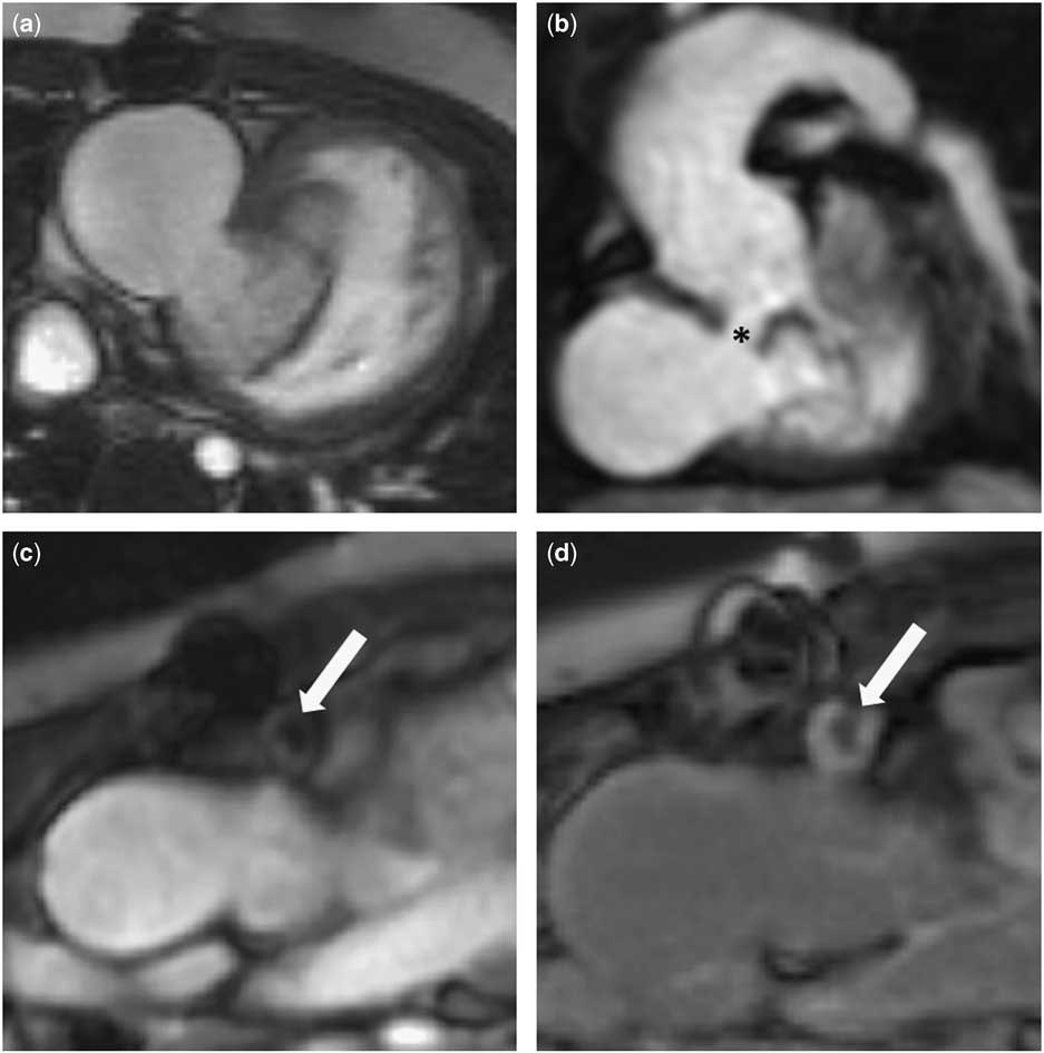
On her most recent clinical assessment, the patient was pink and well with oxygen saturation of 96% in air. There were no signs of cardiac decompensation. Her electrocardiogram and the 24-hour heart rhythm monitoring were free of sustained arrhythmic events. Cardiopulmonary exercise test was negative for ischaemia. The transthoracic echocardiogram showed unobstructed total cavopulmonary connection pathways with no residual fenestration and normal left ventricular systolic function. The interventricular septum was bowing towards the left ventricle, with no significant left ventricular outflow tract obstruction (Fig 1). A CT angiogram was performed to investigate the coronary anatomy because there was suspicion of a coronary fistula, a finding that is commonly seen in this group of patients. The image quality of the CT angiogram, however, was significantly degraded owing to the high heart rate and motion artefacts, but still allowed to demonstrate right coronary dominance with normal origins and course of the major epicardial coronary arteries. No coronary aneurysms or fistulas were seen. Cardiac MRI was performed after the CT scan in order to assess the cardiac anatomy after surgery and as part of the routine long-term follow-up. It showed the hypoplastic and hypertrabeculated right ventricle lying anteriorly to the dominant left ventricle. The hypertrabeculated part communicated with an aneurysmal cavity in the right ventricular outflow tract with a maximum diameter of 43 mm (Fig 2a). In addition, there was a connection between the aneurysm and the left ventricular outflow tract (Fig 2b). Early and late gadolinium enhancement imaging was performed, showing a small thrombus in the aneurysmatic right ventricular outflow tract cavity and extensive hyperenhancement in the hypoplastic right ventricle and the interventricular septum (Fig 2c and d). On the basis of these findings, the patient was commenced on oral anti-coagulant therapy with warfarin. The parent of the patient granted permission to publish the case report.
Transthoracic echocardiography images showing the hypoplastic and hypertrabeculated right ventricle (RV) with the interventricular septum (arrow) bowing towards the left ventricle (LV) and left ventricular outflow tract (LVOT). Retrospectively, the right ventricular outflow tract (RVOT) aneurysm is already seen here (asterisk). AAo=ascending aorta.

Cardiac MRI: ( a ) steady-state free precession (SSFP) short-axis cine image showing the right ventricular outflow tract (RVOT) aneurysm and the hypoplastic right ventricle. ( b ) Non-contrast three-dimensional SSFP magnetic resonance angiography in the coronal plane. Asterisk demonstrates the communication of the aneurysm with the left ventricular outflow tract. ( c and d ) Early and late gadolinium enhancement images showing the hypointense thrombus in the RVOT (arrows).

Discussion
Congenital ventricular septal aneurysms were previously reported in three cases of pulmonary atresia with intact ventricular septum, and they were associated with irregularities of the coronary arteries.Reference Nguyen, Srivastava, Ko and Lai 2 , Reference Patel, Farrell, Harris, Gaynor and Gillespie 3 Acquired ventricular septal aneurysms have also been associated with myocardial ischaemia and infarct of the left ventricle following right ventricle decompression during surgical palliation or progression of existing coronary arterial abnormalities.Reference Patel, Farrell, Harris, Gaynor and Gillespie 3
We describe a patient with pulmonary atresia with intact ventricular septum and an acquired right ventricular outflow tract aneurysm communicating with the left ventricular outflow tract in the presence of a normal coronary anatomy. Early and late gadolinium enhancement imaging showed a small thrombus in the aneurysmatic right ventricular outflow tract, which increased the risk for thromboembolism. The patient was therefore started on oral anti-coagulation with Warfarin.
The cause for the defect between the left ventricular outflow tract and the aneurysm was discussed between surgeons and cardiologists, and it was found that there were some possible explanations, although they remain speculative. First, the right and left ventricular outflow tract communication could be related to partial rupture of the aneurysmal area owing to ischaemia. Cardiac MRI showed areas of myocardial fibrosis in the interventricular septum and right ventricle, but the most recent CT coronary angiogram showed a normal coronary artery anatomy. Second, the defect might be the result of manipulations during previous surgery at the time of ligation of the main pulmonary artery. Finally, we cannot exclude a small congenital ventricular septal defect as the underlying cause.
Septal aneurysms in patients with pulmonary atresia and intact ventricular septum have been previously associated with coronary anomalies.Reference Freedom, Anderson and Perrin 5 However, normal coronary anatomy does not exclude the presence of similar structural abnormalities. This case demonstrates that multimodality imaging is of importance during the long-term follow-up of these patients. It is not only helpful for the assessment of the postoperative anatomy after total cavopulmonary connection but also for detecting complications that may require specific treatment management.
Acknowledgements
None.
Financial Support
This research received no specific grant from any funding agency or from commercial or not-for-profit sectors.
Conflicts of Interest
None.