INTRODUCTION
The domestic cat flea, Ctenocephalides felis (Bouché) is the most abundant ectoparasite of cats and dogs worldwide. The fleas have shown a propensity to develop resistance to insecticides (Bossard et al. Reference Bossard, Dryden and Broce2002), thus becoming arduous to manage and control. In addition to developing new chemical compounds with anti-ectoparasitic activity, other potential avenues to explore include the development of biological control using natural parasites of the flea (Henderson et al. Reference Henderson, Manweiler, Lawrence, Tempelman and Foil1995).
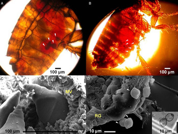
The gregarine protist, Steinina ctenocephali (Ross, Reference Ross1909) (Phylum Apicomplexa) inhabits in the alimentary canal of the cat flea. It colonizes in a newly hatched larva by entry of sporozoites released from ingested oocysts along with the flea feces. The S. ctenocephali develop intracellularly during larval and pupal stages of cat flea, once the cat fleas emerge (as fully grown adult) and start feeding blood, the gregarine move out to lumen of flea gut attaching itself in the intestinal wall, and continues feeding as a trophozoite (Fig. 1A), thus becoming gamonts, which undergo syzygy and secrete a gametocyst wall. The gregarine then produces infective oocysts (Fig. 1D), which are released and propagated via fecal discharge. The fecal matter carrying oocysts also contains undigested blood meal, which serves as food to the flea juveniles, whilst ensuring transmission and perpetuation of the gregarine (Alarcón et al. Reference Alarcón, Huang, Tsai, Chen, Dubey and Wu2011). A wealth of recent work on S. ctenocephali conducted on cat flea populations from Taiwan have addressed diverse topics such as dynamic of infection patterns, life history, morphology, taxonomy and molecular systematics of this gregarine (Alarcón et al. Reference Alarcón, Huang, Tsai, Chen, Dubey and Wu2011, Reference Alarcón, Huang, Dubey and Benitez2013). Interestingly, in such studies S. ctenocephali was the only species of gregarine found associated with this holometabolous insect, which is consistent with the high specificity and monoxenicity usually found among these apicomplexans (Rueckert et al. Reference Rueckert, Chantangsi and Leander2010). Despite passing unnoticed since its description in 1909, recent advances in microscopy and genetics have greatly facilitated the rediscovery of S. ctenocephali and allowed investigations leading to new insights into this organism and its role in the partnership with the cat flea. An important outcome of the recent studies is the realization that flea and gregarine shaped a close relationship over time (Alarcón et al. Reference Alarcón, Huang, Tsai, Chen, Dubey and Wu2011). The gregarine may decrease fitness of cat flea, which may explain the unusually low prevalence of the gregarine in fleas of urban cat and dog populations (Alarcón et al. Reference Alarcón, Huang, Dubey and Benitez2013), but it is unclear how the outcome of this microbe–host interaction affect each partner. Knowing whether the interaction is beneficial (i.e. mutualism), harmful (i.e. parasitic) or without effect on cat flea (i.e. commensalism) will be critical to determine the viability of a biological control programme based on gregarines. In order to investigate this key question, we analysed the effects of S. ctenocephali infection on the survival and development of the cat flea by experimentally manipulating the doses of sporozoites ingested by flea larvae, and monitored the outcome in terms of survival and length of developmental stages. Here, we have shown for the first time that S. ctenocephali rather than negatively affecting the fitness of its host, S. ctenocephali does not change the survival rate of the flea. In addition, we found that S. ctenocephali is capable of manipulating its host life history by accelerating larval development.
Cat flea, Ctenocephalides felis infected with Steinina ctenocephali symbionts. (A) Live female cat flea infected with S. ctenocephali trophozoites observed under stereo zoom microscope (white arrow heads indicate 4 young trophozoites attached to the intestine wall). (B) Live positive artificially infected male flea. A newly emerged flea after feeding blood of cat for 4 days revealed the presence of several freshly/newly transformed sporozoites into early trophozoites (white arrow heads). (C) Scanning electron micrographs (SEM) of trophozoites of S. ctenocephali, partially embedded in the microvilli (MV) brush of cat flea intestinal epithelium. (D) (SEM), lemon-shaped oocysts (arrowheads) were released from ruptured gametocyst (RG). (E) Two lemon-shaped S. ctenocephali oocysts observed from gregarine infected flea feces visualized on a hematocytometer (Hausser Scientific, Horsham).

MATERIALS AND METHODS
Flea collection
Adult infected cat fleas, C. felis were collected from 933 dogs and 197 cats in Taipei Animal Shelter, Taiwan. Fleas were obtained by combing the fur of each cat and dog with a fine flea comb for 10 min. Collected fleas were transferred into plastic tubes, labelled with collection data and maintained individually from each dog and cat, and then taken to the laboratory in the Division of Entomology, National Taiwan University, Taiwan. The whole fleas were prepared as saline solution-mounted slides and quickly examined individually by using a dissecting stereo zoom microscope (Leica Zoom 2000, New York) to confirm S. ctenocephali infection. Finally, gregarine infected fleas were separated and placed into microcells.
Microcells and oocyst collection
Microcells techniques were those used by Thomas et al. (Reference Thomas, Wallenfels and Popiel1996); Hsu and Wu (Reference Hsu and Wu2000); Rust et al. (Reference Rust, Hinkle, Waggoner, Mencke, Hansen and Vaughn2001), and McCoy et al. (Reference McCoy, Broce and Dryden2008). These authors had used basically bandaging-stockinette to prevent the chamber from being dislodged from experimental cats. For this purpose, we modified a commercially available adjustable cat backpack harness, and adapted it as a jacket (Suppl. figure 1, suppl. video 1). Microcells were fitted under the jacket and hold in place by a rubber bracelet. This design is much more efficient and easy to operate, and, moreover, maximum of 12 microcells could be placed simultaneously. Fleas were kept into microcells until gametocysts were dehisced. Under stereomicroscope the absence of gametocyst(s) in the gut was used as indicator that gametocyst(s) had been ruptured and emptied (releasing oocysts) along with the flea feces excreted. Flea feces were removed from the microcells and stored to 4 °C until experiments.
Oocyst manipulation
The concentration of oocysts in feces was determined with a hemacytometer (Bright-Line, Hausser Scientific, Horsham, PA) under BH2 phase contrast microscope (Olympus, Tokyo, Japan) (Fig. 1E). [0·032 gr of feces had a concentration of 270 oocysts/0·001 g (total infective powder, TIP)]. An initial stock of feces containing 100 oocysts mg−1 was prepared (stock infective powder, SIP) from TIP by mixing with porcine blood curd powder (PBC) as diluting solvent. Noticeably, the gregarine-free PBC is an artificial flea larval diet used successfully in the laboratory for optimum full-flea development (Hsu, Reference Hsu2000; Hsu et al. Reference Hsu, Hsu and Wu2002). From SIP and following the procedure described above, we prepared oocyst doses of 50, 25, 12·5, 6·25 oocysts mg−1 and a control only containing PBC free of oocysts.
Flea infection and data recording
The uninfected larvae were obtained from a colony of fleas, which is maintained on cats as hosts, as described by Hsu and Wu (Reference Hsu and Wu2000). To infect flea larvae artificially with S. ctenocephali, we used only 80 wells from a 96-well round-bottom polypropylene tissue culture plates (Costar 3599, Corning Inc., Corning, NY). A total of 16 wells per treatment were loaded individually with 1 mg of infective powder containing 50, 25, 12·5 and 6·25 oocysts mg−1, with a control group included. A single 1-day-old flea egg was placed in each well. The hatching larvae fed on the powder until emergence as adults. A daily record of life history parameters (mortality, instars duration, pupation time, etc.) in each microwell was recorded. To verify gregarine infection, newly emerged fleas (Fig. 1B) were blood fed on cat by 3–5 days using microcells. The experiment was replicated three times, and observations were documented for each replication separately. Although, pseudoreplication is a limitation in an experimental design we used a pseudoreplication in each treatment to minimize uncontrolled source of variance like differences in the amount of powder loaded in each well.
Data analyses
We estimated survival curves of cat flea of different ages by the Kaplan–Meier method using the survfit procedure of the library of survival of the statistical package R. The survival curves of different ages were compared by a log-rank test (survdiff procedure) in the same package. The effect of S. ctenocephali on the developmental time of C. felis were evaluated for different oocyst concentrations by using one-way analysis of variance (ANOVA) followed by the pairwise Tukey's honest significance difference (HSD) test. All analyses were performed in the statistical environment R (R version 2·9·2; Development Core Team).
RESULTS
Steinina ctenocephali is harmless to cat fleas
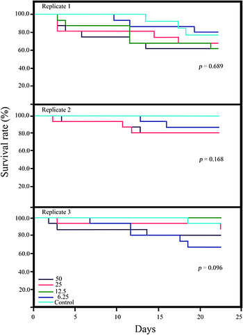
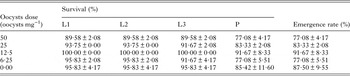
The effects of ingesting different concentrations of gregarine oocysts on the emergence rates of C. felis after three replicates are summarized in Table 1. The survival of fleas exposed to S. ctenocephali since the earliest larval stage, measured as emergence rate of adult fleas was not altered by feeding on different doses of gregarine oocysts spanning 0–50 oocysts mg−1. The adult emergence percentage in exposed fleas ranged from 77 to 91%, a level that falls within the range of previous reports (Hsu et al. Reference Hsu, Hsu and Wu2002) and was essentially similar to the emergence rate in the control group (87·5%) (Table 1). The observed increase in dose 0 is noise but fall within the ranges of the standard error estimate. Besides, no significant difference was observed in survival rate of flea larvae among replicates (P = 0·689, P = 0·168 and P = 0·096, by log-rank test, respectively (Fig. 2). This experiment demonstrates that overall survival through development until adulthood is not affected by gregarine infection, and it also shows that the two feeding methods (i.e. SIP and artificial diet) are optimum diets to reach adulthood.
Survival curves for cat fleas, exposed to different oocyst concentration. Dose-dependent effect of oocysts on the survival rate of cat flea observed for an experimental duration of 25 days. (black – 50), (pink – 25), (green –12·5), (blue – 6·25 oocysts mg−1 and (light blue – control).

Effects of Steinina ctenocephali on survival and emergence rates of Ctenocephalides felis (±s.e. after three replications).

L1, first instar larvae; L2, second instar larvae; L3, third instar larvae; P, pupa.
Steinina ctenocephali accelerates the total development time of the cat flea
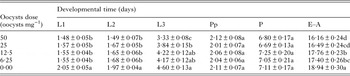
In addition to measuring the effects of gregarine infection on emergence rates, we also looked at the duration of each stage to determine whether the timing of development is affected. We reasoned that by subtracting nutritional resources, the gregarine could alter, presumably delay the progression of the larval stages. Our data show that the total developmental time of fleas exposed to different doses of gregarine oocysts did vary significantly when compared to the control group (Table 2), although in an unexpected way. Fleas fed on feces containing gregarines developed much faster and emerged earlier when compared to the control group (P = 2·2 × 10−16, F = 24·62 for the first instars larvae; P = 1·22 × 10−6, F = 8·84 for the second instar larvae; P = 4·16 × 10−11, F = 15·38 for the third instar larvae and P = 3·50 × 10−13, F = 18·97 for the emergence time, respectively) (Table 2). In contrast, there is no difference in the developmental time for prepupa and pupa between treated and control groups (P = 0·7945, F = 0·42, prepupal stage and P = 0·1384, F = 1·76 pupal stage, respectively).
Effects of Steinina ctenocephali on the development of Ctenocephalides felis (±s.e. after three replications).

Means within a column followed by the same letter are not significantly different (Turkey's HSD test, P < 0·05)
L1, first instar larvae; L2, second instar larvae; L3, third instar larvae; Pp, prepupa; P, pupa; E–A, eggs to adult.
DISCUSSION
Stray dog and cat populations in urban areas pose critical sanitary, economic and social problems. Ectoparasite infestation, which in turn leads to spread of microbial pathogens, allergic conditions and other health concerns, is one of the crucial problems associated with uncontrolled stray domestic animals. Therefore, a deep understanding of the biology of fleas is an important part of a comprehensive effort to mitigate the problems mentioned above. Fleas, like most insects are hosts to a number of microbial eukaryotes that establish symbiotic relationships that are diverse in nature. Among them, gregarines are ubiquitous, but only a few instances of gregarines infecting fleas have been reported (Alarcón et al. Reference Alarcón, Huang, Tsai, Chen, Dubey and Wu2011). A number of studies have addressed different biological and ecological aspects of their interactions (Beard et al. Reference Beard, Butler and Hall1990; De Avelar and Linardi, Reference De Avelar and Linardi2008; Alarcón et al. Reference Alarcón, Huang, Tsai, Chen, Dubey and Wu2011). However, the effects of gregarine infection on the flea, as well as the characteristics of the symbiotic relationship between the partners it is presently unknown. Gregarines are often considered less harmful or even harmless to their hosts. Nevertheless, they divert nutrients toward their own growth, occupy space, and invoke immune reactions. They also cause mechanical injury to gut epithelium providing the port of entry for other microorganisms into the body cavity of the host (Sneller, Reference Sneller1979); altering longevity, and developmental time (Zuk, Reference Zuk2008). Pathogenicity of gregarines has been mostly attributable to their trophozoite stage especially during attachment to host tissue and nutrient acquisition via epimerite (Valigurová, Reference Valigurová2012). In contrast, the effects of trophozoites (Fig. 1C) derived from ingested sporozoites have received little attention. Cryptosporidium, an apicomplexan parasite that is a close relative to gregarine lineages (Templeton et al. Reference Templeton, Enomoto, Chen, Huang, Lancto, Abrahamsen and Zhu2010), is capable of inducing apoptosis in infected cells by increasing membrane permeability (McCole et al. Reference McCole, Eckmann, Laurent and Kagnoff2000), which ultimately results in cell death (Griffiths et al. Reference Griffiths, Moore, Dooley, Keusch and Tzipori1994). The apicomplexan invasion machinery comprehends rhoptries, dense granules, and micronemes, which are participating in recognition of and attachment to the host cell, invasion, and formation of the parasitophorous vacuole where the sporozoite reside (Dubremetz et al. Reference Dubremetz, Garcia-Reguet, Conseil and Fourmaux1998; Singh and Chitnis, Reference Singh and Chitnis2012). Gregarines exhibit similar ultrastructural features, which along with similar modes of attaching to host cells, and a robust phylogenetic association led to propose that gregarine modes of infection could resemble early phases in the evolution of the obligate intracellular parasitism of apicomplexa (Leander, Reference Leander2008). It is thus possible that molecular determinants of pathogenesis and/or cell damage (e.g. cell death, gut epithelium injury) homologous to the apicomplexan invasion machinery are also contributing to the extent of damage caused by the gregarine sporozoite, in addition to the energy expenditure from eliciting host defence.
The gregarine S. ctenocephali passes vertically from flea's parent to progeny via the fecal–oral route, a mode of transmission that has been confirmed by experimental infections with gregarine-infected feces from adult fleas (Beard et al. Reference Beard, Butler and Hall1990; Alarcón et al. Reference Alarcón, Huang, Tsai, Chen, Dubey and Wu2011). Sporozoites of S. ctenocephali are released into the gut of first instar larvae from ingested infective oocysts, developing into sporozoites that feed on the larval intestinal tissue. The effect of different doses of oocysts on the survival and development of cat flea is presented herein for the first time. The doses of S. ctenocephali-invading sporozoites tested are not affecting adult emergence percentage or survival rate of fleas. Our results demonstrate that the S. ctenocephali is not or least parasitic to the off-host stages of the flea sharing symbiotic relationship (Table 1, Suppl. figure 1). To our surprise, we found that high doses of oocysts accelerated the total developmental time of C. felis. In other words, we observed a significant reduction in the number of days spent in the larval stages and the entire time length from eggs to adults when compared with the control group (Table 2). From this and previous findings, we suggest that S. ctenocephali has shaped a very intimate relationship with its host. This conclusion is drawn by mainly two observations. First, Alarcón et al. (Reference Alarcón, Huang, Dubey and Benitez2013) found that S. ctenocephali as well as C. felis peaked simultaneously their populations in summer under optimal conditions of temperature, 20–30 °C and humidity, 53–85% RH, and second, the cycle of S. ctenocephali appears to be coordinated with the host's metamorphosis triggering their exit from the host cells after the first blood meal of newly emerged adult cat flea (Alarcón et al. Reference Alarcón, Huang, Tsai, Chen, Dubey and Wu2011). So, upon an adequate diet a healthy developing larva transformed into adult without being lethally affected by S. ctenocephali at this stage. Therefore, the emergence rates of fleas were even higher than that of previously reported in experimental setting using uninfected fleas from our colony (Hsu and Wu, Reference Hsu and Wu2000; Hsu et al. Reference Hsu, Hsu and Wu2002). Altogether, our data establish with a high degree of confidence that S. ctenocephali is a well-adapted gregarine from the cat flea. We have also unveiled a sophisticated mechanism of the gregarine/cat flea relationship in the line of the manipulative hypothesis (Barnard and Behnke, Reference Barnard and Behnke1990; Poulin, Reference Poulin1994). Our interpretation is that S. ctenocephali has developed the capacity of accelerating, and therefore shortening the development time of the cat flea as a strategy to increase their infection rate (fitness) and transmissibility under favourable environmental conditions to flea development. Noteworthy, L1, L2 and L3 larval stages evidenced a clear alteration of their velocity of development, whereas pre-pupa and pupa did not, leading to speculate that at this point, the intracellular stage of the gregarine is predominantly dormant. A high degree of specialization between host and parasite is a well-known outcome of a long history of coevolution, and it is strikingly illustrated in a coordination of their life cycles (Rothschild and Ford, Reference Rothschild and Ford1964; Prensier et al. Reference Prensier, Dubremetz and Schrevel2008). In some cases, the arms race ensued at the establishment of a symbiotic relationship results in the adoption of manipulative strategies by the parasite. A frequent type of manipulation is the suppression of the immune response in the host, which obviously facilitates the development of the parasite's stages associated with the host. In other cases, the effect of the parasite can lead to gross changes in morphology, behavioural alterations (e.g. increased aggression), reproductive changes (e.g. decreased mating, sterility), etc. Protozoa make only a small fraction of the known cases of host manipulation. Moreover, the most thoroughly studied microbial manipulators are not strictly protozoa but fungi (i.e. Microsporidia), which are known for some spectacular cases of feminizing and sex ratio distortion effects on their hosts (Terry et al. Reference Terry, Smith, Sharpe, Rigaud, Littlewood, Ironside, Rollinson, Bouchon, MacNeil, Dick and Dunn2004). A few examples of manipulators have been described from alveolates, the group of protists that includes gregarines, apicomplexans and ciliates. For example, mosquitoes infected with the ciliate Lambornella clarki Corliss and Coats return to water and display a false oviposition behaviour, which helps the ciliate to infect more mosquito larvae (Egerter et al. Reference Egerter, Anderson and Washburn1986). Some apicomplexan parasites are also known to manipulate their hosts. Plasmodium species impair the blood clotting ability of the mosquito, forcing it to take more but smaller blood meals, resulting in more infected hosts (Koella et al. Reference Koella, Rieu and Paul2002), whereas Toxoplasma can induce elevated aggression and reduces defensive behaviour in mice and rats, increasing the exposure to predators (e.g. cats), which helps the parasite to spread (Webster and Brunton, Reference Webster and Brunton1994; Lagrue and Poulin, Reference Lagrue and Poulin2010). Finally, intracellular stages of gregarines’ closest relatives Cryptosporidium parvum deploy strategies to modulate the apoptotic programme of the host cell to their favour (Heussler et al. Reference Heussler, Kuenzi and Rottenberg2001).
This study shows that the gregarine S. ctenocephali manipulates its host, the cat flea, inducing a notable acceleration of the larval stages. The signal cues behind this acceleration of cat flea development are unknown, but may be hormonal in nature, perhaps triggered by the first blood meal of newly emerged adults (Alarcón et al. Reference Alarcón, Huang, Tsai, Chen, Dubey and Wu2011). Interestingly, early researchers had noticed hormone-associated correlations between both protozoan flagellates and particular gregarines (Cleveland, Reference Cleveland1959; Corbel, Reference Corbel1964; Porchet-Henneré, Reference Porchet-Henneré1969). Indeed, gregarines infecting flies of genus Schneideria Kochetkova are examples of very particular and unique concomitance host's brain hormones and parasites (Nowlin, Reference Nowlin1922; Malavasi, Reference Malavasi1976).
Identifying the molecular basis of the symbiont-mediated manipulation of the cat flea is very important to understand the significance and evolution of this symbiotic relationship in the context of the origins of parasitism in Apicomplexa. In addition, this avenue of research can lead to artificial disruption of the cat flea development, and contribute to spawn a new generation of biological control strategies.
SUPPLEMENTARY MATERIAL
The supplementary material for this article can be found at https://doi.org/10.1017/S0031182016002122
ACKNOWLEDGEMENTS
We sincerely thank W. J. Wu (NTU) for providing logistic support, suggestions and laboratory facilities; the staffs of Animal Shelter, Taipei (Taiwan) for access to flea sampling; to Y. C. Hsu, M. L. Daza for assisting in flea collections; to M. SM. Margarita for facilities in the Universidad de Concepción, Chile, and to CHS, FCIAR for partly facilitating this research by a grant (no. 386345) from the Natural Sciences and Engineering Research Council, Canada. This study complies with the current law of Taiwan (ROC). This work was carried out at the Division of Entomology, National Taiwan University, 1st Roosevelt Road, Taipei, Taiwan (Republic of China).
FINANCIAL SUPPORT
This research was supported by National Science Council, Taiwan under graduate fellowship (no. 96-2313-B002-053-MY3) to the first author.
AUTHOR CONTRIBUTIONS
M. E. A. conceived, designed and performed the experiments; M. E. A., A. K. D., A. J.-F., R. E. B. and C. H. S. analysed the data; M. E. A., A. J.-F., R. E. B. and C. H. S. contributed reagents/materials/analysis tools; M. E. A., A. K. D., A. J.-F. and C. H. S. wrote the paper.