Introduction
Obsessive-compulsive disorder (OCD) is a chronic disabling neuropsychiatric disorder with a lifetime prevalence of 2–3%. It is characterized by the presence of recurrent and persistent thoughts (obsessions) and/or repetitive behaviors or mental acts (compulsions) (DSM-5, 2013). These symptoms are excessive and time-consuming, and cause considerable economic burden and distress for patients and their families.
The pathophysiology of OCD is not yet fully understood. Alexander and colleagues first described the role of cortico-striato-thalamo-cortical (CSTC) circuitry in OCD in 1986 (Alexander et al., Reference Alexander, DeLong and Strick1986), and since then, the structural, functional, and metabolic brain abnormalities of OCD patients in the CSTC loops have been studied using neuroimaging. The CSTC circuits are central to reward processing, action selection, habit formation, and motor control (Harrison et al., Reference Harrison, Pujol, Cardoner, Deus, Alonso, Lopez-Sola, Contreras-Rodriguez, Real, Segalas, Blanco-Hinojo, Menchon and Soriano-Mas2013; Hou et al., Reference Hou, Zhao, Zhang, Song, Wu, Wang, Zhou, Xie, He, Guo, Qu and Li2014; Posner et al., Reference Posner, Marsh, Maia, Peterson, Gruber and Simpson2014). It is currently accepted that the CSTC circuits can be subdivided into three specific loops: the limbic, sensorimotor, and cognitive loops. The limbic loop consists of the orbitofrontal cortex (OFC) and anterior cingulate cortices (ACC), the sensorimotor loop is made up of the sensorimotor cortices, and the cognitive loop includes the dorsolateral prefrontal cortex (DLPFC) and superior frontal gyrus (SFG) (Lehericy et al., Reference Lehericy, Ducros, Van de Moortele, Francois, Thivard, Poupon, Swindale, Ugurbil and Kim2004; Yin and Knowlton, Reference Yin and Knowlton2006; Di Martino et al., Reference Di Martino, Scheres, Margulies, Kelly, Uddin, Shehzad, Biswal, Walters, Castellanos and Milham2008).
The striatum is the main gray matter nucleus in the center of the brain, and it is an important structure for reward learning, inhibition, goal-directed and routine behaviors, and emotion (Balleine and O'Doherty, Reference Balleine and O'Doherty2010). The striatum also plays an important role in the CSTC loops. Functional impairment of the striatum could result in a failure to completely integrate information about the end of a given procedure, possibly leading to ritualized compulsive behaviors (Martos and Braz, Reference Martos and Braz2017). The striatum is heterogeneous, its sub-regions such as pre/post putamen, caudate nucleus, and ventral striatum, receive fiber projection from different cortices (Voorn et al., Reference Voorn, Vanderschuren, Groenewegen, Robbins and Pennartz2004; Crittenden and Graybiel, Reference Crittenden and Graybiel2011). While the ventral striatum, putamen have showed association with decreased or increased whole-brain global brain connectivity in OCD patients, separately (Anticevic et al., Reference Anticevic, Hu, Zhang, Savic, Billingslea, Wasylink, Repovs, Cole, Bednarski, Krystal, Bloch, Li and Pittenger2014).
Di Martino and colleagues have divided the striatum into six regions and explored the differences of functional connectivity (FC) between the seeds and the whole brain. Inferior ventral striatum (VSi) predicted activation within regions implicated in emotional processing, whereas superior ventral striatum (VSs) predicted activity in regions associated with executive function, decision making, and motor planning. The dorsal caudate (DC) receives projections from the DLPFC, and was more highly correlated with the cognitive control. While the putamen has anatomical connectivity (Lehericy et al., Reference Lehericy, Ducros, Van de Moortele, Francois, Thivard, Poupon, Swindale, Ugurbil and Kim2004) and high degree of coactivation with supplementary motor cortex (Postuma and Dagher, Reference Postuma and Dagher2006), is the main motor structure in the striatum. The dorsal putamen positively correlated with primary and supplementary motor cortex and ACC, and the dorsal caudal putamen (DCP) and dorsal rostral putamen (DRP) only show the difference of connection strength with aforementioned regions. The ventral rostral putamen seed positively correlated with regions associated with conflict monitoring and error related processes. Thus, the segmentation of the striatum into sub-regions and using them as regions of interest (ROIs) will add precision to the analysis of whole-brain FC.
The cortical part of the CSTC loop consists of the OFC and ACC. The OFC receives input from multimodal sensory regions and projects to the striatum (Jarbo and Verstynen, Reference Jarbo and Verstynen2015). The OFC can switch between habitual and goal-directed behaviors following outcome evaluation (Gremel and Costa, Reference Gremel and Costa2013). The ACC has been characterized as a part of the executive control cortex, and it includes specific processing modules for emotional information (Paus, Reference Paus2001). The sensorimotor cortices, including the supplementary motor area (SMA) and caudal cingulate motor area, are involved in movement execution, sequence generation, and motor learning. These regions project to the striatum (McFarland and Haber, Reference McFarland and Haber2000) and are components of the sensorimotor CSTC loop. The DLPFC receives fibers from the putamen (Lehericy et al., Reference Lehericy, Ducros, Van de Moortele, Francois, Thivard, Poupon, Swindale, Ugurbil and Kim2004) and, together with the SFG, has been reported to play an important role in a variety of cognitive functions (Li et al., Reference Li, Qin, Liu, Fan, Wang, Jiang and Yu2013). Dysregulation of these circuits contributes to numerous forms of psychopathology.
Previous functional magnetic resonance imaging (fMRI) studies of OCD have shown FC changes of the SMA, such as increased FC with the ACC (Yang et al., Reference Yang, Hu, Tang, Li, Yang, Gong and Huang2019) and caudate (Chen et al., Reference Chen, Juhas, Greenshaw, Hu, Meng, Cui, Ding, Kang, Zhang, Wang, Cui and Li2016) and decreased FC with the putamen (Harrison et al., Reference Harrison, Soriano-Mas, Pujol, Ortiz, Lopez-Sola, Hernandez-Ribas, Deus, Alonso, Yucel, Pantelis, Menchon and Cardoner2009). Studies on OCD patients have reported increased FC between the DLPFC and ventral striatum (Sakai et al., Reference Sakai, Narumoto, Nishida, Nakamae, Yamada, Nishimura and Fukui2011) and decreased FC between the DLPFC and dorsal striatum (Harrison et al., Reference Harrison, Soriano-Mas, Pujol, Ortiz, Lopez-Sola, Hernandez-Ribas, Deus, Alonso, Yucel, Pantelis, Menchon and Cardoner2009). The SFG in OCD patients has shown abnormal in structure and function (Radua and Mataix-Cols, Reference Radua and Mataix-Cols2009; Chen et al., Reference Chen, Juhas, Greenshaw, Hu, Meng, Cui, Ding, Kang, Zhang, Wang, Cui and Li2016). Meta-analyses have also reported structural and functional alterations in the limbic loop of OCD patients (Hu et al., Reference Hu, Du, Chen, Li, Zhou, Zhang, Liu, Lu, Mreedha, Huang and Gong2017; Thorsen et al., Reference Thorsen, Hagland, Radua, Mataix-Cols, Kvale, Hansen and van den Heuvel2018). The dysregulation of regions within these circuits could result in excessive reactions to threats, reduced responses to rewards, and eventually the symptoms of OCD (Ahmari et al., Reference Ahmari, Spellman, Douglass, Kheirbek, Simpson, Deisseroth, Gordon and Hen2013). However, the previous findings on the limbic loop in OCD patients are inconsistent. For example, studies with neuroimaging have reported OFC volumes increased (Tang et al., Reference Tang, Zhu, Gong, Zhu, Wang and Chen2016) and decreased (Atmaca et al., Reference Atmaca, Mermi, Yildirim and Gurok2016), putamen with volumes increased (Rotge et al., Reference Rotge, Langbour, Guehl, Bioulac, Jaafari, Allard, Aouizerate and Burbaud2010) and decreased (Hoexter et al., Reference Hoexter, de Souza Duran, D'Alcante, Dougherty, Shavitt, Lopes, Diniz, Deckersbach, Batistuzzo, Bressan, Miguel and Busatto2012). The results of OCD studies using fMRI are also inconsistent. While most prior resting-state functional magnetic resonance imaging (rs-fMRI) studies support hyperconnectivity in the limbic CSTC loops (Harrison et al., Reference Harrison, Soriano-Mas, Pujol, Ortiz, Lopez-Sola, Hernandez-Ribas, Deus, Alonso, Yucel, Pantelis, Menchon and Cardoner2009; Sakai et al., Reference Sakai, Narumoto, Nishida, Nakamae, Yamada, Nishimura and Fukui2011; Beucke et al., Reference Beucke, Sepulcre, Talukdar, Linnman, Zschenderlein, Endrass, Kaufmann and Kathmann2013) in OCD patients, two recent studies reported opposite results (Posner et al., Reference Posner, Marsh, Maia, Peterson, Gruber and Simpson2014; Bernstein et al., Reference Bernstein, Mueller, Schreiner, Campbell, Regan, Nelson, Houri, Lee, Zagoloff, Lim, Yacoub and Cullen2016). The inconsistency of these findings could be due to the heterogeneity of OCD (Jang et al., Reference Jang, Kim, Jung, Choi, Jung, Lee, Choi, Kang and Kwon2010) or could be related to the influence of antidepressant pharmacotherapy on the brain.
The first-line treatment modality for OCD is selective serotonin reuptake inhibitors (SSRIs). These drugs have been associated with various brain changes, including increasing gray matter (GM) volume in putamen (Hoexter et al., Reference Hoexter, de Souza Duran, D'Alcante, Dougherty, Shavitt, Lopes, Diniz, Deckersbach, Batistuzzo, Bressan, Miguel and Busatto2012), increasing FC between the ACC and ventral striatum (Anand et al., Reference Anand, Li, Wang, Wu, Gao, Bukhari, Mathews, Kalnin and Lowe2005), increasing the amplitude of low frequency fluctuation values in OFC (Zhu et al., Reference Zhu, Qiu, Meng, Cui, Zhang, Huang, Zhang, Li, Gong, Zhang and Lui2015), and increasing small-world efficiency (Shin et al., Reference Shin, Jung, He, Wang, Shim, Byun, Jang, Kim, Lee, Park and Kwon2014). It has also been demonstrated that modulations in serotonergic neurotransmission can mediate neuroplasticity in many of the cortical and subcortical regions involved in OCD (Soumier et al., Reference Soumier, Banasr, Kerkerian-Le Goff and Daszuta2010). These studies support the idea that the therapeutic effects of SSRIs can alter the structure and function of the brain. Previous studies on OCD have relied on samples of patients who were medicated or off medication for 8 weeks (Hou et al., Reference Hou, Wu, Lin, Wang, Zhou, Guo, Gu, He, Ahmed, Hu, Qu and Li2012; Beucke et al., Reference Beucke, Sepulcre, Talukdar, Linnman, Zschenderlein, Endrass, Kaufmann and Kathmann2013; Harrison et al., Reference Harrison, Pujol, Cardoner, Deus, Alonso, Lopez-Sola, Contreras-Rodriguez, Real, Segalas, Blanco-Hinojo, Menchon and Soriano-Mas2013), which may confound the results of studies.
In this study, we aim to evaluate the functional brain abnormalities associated with OCD and the effect of medication. We hypothesize that the FC within the limbic CSTC loop is reduced in drug-naive OCD patients compared with the healthy control (HC) subjects. We recruited a total of 90 participants that included two subgroups of OCD patients (drug-naïve and drug-free) and a group of HC subjects. We measured FC using the temporal correlation of regional BOLD signals in rs-fMRI. To study the pharmacotherapy effects of SSRIs, we examined group differences between the drug-naïve and drug-free groups.
Methods
Subjects
Thirty drug-naïve and 30 drug-free OCD patients (based on the DSM-IV criteria) were recruited between September 2013 and October 2016 from the Shanghai Mental Health Center, China. Thirty age-, gender-, and education level-matched HCs were recruited through local advertisements (see Table 2 for sample characteristics).
All subjects were 18–55 years old, right-handed, and had at least a junior high school education level. All were free of a history of drug or alcohol abuse, craniocerebral injury, serious physical illness, and other contraindications for MRI such as being pregnant or preparing to get pregnant. The subjects were screened using the Mini-International Neuropsychiatric Interview (M.I.N.I.) (Sheehan et al., Reference Sheehan, Lecrubier, Sheehan, Amorim, Janavs, Weiller, Hergueta, Baker and Dunbar1998) to assess whether there was a history of any DSM-IV psychiatric disorder.
The OCD patients had a Yale-Brown Obsessive-Compulsive Scale (Y-BOCS) (Goodman et al., Reference Goodman, Price, Rasmussen, Mazure, Delgado, Heninger and Charney1989) total score of ⩾16. The drug-naïve OCD patients have never taken any kind of psychotropic medication. The drug-free group had exposed to psychotropic medication and to have been off-medication for at least 8 weeks before the MRI scanning. The exclusion criteria for the OCD group included (1) any Axis I psychiatric disorders except OCD (assessed using M.I.N.I.) and (2) had high suicide risk. The HC subjects had no history of psychiatric illness, as determined by the M.I.N.I., and they reported no psychiatric treatment history in any of their first-degree relatives.
This study was approved by the Research Ethics Committee at Shanghai Mental Health Center, Shanghai Jiao Tong University School of Medicine. Written informed consent was obtained from each subject.
Clinical measures
The severity of OCD, depression, and anxiety symptoms was assessed using the Y-BOCS, Beck Depression Inventory-second edition (BDI-II, Beck et al., Reference Beck, Steer, Ball and Ranieri1996), and Beck Anxiety Inventory (BAI, Steer et al., Reference Steer, Rissmiller, Ranieri and Beck1993), respectively. We administered a questionnaire to the OCD patients and their families to collect the general condition of the patients, including the age of onset (age at which the individual sensed the symptoms affected his/her daily life), duration of illness, family history of any psychiatric illness, and medication history of the patients.
Image acquisition
MRI data were acquired using a 3.0T Siemens Verio scanner (Siemens, Erlangen, Germany) with a 12-channel head matrix receive coil. The scanner was located in the Department of Radiology at the Shanghai Mental Health Center, Shanghai, China. Foam pads and earplugs were used to reduce head motion and scanner noise, respectively. Subjects were instructed to close their eyes, be still, relax, avoid falling asleep, and try not think of anything in particular.
The fMRI data were acquired using an echo-planar imaging (EPI) sequence with the following parameters: repetition time (TR) = 3000 ms; echo time (TE) = 30 ms; flip angle (FA) = 85°; field of view = 216 mm; data matrix = 72 × 72; slice thickness = 3.0 mm; inter-slice gap = 0 mm; voxel size = 3.0 mm isotropic (scan time: 8 min, 38 s). For each subject, an additional high-resolution anatomical image was acquired using a T1-weighted magnetization-prepared rapid gradient-echo (3D MP-RAGE) sequence in the sagittal plane (192 slices, TR = 2300 ms, TE = 2.96 ms, FA = 9°, data matrix = 256 × 256 voxels, voxel size = 1 mm isotropic). No participants displayed structural abnormalities upon visual inspection.
Data preprocessing
MRI data analysis was carried out using Data Processing Assistant for Resting-State fMRI software (DPABI_V2.2) (http://www.rfmri.org/dpabi) (Yan et al., Reference Yan, Wang, Zuo and Zang2016), which integrates SPM8 (http://www.fil.ion.ucl.ac.uk/spm/software/spm8) and REST (http://restfmri.net/forum/REST_V1.8). The first 10 volumes were removed to allow for equilibrium of the MR signal. We performed correction for slice timing effects and realignment for head motion. The head motion of all participants during resting-state fMRI acquisition was examined, five HCs and nine OCD patients were excluded for had more than 1.5° of maximal rotation and 1.5 mm of maximal translation. The functional images were spatially normalized into standard space (Montreal Neurological Institute, MNI) using the standard EPI template. The normalized images were checked by visual inspection, and no subject was excluded due to poor registration and normalization. The data were spatially smoothed (Gaussian kernel of 6 mm full width at half-maximum). The time series were further cleaned by regressing out the mean global signal, white matter signal, and cerebrospinal fluid signal. Then the time series were filtered with a band-pass filter (0.01–0.08 Hz).
After preprocessing, we had 29 drug-naïve OCD patients (DN group), 22 drug-free OCD patients (DF group), and 25 HC group that were included in the analysis.
Statistical analysis
Regions of interest and seed generation
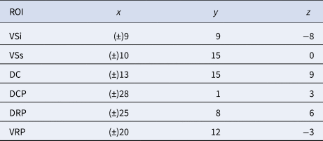
Based on previous work, we included 12 ROIs in this study. For each hemisphere, six MNI coordinates were specified and a spherical mask of 4 mm radius were created around each center voxel (Di Martino et al., Reference Di Martino, Scheres, Margulies, Kelly, Uddin, Shehzad, Biswal, Walters, Castellanos and Milham2008) (see Table 1).
Coordinates for striatal ROI

ROI, regions of interest; VSi, inferior ventral striatum; VSs, superior ventral striatum; DC, dorsal caudate; DCP, dorsal caudal putamen; DRP, dorsal rostral putamen; VRP, ventral rostral putamen.
Coordinates for right and left hemisphere seeds defined in the MNI stereotaxic space.
To create correlation maps for the seed ROIs, the Pearson correlation coefficients were computed between the seed ROIs and voxels in the rest of the brain. We then applied the Fisher's r-to-z transformation to improve the Gaussianity of the distribution. A one sample t test (base = 0) on the HC data contained in the zFC.nii file was used to generate whole-brain rsFC maps. We calculated one sample t test (base = 0) for the HC, DF, and DN groups based on the different ROIs to generate separate hyperconnectivity and hypoconnectivity maps for each ROI. These maps are named i1, i2, and i3 (hyperconnectivity/hypoconnectivity). Finally, we used the REST Image Calculator (i = i1 + i2 + i3) to compute the masks based on an analysis of variance (ANOVA).
For each voxel, a one-way ANOVA was performed with group as an independent variable and the FC as a dependent variable. Age, gender, and duration of illness were included as covariates. Multiple comparisons for the one-way ANOVA were corrected using Monte Carlo stimulations. The statistical threshold was set at p corrected < 0.05, with the multiple comparison correction performed using Alphasim (individual voxel p uncorrected = 0.005, number of iterations = 1000). For each Alphasim correction, the amount of Gaussian smoothing was re-estimated before the computing of each corresponding threshold of cluster size (see Table 4).
We then extracted the FC of significant group difference and performed further analyses in SPSS 19.0. The differences in demographic and clinical characteristics between the OCD and HC groups were detected with two-sample t tests (continuous data) or a χ2 test (categorical data), as appropriate. Correlations between the severity of OCD symptoms and FC were examined. A difference was considered statistically significant at p < 0.05.
Results
Subjects
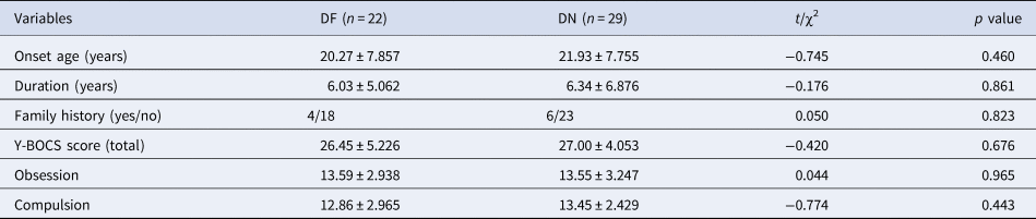
The characteristics and clinical assessments for OCD patients and HCs are summarized in Table 2. The mean duration of off-medication before the MRI scan was 12 months (s.d. = 43.09; range: 4–39 months) for OCD patients in the drug-free group, excluding the missing values. There were no significant differences in age, gender, or education among the three groups (all p > 0.05) (see Table 2). There were no significant differences in the age of onset, duration of illness, or severity of symptoms (Y-BOCS) between the DF and DN groups (all p > 0.05) (see Table 3).
Demographic and clinical data of OCD and HC (mean ± SD)

OCD, obsessive-compulsive disorder; DF, drug-free OCD patients; DN, drug-naïve OCD patients; HCs, healthy controls; BDI-II, Beck Depression Inventory-II; BAI, Beck Anxiety Inventory.
Comparison of clinical data between DF and DN groups
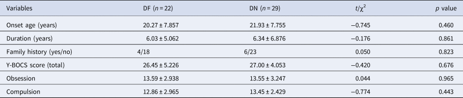
OCD, obsessive-compulsive disorder; DF, drug-free OCD patients; DN, drug-naïve OCD patients; Family history, family history of any psychiatric illness; Y-BOCS, the Yale-Brown Obsessive Compulsive Scale.
Functional connectivity
Functional connectivity in healthy controls
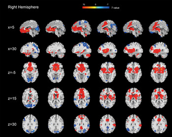
The 25 HCs were included in the ROI-based FC analysis. We conducted a one sample t test (base = 0), and the data from the HC group in the zFC.nii file were used to generate the whole-brain FC maps for the 12 striatal seeds (see Fig. 1). Only the map generated from the right striatal seeds in HC is shown, but similar patterns were obtained for the left side (see online Supplementary material).
Right striatal seeds to whole brain connectivity analysis in HCs (p < 0.05 FDR corrected). VSi, inferior ventral striatum; VSs, superior ventral striatum; DC, dorsal caudate; DCP, dorsal caudal putamen; DRP, dorsal rostral putamen; VRP, ventral rostral putamen.

In the HC group, the ventral striatum was positively correlated with the OFC, ACC, temporal lobe, hippocampus, basal ganglia, and was negatively correlated with the cuneus, occipital areas, and lingual gyrus. In contrast, activity in the putamen was positively correlated with the ACC, basal ganglia, insula, temporal lobe and negatively correlated with the posterior cingulate cortex in the HC group.
Functional connectivity of the three groups
Seed-based functional connectivity analyses
First, we conducted a comparison of FCs between the OCD (DN and DF groups) and HC group, the results are presented in the online Supplementary material (eTable 9). Then we conducted an ANOVA of three groups.
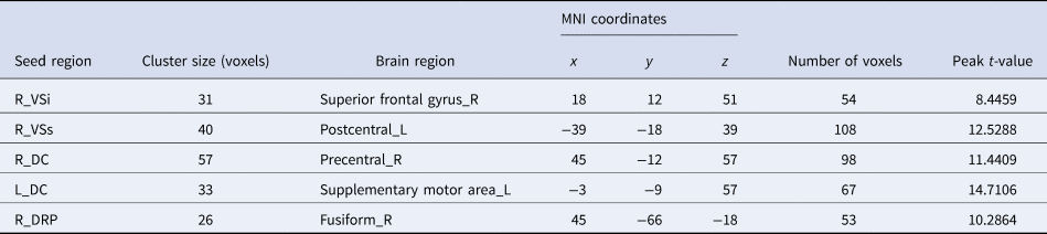
The group differences in positive FC mainly included the OFC, medial SFG, middle temporal gyrus (MTG), thalamus, and SMA (Table 4). The group differences in negative FC mainly included the SFG, postcentral gyrus, precentral gyrus, SMA, and fusiform gyrus (see Table 5).
Brain regions demonstrating positive FC in the ANOVA

ROI, regions of interest; VSi, inferior ventral striatum; VSs, superior ventral striatum; DC, dorsal caudate; DCP, dorsal caudal putamen; DRP, dorsal rostral putamen; VRP, ventral rostral putamen. L, left; R, right; MNI, Montreal Neurological Institute.
Note: List of the brain regions showing a significant (p corrected <0.05 with Alphasim correction, individual voxel p uncorrected <0.005) positive FC with the seed regions among three groups.
Brain regions demonstrating negative FC in the ANOVA
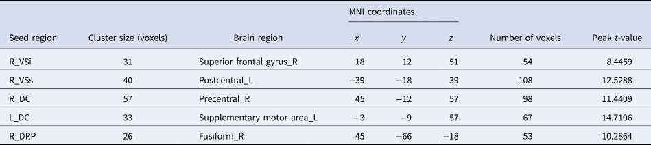
ROI, regions of interest; VSi, inferior ventral striatum; VSs, superior ventral striatum; DC, dorsal caudate; DCP, dorsal caudal putamen; DRP, dorsal rostral putamen; VRP, ventral rostral putamen. L, left; R, right; MNI, Montreal Neurological Institute.
Note: List of the brain regions showing a significant (p corrected <0.05 with Alphasim correction, individual voxel p uncorrected <0.005) negative FC with the seed regions among three groups.
Post-hoc analyses
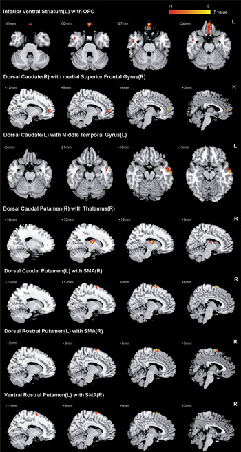
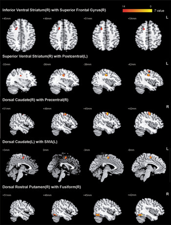
We extracted the FC of significant group difference in the ANOVA analysis (Fig. 2 and 3) and used SPSS to complete the post-hoc analyses. A Bonferroni correction (p < 0.05, three comparisons) was then performed for the post-hoc pairwise comparisons, and even with more strict multiple comparison corrections, our main results still exist.
Positive FC among three groups in ANOVA (p < 0.005). L, left; R, right.

Negative FC among three groups in ANOVA (p < 0.005). L, left; R, right.

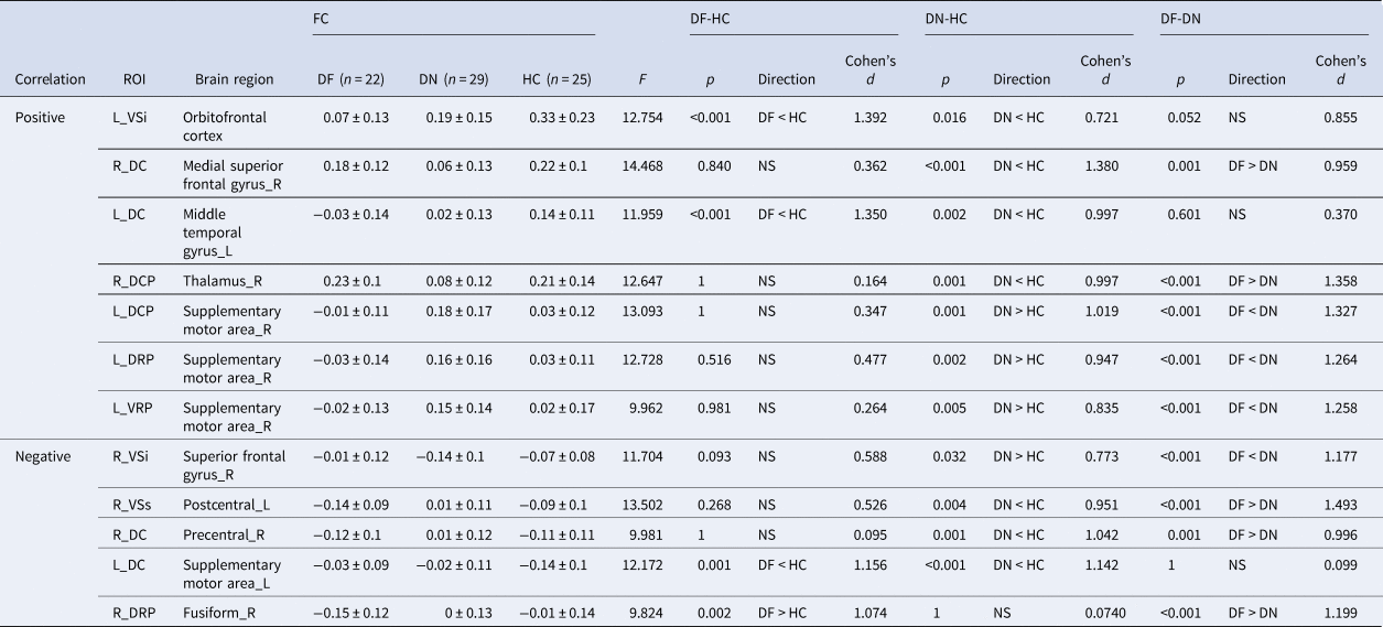
Brain regions with positive functional connectivity: Table 6 shows the seed regions with positive FC in the groups. Compared with the HC group, the DN group had decreased FC between the left VSi with OFC, the right DC with medial SFG, the left DC with left MTG, and the right DCP with right thalamus. FC was increased between the left dorsal and ventral putamen and right SMA.
Group-comparison results of post-hoc analyses

ROI, regions of interest; VSi, inferior ventral striatum; VSs, superior ventral striatum; DC, dorsal caudate; DCP, dorsal caudal putamen; DRP, dorsal rostral putamen; VRP, ventral rostral putamen. DF, drug-free, DN, drug-naïve, HC, healthy controls; FC, functional connectivity; L, left; R, right.
Note: Bonferroni correction (p < 0.05, three comparisons) was performed for the post-hoc analyses.
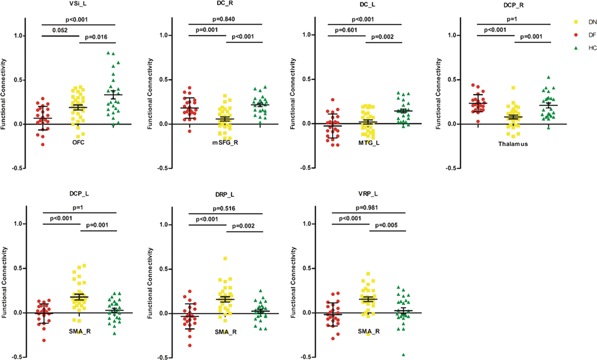
Compared with the HC group, the DF group had decreased FC between the left VSi and OFC, the left DC with left MTG. Compared with the DN group, the DF group had increased FC between the right DC with medial SFG, the right DCP with right thalamus. The FC was decreased between the left dorsal and ventral putamen with right SMA. The positive FC values in every subject are presented in Fig. 4.
Comparison of positive FC among three groups. The y axis shows the values extracted from the significant positive FC. VSi, inferior ventral striatum; DC, dorsal caudate; DCP, dorsal caudal putamen; DRP, dorsal rostral putamen; VRP, ventral rostral putamen.; OFC, orbitofrontal cortex; mSFG, medial superior frontal gyrus; MTG, middle temporal gyrus; SMA, supplementary motor area; SFG, superior frontal gyrus. p < 0.05, Bonferroni corrected.

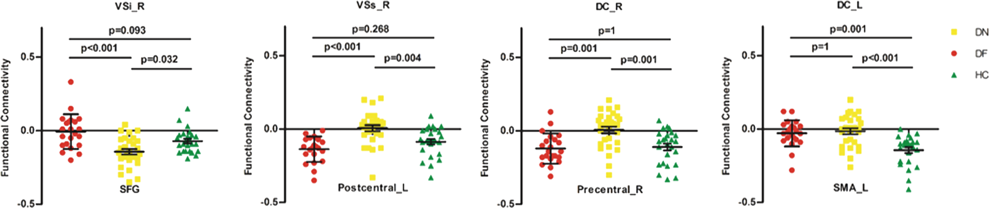
Brain regions with negative functional connectivity: The lower half of Table 6 shows the seed regions with negative FC in the groups. Compared with the HC group, the DN group had increased FC between the right VSi with right SFG. The FC was decreased between the right VSs with the left postcentral gyrus, the right DC with right precentral gyrus, and the left DC with left SMA.
The FC between the left DC with left SMA was decreased in the DF group compared with the HC group. In contrast, the FC between the right DRP and the right fusiform was increased.
Compared with the DN group, the DF group had decreased FC between the right VSi with right SFG. The FC was increased between the right VSs with right postcentral gyrus, the right DC and right precentral gyrus, and the right DRP and right fusiform gyrus. The negative FC values in every subject are presented in Fig. 5.
Comparison of negative FC among three groups. The y axis shows the values extracted from the significant negative FC. VSi, inferior ventral striatum; VSs, superior ventral striatum; DC, dorsal caudate; SFG, superior frontal gyrus; SMA, supplementary motor area. p < 0.05, Bonferroni corrected.

Post-hoc analyses of clinical scales: Both drug-naïve and drug-free OCD patients showed higher scores of depression and anxiety scales relative to HC group. There was no significant difference between drug-naïve and drug-free groups in these two scales (see Table 7).
The post-hoc results of depression and anxiety scales

BDI-II, Beck Depression Inventory-second edition; BAI, Beck Anxiety Inventory; DF, drug-free, DN, drug-naïve, HC, healthy controls.
Note: Bonferroni correction (p < 0.05, three comparisons) was performed for the post-hoc analyses.
Association of connectivity with OCD symptom severity
We found no correlations among FC and age of onset, duration, or symptom severity (Y-BOCS and its sub-scale scores) for the OCD groups (DN, DF, DN + DF) (p > 0.05).
Discussion
We examined resting-state FC alternations in patients with OCD. In both drug-naïve and drug-free groups, we identified FC alterations, mainly in the limbic CSTC loop (the OFC and thalamus), the sensorimotor CSTC loop (the SMA), and the cognitive CSTC loop (the SFG). Moreover, in some brain regions, we did not find significant difference of FC between the drug-free OCD group and HC. These findings suggest that OCD patients have functional abnormalities within brain regions of CSTC loops, and exposure to antidepressants such as SSRIs may affect the function of some brain regions.
In the drug-naïve OCD group, FC was decreased between the left VSi with OFC, and the DCP with thalamus, relative to the HCs. These findings are consistent with previous studies of drug-naïve OCD patients (Posner et al., Reference Posner, Marsh, Maia, Peterson, Gruber and Simpson2014; Yang et al., Reference Yang, Hu, Tang, Li, Yang, Gong and Huang2019), and as opposed to studies of medicated subjects (Harrison et al., Reference Harrison, Soriano-Mas, Pujol, Ortiz, Lopez-Sola, Hernandez-Ribas, Deus, Alonso, Yucel, Pantelis, Menchon and Cardoner2009, Reference Harrison, Pujol, Cardoner, Deus, Alonso, Lopez-Sola, Contreras-Rodriguez, Real, Segalas, Blanco-Hinojo, Menchon and Soriano-Mas2013). Recently, Jung and colleagues found decreased small-world efficiency of the limbic CSTC loop in OCD patients (Jung et al., Reference Jung, Yucel, Yun, Yoon, Cho, Parkes, Kim and Kwon2017), which also supports our study. Clinically, striatal dysfunction has been shown to lead to inefficient thalamic gating, resulting in abnormal activation of OFC (Del Casale et al., Reference Del Casale, Kotzalidis, Rapinesi, Serata, Ambrosi, Simonetti, Pompili, Ferracuti, Tatarelli and Girardi2011), which has been related to goal-directed dysfunction in OCD (Gillan et al., Reference Gillan, Apergis-Schoute, Morein-Zamir, Urcelay, Sule, Fineberg, Sahakian and Robbins2015). OCD patients often exaggerate the adverse consequences of threats, continuously focusing on their obsession, and cannot make accurate judgments about the negative emotions caused by the compulsive behaviors. With regard to the ACC, we did not find evident changes in its FC with the striatum, which is consistent with some previous findings (Sakai et al., Reference Sakai, Narumoto, Nishida, Nakamae, Yamada, Nishimura and Fukui2011; Bernstein et al., Reference Bernstein, Mueller, Schreiner, Campbell, Regan, Nelson, Houri, Lee, Zagoloff, Lim, Yacoub and Cullen2016). Differences in data processing methods and symptom dimensions of OCD might lead to difference in findings.
The FC observed between the DCP with the thalamus did not differ between the DF and HC groups. Emerging longitudinal studies have indicated that pharmacotherapy alters the structure and function of several brain regions, including the putamen and thalamus (Hoexter et al., Reference Hoexter, de Souza Duran, D'Alcante, Dougherty, Shavitt, Lopes, Diniz, Deckersbach, Batistuzzo, Bressan, Miguel and Busatto2012; Atmaca et al., Reference Atmaca, Mermi, Yildirim and Gurok2016; Bernstein et al., Reference Bernstein, Cullen, Harris, Conelea, Zagoloff, Carstedt, Lee and Mueller2019), our results indirectly suggest that exposure to stable SSRIs does increase the FC within the limbic CSTC loop, in agreement with a previous work (Heller et al., Reference Heller, Johnstone, Light, Peterson, Kolden, Kalin and Davidson2013). However, acute and chronic treatment with antidepressants appears to cause opposite changes of FC. Studies of healthy subjects have reported widespread decreased connectivity in most cortical and subcortical areas as acute influence (i.e. scanning after hours of antidepressant intake) (Schaefer et al., Reference Schaefer, Burmann, Regenthal, Arelin, Barth, Pampel, Villringer, Margulies and Sacher2014; Klaassens et al., Reference Klaassens, Rombouts and Winkler2017), while longitudinal treatment studies (scanning after months of antidepressant intake) of OCD patients supported increased connectivity in the limbic loop (Anand et al., Reference Anand, Li, Wang, Wu, Gao, Bukhari, Mathews, Kalnin and Lowe2005; Heller et al., Reference Heller, Johnstone, Light, Peterson, Kolden, Kalin and Davidson2013). Animal study has also reported that acute and chronic treatment with antidepressants can lead to opposite endothelial nitric oxide mRNA expression in most brain regions, which can modulates major neurotransmitters such as norepinephrine, serotonin, dopamine, and glutamate (Yoshino et al., Reference Yoshino, Ochi, Yamazaki, Nakata, Iga and Ueno2017). The initial treatment of antidepressants would increase the risk of suicidal behavior in children and adolescent patients with major depression, supporting that effect of acute and chronic treatment with antidepressants may be opposite. Future longitudinal studies are needed to reveal the pharmacotherapeutic mechanisms in these loops.
We also found increased positive FC between the dorsal and ventral putamen with SMA in the DN relative to the HC group. As one of the major components of the sensorimotor CSTC loop, the SMA rapidly evaluates successful and erroneous actions and thus mediates action monitoring (Bonini et al., Reference Bonini, Burle, Liegeois-Chauvel, Regis, Chauvel and Vidal2014). Repetitive transcranial magnetic stimulation experiments have shown that the stimulation of the SMA can improve the symptom of OCD (Hawken et al., Reference Hawken, Dilkov, Kaludiev, Simek, Zhang and Milev2016). Prior functional studies have reported that compared with HCs, OCD patients had decreased connectivity (Harrison et al., Reference Harrison, Soriano-Mas, Pujol, Ortiz, Lopez-Sola, Hernandez-Ribas, Deus, Alonso, Yucel, Pantelis, Menchon and Cardoner2009) or no significant difference (Chen et al., Reference Chen, Juhas, Greenshaw, Hu, Meng, Cui, Ding, Kang, Zhang, Wang, Cui and Li2016) between putamen and SMA. Inconsistencies of these abnormalities may be partially interpreted as the difference in medication status. A reduced number of striatal cholinergic interneurons (SCIN) are involved in OCD pathophysiology (Xu et al., Reference Xu, Kobets, Du, Lennington, Li, Banasr, Duman, Vaccarino, DiLeone and Pittenger2015), and works in animals have shown that the ablation of SCINs had no effect on the corticostriatal connectivity across the medial striatum, but act on the over-recruitment of lateral striatal regions involved in triggering and selecting motor responses, resulting in abnormal repetitive behaviors (Martos and Braz, Reference Martos and Braz2017). The hyperconnectivity we observed between lateral striatal regions (mainly the putamen) and SMA could contribute in part to the repetitive behaviors in OCD. In addition, the DF group did not differ significantly from the HC group in terms of FC between the putamen and SMA, suggesting that exposure to medication may alter this connectivity.
Consistent with prior findings (Posner et al., Reference Posner, Marsh, Maia, Peterson, Gruber and Simpson2014; Chen et al., Reference Chen, Juhas, Greenshaw, Hu, Meng, Cui, Ding, Kang, Zhang, Wang, Cui and Li2016; Vaghi et al., Reference Vaghi, Vertes, Kitzbichler, Apergis-Schoute, van der Flier, Fineberg, Sule, Zaman, Voon, Kundu, Bullmore and Robbins2017), we detected reduced FC between the DC with the MTG and the SFG within the cognitive CSTC loop. The MTG and SFG are related to several cognitive processes, including language and semantic memory processing and multimodal sensory integration (Visser et al., Reference Visser, Jefferies, Embleton and Lambon Ralph2012; Li et al., Reference Li, Qin, Liu, Fan, Wang, Jiang and Yu2013). Abnormal FC among these regions could reflect underlying problems in the processing and integration of cognitive information. Meanwhile, the FC between the DC and SFG did not differ between the DF and HC groups. A longitudinal study has indicated that pharmacotherapy alters the function of the SFG (Shin et al., Reference Shin, Jung, He, Wang, Shim, Byun, Jang, Kim, Lee, Park and Kwon2014), also supporting our results. However, we did not find abnormal FC between the DLPFC and striatum, which has been reported in some rs-fMRI studies (Harrison et al., Reference Harrison, Soriano-Mas, Pujol, Ortiz, Lopez-Sola, Hernandez-Ribas, Deus, Alonso, Yucel, Pantelis, Menchon and Cardoner2009; Sakai et al., Reference Sakai, Narumoto, Nishida, Nakamae, Yamada, Nishimura and Fukui2011). The DLPFC is thought to play a crucial role in cognitive function (Li et al., Reference Li, Qin, Liu, Fan, Wang, Jiang and Yu2013). Differences in data processing methods and confounds from comorbid disorders may have caused our failure to detect the alternation in the DLPFC.
In the OCD groups, we also found abnormal FCs in the postcentral, precentral, and fusiform. This finding suggests that apart from the CSTC circuitry, there are other brain areas involved in the pathophysiology models of OCD (Menzies et al., Reference Menzies, Chamberlain, Laird, Thelen, Sahakian and Bullmore2008). We found no correlations among the measures of clinical symptoms and FC. The connectivity between the ventral striatum and OFC has been reported to predict the severity of OCD (Harrison et al., Reference Harrison, Soriano-Mas, Pujol, Ortiz, Lopez-Sola, Hernandez-Ribas, Deus, Alonso, Yucel, Pantelis, Menchon and Cardoner2009), but other work has found no association between the FC and clinical symptoms of OCD (Sakai et al., Reference Sakai, Narumoto, Nishida, Nakamae, Yamada, Nishimura and Fukui2011). A potential explanation for this discrepancy is that functional abnormalities could be related to underlying vulnerabilities in OCD (Chamberlain et al., Reference Chamberlain, Menzies, Hampshire, Suckling, Fineberg, del Campo, Aitken, Craig, Owen, Bullmore, Robbins and Sahakian2008), while different symptom severities might be influenced by mood and environmental factors.
There are several limitations of this study. The intelligence of the participants might not be adequately represented by their educational level, so future studies should assess the intelligence quotient. Further, the ROI-based resting-state FC MRI studies require the a priori selection of ROIs. We attempted to minimize the potential bias in seed selection by limiting our analyses to six seeds in each hemisphere that were based on previously defined striatal regions (Di Martino et al., Reference Di Martino, Scheres, Margulies, Kelly, Uddin, Shehzad, Biswal, Walters, Castellanos and Milham2008). The heterogeneity of symptoms could also influence our findings (Saxena et al., Reference Saxena, Brody, Maidment, Smith, Zohrabi, Katz, Baker and Baxter2004), and the degree of connectivity abnormalities with respect to different OCD symptom dimensions requires more investigation. Finally, FC can assess the temporal coherence between two brain regions, but it does not allow us to draw conclusions about directionality. These issues certainly merit future investigation.
Conclusion
We performed a cross-sectional study to examine FCs among drug-naïve and drug-free OCD patients compared with HCs. The drug-naïve OCD patients had reduced FC within the limbic CSTC loop, and our findings suggest that antidepressant (mainly SSRIs) may differentially modulate the FC among brain regions. These findings provide novel insights into how pharmacotherapy shapes neurofunctional circuits in OCD and might also account for the inconsistency in previous work.
Supplementary material
The supplementary material for this article can be found at https://doi.org/10.1017/S0033291719002988.
Acknowledgements
The authors thank all the subjects who took part in the study.
Financial support
This research was supported by the National Natural Science Foundation of China Grant (81671340; 81971682; 81571756); the Shanghai Municipal Education Commission-Gao Feng Clinical Medicine Grant (20161321; 20171929); the Municipal Human Resources Development Program for Outstanding Leaders in Medical Disciplines in Shanghai (2017BR058), The National Key R&D Program of China (2018YFC2001600), Hundred-Talent Fund from Shanghai Municipal Commission of Health (2018BR17), and Research Funds from Shanghai Mental Health Center (13dz2260500).
Conflict of interest
None.
Ethical standards
The authors assert that all procedures contributing to this work complied with the ethical standards of the relevant national and institutional committees on human experimentation and with the Helsinki Declaration of 1975, as revised in 2008.