




No CrossRef data available.
Article contents
Molecular cloning and partial characterization of a nematode-specific 24 kDa protein from Ascaris suum
Published online by Cambridge University Press: 13 December 2004
Abstract
The cloning and molecular characterization of a cDNA encoding Ascaris suum 24 kDa antigen (As24) are described. The cDNA sequence consists of 853 bp with an open reading frame coding for a protein of 147 amino acids with an inferred signal peptide of 19 amino acids. The predicted molecular mass and pI were 16 kDa and 8·35 respectively. The endogenous protein in adult A. suum was 24 kDa with the expected pI. A search of the public databases revealed over 50% homology with proteins from filarial parasites but not to other known proteins, suggesting that As24 is a nematode-specific protein. Immunohistochemical studies using polyclonal antibodies raised against Escherichia coli-expressed recombinant As24 demonstrated that the endogenous As24 proteins were intensely localized in unembryonated eggs within the uterus, uterine and gut epithelium, muscle tissues and in the hypodermis of an adult female A. suum. Endogenous As24 was expressed throughout A. suum development and was detected in the excretory/secretory products by immunoblot analysis. Importantly, a homologous protein(s) was detected in Ascaris from human and Toxocara canis from dog, suggesting that As24 is a nematode-specific protein.
Keywords
Information
- Type
- Research Article
- Information
- Copyright
- © 2004 Cambridge University Press
INTRODUCTION
The genus Ascaris (Nematoda: Ascaridida) includes two pathogenic species namely Ascaris lumbricoides and Ascaris suum which infect humans and domestic pigs, respectively. A. lumbricoides is the commonest roundworm and is estimated to infect a quarter of the world's human population (Chan, 1997). A. suum is an economically important parasite distributed world-wide and is biochemically well characterized. Recent studies have shown that the pig ascarid is morphologically and antigenically indistinguishable from the human worm and can develop in human hosts (Anderson, Romero-Abal & Jaenike, 1993; Peng et al. 1998; Abebe et al. 2002), suggesting its zoonotic significance. Studies of A. suum are believed to provide important information about the biology of other ascarids in general (Crompton, 2001).
Numerous data have shown that pigs develop strong immunity against A. suum infection following repeated inoculations with UV-attenuated or normal infective eggs (Kelly & Nayak, 1964; Urban & Tromba, 1982; Jungersen et al. 1999; Serrano et al. 2001); however, this may lead to an occasional development of patent infection. Immunization with non-infective material would, therefore, be more practical. Crude antigens from different life-cycle stages of A. suum have been reported to induce host-protective immunity (Urban & Romanowski, 1985; Lukes, 1992; Hill et al. 1994; Serrano et al. 2001). These findings suggest that immunogenic molecules expressed by various developmental stages of A. suum can mediate a protective response(s) against Ascaris infection. However, there are few data that would enable us to elucidate Ascaris-specific potent immunoreactive protein antigens which could become potential targets for a vaccine and/or chemotherapeutic agents.
Recently, four antigenic proteins from A. suum, designated as As37, As14, As16 and AsPPase, have been characterized (Tsuji et al. 2001, 2002, 2003; Islam et al. 2003), which are expressed in all life-cycle stages of the parasites with homologues in Ascaris from humans and Toxocara canis from dogs, and suggested that the A. suum-mouse infection model can be used for identifying immune-protective molecules. Preliminary published studies have also indicated that these molecules may serve as potential vaccine antigens for human and pig ascariasis.
To identify and characterize Ascaris-expressed potential protein antigens, the present study was, therefore, undertaken to describe the molecular cloning and partial characterization of a cDNA encoding a 24 kDa protein from A. suum (As24). Recombinant As24 proteins expressed in Escherichia coli were used for polyclonal antibody production to localize endogenous As24 in adults of Ascaris by immunohistochemical analysis. Attempts were also made to demonstrate expression of the endogenous form of As24 in various developmental stages including excretory/secretory (ES) products of both larval and adult of Ascaris of pig origin. Interestingly, As24 homologous protein was identified in Ascaris from human and T. canis from dog. Our partial characterization of As24 suggests it may be a potential target as a vaccine antigen for human and pig ascariasis.
MATERIALS AND METHODS
Parasites
Adults of Ascaris were obtained from infected pigs at a slaughter house in Shimotsuma, Japan. Unembryonated and embryonated infective eggs, 3rd-stage larvae (L3) from eggs, the lung-stage L3 and ES products from the larval stages and adult worms were collected as previously described (Geenen et al. 1999; Tsuji et al. 2000, 2001). RNA was isolated from L3 using an RNA isolation kit (Clontech). Poly (A)+ mRNA was prepared from total RNA employing a polytract mRNA isolation kit (Clontech), and first-strand cDNA synthesis was performed by using a cDNA synthesis kit and an oligo (dT)15 primer (Amersham Pharmacia Biotech). A L3 cDNA library was constructed in UniZap XR vector (Stratagene) according to the manufacturer's instructions. Adults of Ascaris were recovered from a naturally infected human in Bangladesh, and T. canis from an infected dog in Miyazaki, Japan. Protein concentrations of PBS-soluble parasite antigens and ES products were measured using the Micro-BCA protein assay reagent (Pierce).
Production of pig immune serum
Immune serum was obtained from pigs subjected to repeated, drug-abbreviated infection with embryonated infective eggs of Ascaris of pig origin (Kasuga-Aoki et al. 2001). Briefly, female cross-bred pigs (Whitelarge×Landrace×Duroc) at 6 weeks of age were each inoculated orally with 2×10⁴, 1×10⁵, 1×10⁵ and 1×10⁵ eggs every 2 weeks. Medicated feed containing 3 mg/kg fenbendazole was given to the infected pigs on days 6 to 8 after each inoculation. The pigs were bled and killed on day 14 after the final infection, and the serum collected was stored at −20 °C until use.
cDNA cloning of As24
An L3 cDNA library constructed in the UniZap XR vector (Stratagene) was used for immunoscreening. The library was screened with a 1[ratio ]200 dilution of the pig immune serum, as described by Sambrook, Fritsch & Maniatis (1989). Several clones were obtained upon immunoscreening of 5×10⁵ plaques. The initial cDNA clone obtained for the A. suum 24 kDa antigen, was partial and lacked its 5′ end. To obtain the missing 5′ region of the message, the first-strand A. suum L3 cDNA was amplified by PCR using the nematode splice leader sequence (Blaxter & Liu, 1996) as the sense SL1 primer (5′-GGTTTAATTACCCAAGTTTGA-3′) and an antisense primer (5′-GTGTTCTGGCTTGTCCCAATCTTC-3′) derived from the initial clone. The PCR fragments were ligated into pTrcHisB™ vector (Invitrogen) according to the manufacturer's protocol.
DNA sequence analysis
The nucleotide sequences of the cDNAs were determined by the Sanger dideoxy chain termination method using a PRISM Ready Dye Terminator Cycle sequencing kit (Perkin-Elmer) in an automated sequencer (373A DNA sequencer, Applied Biosystems). The GENETYX-WIN DNA sequence analysis software system (Software Inc.) and the BLAST Program at the network server of the National Center for Biotechnology Information were used to analyse the nucleotide sequence and deduce the amino acid sequences in determining similarities with previously reported sequences in GenBank™. A primary sequence motif was identified using the PROSITE (Bairoch, 1992) network server at EMBL. Analysis of the signal sequence (Nielsen et al. 1997) was performed using SignalP V1.1 at the Center for Biological Sequence Analysis (http://www.cbs.dtu.dk/services/SignalP/index.html).
Expression and purification of recombinant As24 fusion protein
The entire coding region of As24 was amplified by PCR using a sense primer (5′-CCGAGCTCGAGAATGCAGGCAATCTTATCATGCATT) which contained a XhoI (Promega) site upstream from the start codon and an antisense primer (5′-CCGAATTCTCACTTAGGTGACTTGTAATGCAGGACA-3′) which contained an EcoRI (Promega) site immediately downstream of amino acid residue 147. The PCR fragments digested with XhoI and EcoRI were ligated into the plasmid expression vector pTrcHisB™ (Invitrogen), which had also been digested with the same enzymes, according to the manufacturer's instructions. The resultant plasmid was transformed into E. coli TOP10F′ strain (Invitrogen). Expression of the fusion protein was induced with isopropyl-β-D-thiogalactoside (IPTG). The fusion protein was purified using ProBond™ resin (Invitrogen) under non-denaturing conditions and subsequently eluted with a stepwise gradient of imidazole (50–500 mM). The eluted fractions were further purified by using AKTA equipped with a HiTrap™ chelating HP column (Amersham Pharmacia Biotech), as described in the manufacturer's protocol. The HPLC-purified fractions were concentrated by a Centrisart I (with a molecular weight cut-off 20000; Sartorius) and then dialysed extensively at 4 °C with several successive changes of 20 mM Tris–HCl, pH 7·5, and a decreasing concentration of NaCl in a Slide-A-Lyzer Dialysis Cassette (Pierce). The purification process was monitored by 12·5% SDS–PAGE and immunoblotting using a T7 Tag monoclonal antibody (Novagen) specific for the fusion peptide of the recombinant protein. Protein concentrations were measured using the Micro-BCA protein assay reagent (Pierce).
Generation of mouse polyclonal antibodies
Male BALB/c mice (Japan SLC) were immunized with a subcutaneous injection of 50 μg of recombinant protein As24 (rAs24) mixed with Freund's Complete Adjuvant (Difco Laboratories), followed by 2 booster immunizations (2 weeks apart) using the same route and dose in the same adjuvant. Mice were bled 2 weeks after the second booster immunization. The antisera from the mice were stored at −20 °C. Animal studies were approved by the National Institute of Animal Health Animal Care and Use Committee (Approval no. 23).
Two-dimensional electrophoresis
Extracts of adult Ascaris of pig origin were treated with an equal volume of urea mixture consisting of 9 M urea, 4% Nonidet P-40, 0·8% ampholine (pH 3·5~10·0; Pharmacia) and 2% 2-mercaptoethanol, and then subjected to two-dimensional (2D) PAGE. Non-equilibrium pH gradient electrophoresis was performed (O'Farrell, Goodman & O'Farrell, 1977) in the first dimension using a rectangular gel electrophoresis apparatus (AE-6050 A; ATTO). After electrophoresis at 400 V for 2 h, the gels were incubated in the equilibration buffer for 10 min on a shaker. Electrophoresis in the second dimension was performed on 12·5% SDS–PAGE gels under reducing conditions. The proteins were either stained using a silver staining kit (Dai-ichi Pure Chemicals) or transferred to nitrocellulose membranes for immunoblotting.
Immunoblot analysis
Immunoblot analysis was performed as previously described (Islam et al. 2003). Parasite antigens or rAs24 separated by 1 or 2D electrophoresis were transferred onto nitrocellulose membranes, and the membranes were incubated for 30 min with 5% skim milk without Tween 20. For the detection of native As24, the membranes were incubated with mouse anti-rAs24 sera. To determine the antigenicity of rAs24, pig sera from animals with drug-abbreviated infection, or sera from mice and rabbits repeatedly inoculated with A. suum embryonated infective eggs were used. After the membranes were washed with TBS-Tween 20, they were incubated with alkaline–phosphatase-conjugated affinity purified goat anti-mouse, pig or rabbit IgG (ICN Pharmaceutical) as a secondary antibody. After the membranes were washed, the proteins bound to the secondary antibody were detected with 5-bromo-4-chloro-3-indolylphosphate and nitroblue tetrazolium (NBT/BCIP; Gibco/BRL).
Immunohistochemistry
Adult females of Ascaris of human or pig origin were fixed in 4% paraformaldehyde in 0·1 M phosphate buffer (pH 7·2), overnight and embedded in paraffin. Thin transverse sections were made from paraffin-embedded fixed worms. The sections on glass slides were deparaffinized and dehydrated using a graded series of alcohol. The slides were blocked for 30 min in 1% H2O2 in PBS containing 10% ethanol to inactivate endogenous peroxidases. For immunolocalization, the slides were blocked using PBS containing 10% (v/v) goat serum (Wako) for 30 min at room temperature. They were then flooded with anti-rAs24 sera diluted 1[ratio ]50 in PBS/E. coli lysate, overnight at 4 °C. Subsequently, the slides were rinsed thoroughly with PBS, and the antibody binding was resolved with a peroxidase-labelled anti-mouse IgG and the substrate 3′,3′-diaminobenzidine tetrahydrochloride (Sigma Fast™ tablets, Sigma). After colour development, the slides were dehydrated in a graded series of alcohol and cleared in xylene. The slides were then covered with cover-slips and examined using a microscope (Axiophot; Carl Zeiss).
RESULTS
Identification of cDNA encoding A. suum 24 kDa protein
A clone whose predicted amino acid sequence showed no homology to mammalian proteins was selected for isolation by immunoscreening of an A. suum L3 cDNA library with serum obtained from pigs immunized with A. suum embryonated infective eggs. Sequence analysis revealed that As24 cDNA is 853 bp long (Accession no. AB164681). The ATG initiation codon is predicted to be at 36 to 38 bases and is followed by a region encoding a hydrophobic sequence of 19 amino acids, which may function as a signal peptide. The stop codon is predicted to be at nucleotide position 477–479 with an open reading frame (ORF) extending from position 36 to position 474, coding for 147 amino acids with a predicted molecular mass of 16137 Da and an isoelectric point (pI) of 8·35. Removal of the signal peptide resulted in a putative mature protein with a molecular mass of 14114 Da and a pI of 8·35. No potential site for N-glycosylation was predicted in the putative polypeptide encoded by As24 cDNA. A BLASTX (NCBI, National Institute of Health, http://www.ncbi.nlm.nih.gov/BLAST/) analysis of the translation product deduced from the ORF revealed that As24 shared over 50% similarity with filarial proteins and not with other known proteins, suggesting that As24 is a nematode-specific protein. GenBank™ analysis showed that the deduced amino acid sequence of As24 had the highest homology (55%) with the Brugia pahangi (GenBank™ accession no. CAA10209) (Hunter et al. 1999) Alt-1 protein (34% identity). The deduced amino acid sequence of As24 showed 54% similarity (31% identity) to the sequence of 24 kDa secreted protein of Brugia malayi (AAD11970) (Frank et al. 1999), 53% similarity (31% identity) to the sequence of 22–24 kDa ES 22U protein of Dirofilaria immitis (Di22U) (AAD11968) (Frank et al. 1999), 54% similarity (30% identity) to the sequence of 22 upper protein of Onchocerca volvulus (AAD11969) (Frank et al. 1999) and 50% similarity (32% identity) to the sequence of the putative secreted or extracellular protein precursor, nematode-specific 22 kDa, of C. elegans (NP_492398) (Frank et al. 1999). A comparison of the deduced amino acid sequence of As24 with the amino acid sequence of selected homologous molecules is shown in Fig. 1.

Fig. 1. Comparison of the deduced As24 amino acid sequence (GenBank Accession numbers are indicated in parentheses) with the amino acid sequences of selected homologous molecules. Sequences are from the C. elegans (NP_492398), D. immitis (AAD11968), O. volvulus (AAD11969) and B. pahangi (CAA10209). Identical residues are marked with asterisks, and conserved residues are marked with a dot. Gaps, marked by hyphens, are inserted for better alignment. The predicted signal peptides are shown as underlined. Ce, C. elegans; Di, D. immitis; Ov, O. volvulus; Bp, B. pahangi. The number of amino acid residues is indicated at the right side.
Characterization of recombinant As24
The gene encoding As24 was amplified by PCR with A. suum L3 cDNA. Recombinant As24 was over expressed in E. coli strain TOP10F′ using pTrcHisB™ vector, and was found to migrate on SDS–PAGE as a 28 kDa fusion protein with a polyhistidine tag (Fig. 2). This unexpected mobility of prokaryotically expressed As24 to 28 kDa instead of the calculated molecular mass of 16 kDa on SDS–PAGE analysis is not due to N-linked glycosylation, since the amino acid sequence of As24 does not contain potential predicted N-glycosylation sites. However, it is assumed that the presence of highly charged amino acids in the N-terminus of As24 may be responsible for this aberrant migration in gels (Tsuji et al. 1998). Recombinant As24 was purified by HPLC using HiTrap™ chelating HP column under non-denaturing conditions. Approximately 1 mg of fusion protein rAs24 was obtained from 1 litre of bacterial culture, and its purity was verified by SDS–PAGE (Fig. 2). The HPLC-purified fractions were used for the production of polyclonal antibodies in mice, immunohistochemical analysis and for the examination of reactivity with various immune sera.

Fig. 2. Expression and purification of histidine-tagged recombinant Ascaris suum 24 kDa protein. The SDS-polyacrylamide gel (12·5%) was loaded with As24 transformed E. coli lysate after IPTG induction (Lane 2), 50 mM imidazole fraction (Lane 3) and purified rAs24 after dialysis (Lane 4). The protein was stained with Coomassie blue. Molecular weight markers in kDa are indicated on the left (Lane 1).
Identification of endogenous As24
Immunoblot analysis of 2D gels identified endogenous As24 protein in adult females of Ascaris. Anti-rAs24 mouse sera reacted strongly with 2 protein spots in PBS-soluble adult worm extracts (having 26 kDa and 24 kDa with a same pI of 8·35) (Fig. 3). The endogenous protein spot representing the 24 kDa spot was excised from the immunoblotted polyvinylidene difluoride membrane and its N-terminal residues were determined to confirm that the endogenous form corresponds to the recombinant protein representing As24 cDNA (Tsuji et al. 2002). Analysis revealed that endogenous As24 was identical in sequence to the putative mature protein encoded by As24 cDNA except for a signal peptide (data not shown). This confirmed that As24 cDNA encodes a protein of 24 kDa in A. suum. Thus, the protein was designated as As24. In addition, an endogenous As24 was identified on silver-stained 2D gels which displayed more than 200 visible protein spots (data not shown).

Fig. 3. Two-dimensional immunoblot for the detection of endogenous Ascaris suum 24 kDa protein in adult female worms. Female worm extract (50 μg) was separated by 2D non-equilibrium pH-gradient gel electrophoresis, and the proteins were transferred to a nitrocellulose membrane. The endogenous As24 protein bound to the anti-rAs24 mouse serum was detected by alignment of the stained gel and immunoblot membrane after visualization with NBT/BCIP.
Detection of endogenous As24 in various developmental stages of A. suum with homologues in other ascarids
One-dimensional (1D) immunoblot analysis was performed to examine the expression of the endogenous As24 at various stages of development in Ascaris of pig origin. Anti-rAs24 mouse serum reacted with endogenous proteins (24 kDa) in extracts prepared from adult males and females, embryonated infective eggs, L3s and lung-stage L3s (Fig. 4A). Interestingly, endogenous As24 (22 kDa) was detected in ES products derived from both larval and adult of Ascaris of pig origin (Fig. 4A), indicating that the secreted form was cleaved. These findings suggest that endogenous As24 proteins were expressed in all life-cycle stages of A. suum. We also performed 1D immunoblot analysis of Ascaris from human and T. canis from dog using anti-rAs24 mouse serum. Anti-mouse serum reacted with a PBS-soluble protein from Ascaris of human expressed at 24 kDa, the same weight as that of endogenous As24. A 24 kDa-immunoreactive band was also detected in PBS-soluble extract from T. canis. Almost an identical molecular mass with nearly equal intensities of the immunoreactive bands to endogenous As24 detected in the Ascaris and T. canis suggest the presence of an As24 homologue in both roundworms (Fig. 4B). Sera from mice prior to immunization did not react with any of the antigens present in the parasite extracts (data not shown).

Fig. 4. One-dimensional immunoblot for the identification of endogenous Ascaris suum 24 kDa protein and its homologues. (A) Aliquots of 100 μg of protein equivalents of each parasite extract were electrophoresed on a 12·5% SDS-polyacrylamide gel and blotted onto nitrocellulose membrane. The endogenous As24 proteins bound to the anti-mouse As24 serum were detected by NBT/BCIP. Lane 1, A. suum adult male; Lane 2, adult female; Lane3, embryonated egg; Lane 4, infective L3; Lane 5, lung-stage L3; Lane 6, male ES; Lane 7, female ES; Lane 8, larval ES. (B) As24 homologues in ascarid roundworms. Aliquots of 60 μg of protein equivalents of each parasite extract were electrophoresed on a 12·5% SDS-polyacrylamide gel and blotted onto nitrocellulose membrane. The As24 homologues bound to the anti-serum were detected as mentioned above. Lane 1, A. lumbricoides; Lane 2, T. canis; Lane 3, A. suum; Lane 4, recombinant As24.
The reactivity of rAs24 with sera from pigs with flubendazole-abbreviated infection was also examined by immunoblot analysis. The pig sera reacted weakly with rAs24, suggesting that rAs24 is antigenic in the natural host pig (data not shown). Pig pre-immune sera, however, did not show any reactivity with rAs24. Sera from rabbits and mice repeatedly inoculated with A. suum embryonated infective eggs also reacted weakly with rAs24, whereas pre-immune rabbit and mice sera did not react with rAs24 (data not shown).
Immunohistochemical localization of endogenous As24
Endogenous As24 proteins that reacted with mouse polyclonal antibodies against rAs24 were localized to various tissues, such as the non-embryonated eggs within the uterus, uterine and gut epithelium, the muscle tissues and to the hypodermis of an adult female of Ascaris (Fig. 5B–D). However, no labelling was detectable in sections probed with mouse pre-immune sera (Fig. 5A). The ubiquitous presence of As24 homologous proteins in various organs of Ascaris from human was also seen (data not shown).

Fig. 5. Localization of endogenous Ascaris suum 24 kDa protein in adult female worm by immunohistochemistry. Adult A. suum female worms were fixed in 4% paraformaldehyde, 0·1 M PBS and embedded in paraffin. The sections were exposed to either mouse anti-rAs24 serum (1[ratio ]50, A) or mouse pre-immune serum (1[ratio ]50, B). (C and D) Both are magnified areas of (A). Arrows indicate antibody-labelled regions. cu, Cuticle; hyp, hypodermis; dch, dorsal hypodermal chord; mu, muscle; ov, ovary; ut, uterus.
Nucleotide sequence Accession number
The nucleotide sequence data reported in the present study are cited in the DDBJ/EMBL/GenBank databases under Accession number AB164681.
DISCUSSION
Numerous studies have indicated that A. suum larval antigens can induce host-protective immunity against ascariasis (Kelly & Nayak, 1964; Urban & Romanowski, 1985; Hill et al. 1994; Tsuji et al. 2001). We raised antiserum in pigs repeatedly inoculated with A. suum embryonated eggs containing L3 to identify and isolate the immunoreactive antigens. Pig antiserum was found to react with several recombinant clones in an A. suum L3 cDNA library. GenBank analysis revealed that the nucleotide and deduced amino acid sequences shared no similarity with mammalian proteins, indicating their suitability for further analysis. We therefore raised an antibody in mice using recombinant protein derived from the cDNA, and tested its ability to bind the antigens extracted from A. suum L3 in immunoblot analysis. The results clearly showed that sera from immunized mice reacted with A. suum L3-derived antigens of 24 kDa, designated As24.
A search of the protein data bases revealed that As24 amino acid sequence shares >50% homology with antigens from filarial parasites and to a nematode-specific protein precursor of C. elegans but not to other known proteins. It has been reported that protein Di22U and its homologues in other filarial parasites, and also the model nematode C. elegans strictly conserved 16 cysteine residues, 8 of these located towards the N-terminus and 8 towards the C-terminus (Frank et al. 1999). Although Di22U protein and its homologues have not been fully characterized, we have speculated that the conservation of these cysteine residues may relate to the stabilization of the tertiary structure via intramolecular disulphide bonds or to the binding of metal ions. Notably, As24 sequence has shown a similar pattern of conservation of cysteine residues (Fig. 1). As24 has 8 conserved cysteine residues, 5 of which are located towards the N-terminus and 3 in the middle. These observations indicate that As24 protein may have similar functional characteristics to those reported from human filarial parasites, O. volvulus and B. malayi (Frank et al. 1999). Further biochemical characterization of As24 is needed to establish the functional significance of this protein in Ascaris parasites. The As24 sequence shared the highest homology with the amino acid sequence of the ALT-1 protein of the filarial nematode B. pahangi (Hunter et al. 1999). Also, recent studies have shown that abundant larval transcript (ALT) represents a highly immunoreactive antigen identified from the related parasite B. malayi (Gregory, Blaxter & Maizels, 1997). Immunization with this antigen revealed that ALT gave the highest protection among recombinant antigens yet cloned from filarial nematodes (Gregory et al. 2000). Further, ALT has no similarity to mammalian proteins, suggesting that it is a parasite-specific molecule. In the present study, we identified As24 homologues in ascaridoids A. lumbricoides and T. canis, respectively, suggesting that it is a nematode-specific protein. Identification of parasite-specific protein antigens with no similarity to host proteins is an important step towards the development of parasite vaccine candidates since they would not induce an immune response which should cross-react with host proteins (Abraham et al. 2001; Tsuji et al. 2003). Currently, studies are being undertaken to determine the immunogenic/protective effect using the A. suum-mouse infection model.
Immunoblot analysis showed that protein As24 was expressed in all life-cycle stages of Ascaris, indicating its importance during the development of the parasite. It is interesting to note that As24 is present in the ES products derived from both larval and adult A. suum, suggesting that the protein is released from the surface of the parasite. Studies have indicated that ES products from parasitic stages of nematodes may alter the host physiology and suppress host immune responses (Deehan, Harnett & Harnett, 1997; Pastrana et al. 1998; Harnett & Harnett, 1999). Furthermore, ES may be involved in invasion or parasitism, and it has been suggested that ES products may be involved in parasite survival (Hawdon et al. 1993; Whelan et al. 2000). In the present study, immunohistochemical analysis revealed that As24 is localized to non-embryonated eggs within the uterus, uterine and gut epithelium and to the hypodermis of the adult female worm, suggesting that the protein may be involved in cellular functions in those organs. As the hypodermis of nematode parasites is an important dynamic structure that synthesizes and deposits the components of the new cuticle and thus plays a crucial role in the moulting process (Lee, 2002), the intense expression of As24 in the hypodermal tissues suggests an involvement in this process. Further studies are required to determine the in vivo functions of As24 in the synthesis of hypodermal components and/or in the moulting process in Ascaris. Based on the present results, we conclude that As24 is a nematode-specific antigenic protein and may serve as a potential vaccine antigen for human and pig ascariasis.
We would like to thank Y. Ando and T. Fujisawa for excellent technical assistance. This study was supported in part by a grant from the Zoonosis Control Project (ZCP22) of the Ministry of Agriculture, Forestry and Fishery and by a grant from the Program for Promotion of Basic Research activities for Innovative Biosciences (PROBRAIN).
References
REFERENCES
ABEBE, W.,
TSUJI, N.,
KASUGA-AOKI, H.,
MIYOSHI, T.,
ISOBE, T.,
ARAKAWA, T.,
MATSUMOTO, Y. &
YOSHIHARA, S. (
2002).
Lung-stage protein profile and antigenic relationship between Ascaris lumbricoides and Ascaris suum.
Journal of Parasitology
88,
826–828.Google Scholar
ABRAHAM, D.,
LEON, O.,
LEON, S. &
LUSTIGMAN, S. (
2001).
Development of a recombinant antigen vaccine against infection with the filarial worm Onchocerca volvulus.
Infection and Immunity
69,
262–270.CrossRefGoogle Scholar
ANDERSON, T. J.,
ROMERO-ABAL, M. E. &
JAENIKE, J. (
1993).
Genetic structure and epidemiology of Ascaris populations: patterns of host affiliation in Guatemala.
Parasitology
107,
319–334.CrossRefGoogle Scholar
BAIROCH, A. (
1992).
PROSITE: a dictionary of sites and patterns in proteins.
Nucleic Acids Research
20,
2013–2018.CrossRefGoogle Scholar
BLAXTER, M. &
LIU, L. (
1996).
Nematode spliced leaders–ubiquity, evolution and utility.
International Journal for Parasitology
26,
1025–1033.Google Scholar
CHAN, M. S. (
1997).
The global burden of intestinal nematode infections – fifty years on.
Parasitology Today
13,
438–443.CrossRefGoogle Scholar
CROMPTON, D. W. T. (
2001).
Ascaris and ascariasis.
Advances in Parasitology
48,
285–375.CrossRefGoogle Scholar
DEEHAN, M. R.,
HARNETT, M. M. &
HARNETT, W. (
1997).
A filarial nematode secreted product differentially modulates expression and activation of protein kinase C isoforms in B lymphocytes.
Journal of Immunology
159,
6105–6111.Google Scholar
FRANK, G. R.,
WISNEWSKI, N.,
BRANDT, K. S.,
CARTER, C. R. D.,
JENNINGS, N. S. &
SELKIRK, M. E. (
1999).
Molecular cloning of the 22–24 kDa excretory–secretory 22U protein of Dirofilaria immitis and other filarial nematode parasites.
Molecular and Biochemical Parasitology
98,
297–302.CrossRefGoogle Scholar
GEENEN, P. L.,
BRESCIANI, J.,
BOES, J.,
PEDERSEN, A.,
ERIKSEN, L.,
FAGERHOLM, H. P. &
NANSEN, P. (
1999).
The morphogenesis of Ascaris suum to the infective third-stage larvae within the egg.
Journal of Parasitology
85,
616–662.CrossRefGoogle Scholar
GREGORY, W. F.,
ATMADJA, A. K.,
ALLEN, J. E. &
MAIZELS, R. M. (
2000).
The abundant larval transcript-1 and -2 genes of Brugia malayi encode stage-specific candidate vaccine antigens for filariasis.
Infection and Immunity
68,
4174–4179.CrossRefGoogle Scholar
GREGORY, W. F.,
BLAXTER, M. L. &
MAIZELS, R. M. (
1997).
Differentially expressed, abundant trans-spliced cDNAs from larval Brugia malayi.
Molecular and Biochemical Parasitology
87,
85–95.CrossRefGoogle Scholar
HARNETT, W. &
HARNETT, M. M. (
1999).
Phosphorylcholine: friend or foe of the immune system?
Immunology Today
20,
125–129.Google Scholar
HAWDON, J. M.,
JONES, B. M.,
HOFFMAN, D. R. &
HOTEZ, P. J. (
1993).
Cloning and characterization of Ancylostoma-secreted protein. A novel protein associated with the transition to the parasitism by infective hookworm larvae.
Journal of Biological Chemistry
271,
6672–6678.Google Scholar
HILL, D. E.,
FETTERER, R. H.,
ROMANOWSKI, R. D. &
URBAN Jr, J. F. (
1994).
The effect of immunization of pigs with Ascaris suum cuticle components on the development of resistance to parenteral migration during a challenge infection.
Veterinary Immunology and Immunopathology
42,
161–169.CrossRefGoogle Scholar
HUNTER, S. J.,
MARTIN, S. A. M.,
THOMPSON, F. J.,
TETLEY, L. &
DEVANEY, E. (
1999).
The isolation of differentially expressed cDNA clones from the filarial nematode Brugia pahangi.
Parasitology
119,
189–198.CrossRefGoogle Scholar
ISLAM, M. K.,
MIYOSHI, T.,
KASUGA-AOKI, H.,
ISOBE, T.,
ARAKAWA, T.,
MATSUMOTO, Y. &
TSUJI, N. (
2003).
Inorganic pyrophosphatase in the roundworm Ascaris and its role in the development and molting process of the larval stage parasites.
European Journal of Biochemistry
270,
2814–2826.CrossRefGoogle Scholar
JUNGERSEN, G.,
ERIKSEN, L.,
ROEPSTORFF, A.,
LIND, P.,
MEEUSEN, E. N.,
RASMUSSEN, T. &
NANSEN, P. (
1999).
Experimental Ascaris suum infection in the pig: protective memory response after three immunizations and effect of intestinal adult worm population.
Parasite Immunology
21,
619–630.CrossRefGoogle Scholar
KASUGA-AOKI, H.,
TSUJI, N.,
SUZUKI, K.,
ARAKAWA, T.,
MATSUMOTO, Y. &
ISOBE, T. (
2001).
Identification of larval stage antigens of Ascaris suum recognized with immune sera from pigs.
Journal of Veterinary Medical Science
63,
683–685.CrossRefGoogle Scholar
KELLY, G. &
NAYAK, P. D. (
1964).
Acquired immunity to migrating larvae of Ascaris suum induced in pigs by repeated oral inoculation of infective eggs.
Journal of Parasitology
50,
499–503.CrossRefGoogle Scholar
LEE, D. L. (
2002).
Cuticle, moulting and exsheathment. In
The Biology of Nematodes
(ed. Lee, D. L.), pp.
171–209.
Taylor and Francis,
London.CrossRef
LUKES, S. (
1992).
Ascaris suum-vaccination of mice with liposome encapsulated antigen.
Veterinary Parasitology
43,
105–113.CrossRefGoogle Scholar
NIELSEN, H.,
ENGELBRECHT, J.,
BRUNAK, S. &
von HEIJNE, G. (
1997).
Identification of prokaryotic and eukaryotic signal peptides and prediction of their cleavage sites.
Protein Engineering
10,
1–6.CrossRefGoogle Scholar
O'FARRELL, P. Z.,
GOODMAN, H. M. &
O'FARRELL, P. H. (
1977)
High resolution two dimensional electrophoresis of basic as well as acidic proteins.
Cell
12,
1133–1141.CrossRefGoogle Scholar
PASTRANA, D. V.,
RAGHAVAN, N.,
FITZGERAL, P.,
EISINGER, S. W.,
METZ, C.,
BUCALA, R.,
SCHLEIMER, R. P.,
BICKEL, C. &
SCOTT, A. L. (
1998).
Filarial nematode parasites secrete a homologue of the human cytokine macrophage migration inhibitory factor.
Infection and Immunity
66,
5955–5963.Google Scholar
PENG, W.,
ANDERSON, T. J.,
ZHOU, X. &
KENNEDY, M. W. (
1998).
Genetic variation in sympatric Ascaris populations from humans and pigs in China.
Parasitology
117,
355–361.CrossRefGoogle Scholar
SAMBROOK, J.,
FRITSCH, E. F. &
MANIATIS, T. (
1989).
Molecular Cloning: A Laboratory Manual, 2nd Edn.
Cold Spring Harbor Laboratory Press,
Cold Spring Harbor, New York.
SERRANO, F. J.,
REINA, D.,
FRONTERA, E.,
ROEPSTORFF, A. &
NAVARRETE, I. (
2001).
Resistance against migrating Ascaris suum larvae in pigs immunized with infective eggs or adult worm antigens.
Parasitology
122,
699–707.CrossRefGoogle Scholar
TSUJI, N.,
KASUGA-AOKI, H.,
ISOBE, T.,
ARAKAWA, T. &
MATSUMOTO, Y. (
2002).
Cloning and characterization of a highly immunoreactive 37 kDa antigen with multi-immunoglobulin domains from the swine roundworm Ascaris suum.
International Journal for Parasitology
32,
1739–1746.CrossRefGoogle Scholar
TSUJI, N.,
KASUGA-AOKI, H.,
ISOBE, T. &
YOSHIHARA, S. (
2000).
Cloning and characterization of a peroxiredoxin from the swine roundworm Ascaris suum.
International Journal for Parasitology
30,
125–128.CrossRefGoogle Scholar
TSUJI, N.,
MORALES, T. H.,
OZOLS, V. V.,
CARMODY, A. B. &
CHANDRASHEKAR, R. (
1998).
Molecular characterization of a calcium binding protein from the filarial parasite Dirofilaria immitis.
Molecular and Biochemical Parasitology
97,
69–79.CrossRefGoogle Scholar
TSUJI, N.,
SUZUKI, K.,
KASUGA-AOKI, H.,
ISOBE, T.,
ARAKAWA, T. &
MATSUMOTO, Y. (
2003).
Mice intranasally immunized with a recombinant 16-kilodalton antigen from roundworm Ascaris parasites are protected against larval migration of Ascaris suum.
Infection and Immunity
71,
5314–5323.CrossRefGoogle Scholar
TSUJI, N.,
SUZUKI, K.,
KASUGA-AOKI, H.,
MATSUMOTO, Y.,
ARAKAWA, T.,
ISHIWATA, K. &
ISOBE, T. (
2001).
Intranasal immunization with recombinant Ascaris suum 14-kDa antigen coupled with cholera toxin B subunit induces protective immunity to A. suum infection in mice.
Infection and Immunity
69,
7285–7292.CrossRefGoogle Scholar
URBAN Jr, J. F. &
TROMBA, F. G. (
1982).
Development of immune responsiveness to Ascaris suum antigens in pigs vaccinated with ultraviolet-attenuated eggs.
Veterinary Immunology and Immunopathology
3,
379–409.Google Scholar
URBAN Jr, J. F. &
ROMANOWSKI, R. D. (
1985).
Ascaris suum: protective immunity in pigs immunized with products from eggs and larvae.
Experimental Parasitology
60,
245–254.CrossRefGoogle Scholar
WHELAN, M.,
HARNETT, M. M.,
HOUSTON, K. M.,
PATEL, V.,
HARNETT, W. &
RIGLEY, K. P. (
2000).
A filarial nematode-secreted product signals dendritic cells to acquire a phenotype that drives development of Th2 cell.
Journal of Immunology
164,
6453–6460.CrossRefGoogle Scholar
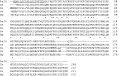
Fig. 1. Comparison of the deduced As24 amino acid sequence (GenBank Accession numbers are indicated in parentheses) with the amino acid sequences of selected homologous molecules. Sequences are from the C. elegans (NP_492398), D. immitis (AAD11968), O. volvulus (AAD11969) and B. pahangi (CAA10209). Identical residues are marked with asterisks, and conserved residues are marked with a dot. Gaps, marked by hyphens, are inserted for better alignment. The predicted signal peptides are shown as underlined. Ce, C. elegans; Di, D. immitis; Ov, O. volvulus; Bp, B. pahangi. The number of amino acid residues is indicated at the right side.

Fig. 2. Expression and purification of histidine-tagged recombinant Ascaris suum 24 kDa protein. The SDS-polyacrylamide gel (12·5%) was loaded with As24 transformed E. coli lysate after IPTG induction (Lane 2), 50 mM imidazole fraction (Lane 3) and purified rAs24 after dialysis (Lane 4). The protein was stained with Coomassie blue. Molecular weight markers in kDa are indicated on the left (Lane 1).
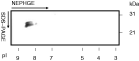
Fig. 3. Two-dimensional immunoblot for the detection of endogenous Ascaris suum 24 kDa protein in adult female worms. Female worm extract (50 μg) was separated by 2D non-equilibrium pH-gradient gel electrophoresis, and the proteins were transferred to a nitrocellulose membrane. The endogenous As24 protein bound to the anti-rAs24 mouse serum was detected by alignment of the stained gel and immunoblot membrane after visualization with NBT/BCIP.
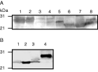
Fig. 4. One-dimensional immunoblot for the identification of endogenous Ascaris suum 24 kDa protein and its homologues. (A) Aliquots of 100 μg of protein equivalents of each parasite extract were electrophoresed on a 12·5% SDS-polyacrylamide gel and blotted onto nitrocellulose membrane. The endogenous As24 proteins bound to the anti-mouse As24 serum were detected by NBT/BCIP. Lane 1, A. suum adult male; Lane 2, adult female; Lane3, embryonated egg; Lane 4, infective L3; Lane 5, lung-stage L3; Lane 6, male ES; Lane 7, female ES; Lane 8, larval ES. (B) As24 homologues in ascarid roundworms. Aliquots of 60 μg of protein equivalents of each parasite extract were electrophoresed on a 12·5% SDS-polyacrylamide gel and blotted onto nitrocellulose membrane. The As24 homologues bound to the anti-serum were detected as mentioned above. Lane 1, A. lumbricoides; Lane 2, T. canis; Lane 3, A. suum; Lane 4, recombinant As24.
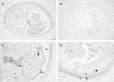
Fig. 5. Localization of endogenous Ascaris suum 24 kDa protein in adult female worm by immunohistochemistry. Adult A. suum female worms were fixed in 4% paraformaldehyde, 0·1 M PBS and embedded in paraffin. The sections were exposed to either mouse anti-rAs24 serum (1[ratio ]50, A) or mouse pre-immune serum (1[ratio ]50, B). (C and D) Both are magnified areas of (A). Arrows indicate antibody-labelled regions. cu, Cuticle; hyp, hypodermis; dch, dorsal hypodermal chord; mu, muscle; ov, ovary; ut, uterus.
 You have
Access
- 18
- Cited by