Introduction
Bicuspid aortic valve is one of the most common CHDs and is seen in 0.8% of all life-births. Reference Sillesen, Vøgg and Pihl1 Bicuspid aortic valve appears in isolation or in conjunction with other defects and is, in addition, frequently seen with associated abnormalities of the aorta. Reference Sillesen, Vøgg and Pihl1–Reference Çelik, Mahtab and Bogers3
The normal tricuspid aortic valve is composed of three equally sized cusps, whereas the bicuspid aortic valve is composed of two functional cusps due to the absence or underdevelopment of a commissure and the adjacent cusps. Reference Mordi and Tzemos2,Reference Saha, Bastiaenen, Hayward and McEwan4 Several classification systems for bicuspid aortic valves based on the morphology of the cusps exist. Reference Sievers and Schmidtke5,Reference Michelena, Della Corte and Evangelista6 Phenotyping bicuspid aortic valve disease may play a role in predicting possible future complications, as some types are more prone to develop complications than others. Reference Mordi and Tzemos2,Reference Niaz, Fernandes, Sanders, Michelena and Hagler7
The aetiology of bicuspid aortic valve is multifactorial, with a notable genetic component. Reference Sillesen, Vøgg and Pihl1,Reference Modica, Sollazzo and Bianco8 It is recognised that neural crest cells are of importance in the development of the outflow tract and aortic arch. Reference Modica, Sollazzo and Bianco8,Reference Kappetein, Gittenberger-de Groot, Zwinderman, Rohmer, Poelmann and Huysmans9 Kappetein et al. suggested that the bicuspid aortic valve could result from deficient signalling of the neural crest cells migrating to the heart from the neural tube. Reference Kappetein, Gittenberger-de Groot, Zwinderman, Rohmer, Poelmann and Huysmans9 Furthermore, Nakamura et al. demonstrated that neural crest cells contribute to the formation of the aortic valve. Reference Nakamura, Colbert and Robbins10 Most interestingly, Nakamura et al. also found that neural crest cells are involved in the maturation of the proximal conduction system of the heart, including the bundle of His. Reference Nakamura, Colbert and Robbins10 Therefore, if a deficiency in neural crest cells may cause a bicuspid aortic valve, it is possible that it may also increase the risk of conduction abnormalities.
Bicuspid aortic valve is a well-recognised risk factor for valve calcification, degeneration, and development of valvular dysfunction. Reference Niaz, Fernandes, Sanders, Michelena and Hagler7,Reference Hamdan, Nassar and Schwammenthal11 It is often not detected before symptom onset in adulthood, with clinical presentation varying according to valve impairment and aortic disease. Reference Niaz, Fernandes, Sanders, Michelena and Hagler7 Aortic valve stenosis and/or aortic regurgitation, Reference Mordi and Tzemos2,Reference Saha, Bastiaenen, Hayward and McEwan4 the most common complications to bicuspid aortic valve, may lead to hypertrophy of the left ventricle and, ultimately, heart failure. Reference Saha, Bastiaenen, Hayward and McEwan4 Individuals diagnosed with bicuspid aortic valve exhibit an approximate 50% risk of requiring valve replacement with or without concomitant aortic surgery. Reference Kusner, Brown, Gleason and Edelman12
Electrocardiographic abnormalities are frequently seen in adults with bicuspid aortic valve, but are generally considered to reflect secondary changes, i.e., hypertrophy of the left ventricle and atrioventricular conduction abnormalities such as heart block due to calcific aortic stenosis or aortic regurgitation. Reference Saha, Bastiaenen, Hayward and McEwan4,Reference Friedman, Zaman, Haft and Melendez13,Reference Karbasi-Afshar, Jonaidi-Jafari, Saburi and Khosravi14 There is limited knowledge on possible conduction abnormalities and electrocardiographic characteristics in pre-symptomatic patients with bicuspid aortic valve.
This study aimed to investigate the electrocardiographic findings in unselected newborns with bicuspid aortic valve and investigate whether early, subtle signs of atrioventricular conduction abnormalities are present early in life.
Materials and methods
Study design
This study was cross-sectional, with prospectively collected data from newborns enrolled in the Copenhagen Baby Heart Study; a multicentre, population-based cohort study with prenatal inclusion. Reference Sillesen, Raja and Pihl15,Reference Vøgg, Basit and Raja16 From April 2016 to October 2018, all expectant parents at the three largest materny wards in Copenhagen were offered inclusion. Reference Sillesen, Raja and Pihl15
A systematic cardiac examination was conducted preferably within 30 days of birth on all enrolled newborns, including transthoracic echocardiography and an eight-lead electrocardiogram. Reference Vøgg, Basit and Raja16 In addition, pre-, peri-, and postnatal information was collected for both mothers and newborns, and umbilical cord blood samples were obtained at birth and analysed subsequently. Reference Vøgg, Basit and Raja16
Approvals
The Copenhagen Baby Heart Study followed the Declaration of Helsinki and was approved by the regional committee of the Capital City Region of Denmark (H-16001518) and the Danish Data Protection Agency (I-Suite no.: 04546, ID-no. HGH-2016-53). The clinical trial registration for the Copenhagen Baby Heart Study is clinicaltrials.gov identifier NCT02753348. Reference Sillesen, Raja and Pihl15 Parents gave written consent prior to inclusion. Reference Sillesen, Raja and Pihl15
Study population
All newborns diagnosed with a bicuspid aortic valve in the Copenhagen Baby Heart Study, who had a valid electrocardiogram obtained within 30 days after birth, were included in this study.
The newborns with bicuspid aortic valve were matched 1:4 with newborns from the Copenhagen Baby Heart Study with a structurally normal heart. Matching criteria were sex, gestational age (+/- 21 days), and age (+/- 1 days), length (+/- 0.02 m), and weight (+/- 0.3 kg) at examination.
Echocardiography
The transthoracic echocardiogram consisted of standard sub-xiphoid, apical, left parasternal, and suprasternal views acquired with cardiac sector transducers 12S-D and 6S-D. Measurements were obtained in accordance with the American Society of Echocardiography guidelines for paediatric echocardiography. Reference Sillesen, Raja and Pihl15 Sonographers were trained by specialists in paediatric echocardiography, and all echocardiograms suspected to be abnormal were reviewed by a cardiologist. Reference Sillesen, Raja and Pihl15 In cases where bicuspid aortic valve was identified, the diagnosis was confirmed by a second cardiologist. Reference Sillesen, Vøgg and Pihl1 A more comprehensive description of the cardiac examination has previously been published. Reference Sillesen, Vøgg and Pihl1,Reference Sillesen, Raja and Pihl15 All instances of bicuspid aortic valve were classified using the system established by Sievers and Schmidtke, which classifies the valve according to number of raphes and orientation of raphe(s) or cusps. Reference Sillesen, Vøgg and Pihl1,Reference Sievers and Schmidtke5
Electrocardiograms
On the same day, the echocardiography was performed, an eight-lead electrocardiogram (I, II, III, aVF, aVR, aVL, V1, and most often V6) was recorded using the MAC 5500 HD system with a paper speed of 25 mm/sec, sensitivity at 10 mm/mV, sample rate of 500 Hz, and bandwidth filter of 0.16–150 Hz (General Electric ECG System, Milwaukee, USA) while the newborn was relaxed or asleep. Reference Sillesen, Raja and Pihl15,Reference Pærregaard, Hvidemose and Pihl17 Digital recordings were used for all electrocardiograms, with an automated analysis performed using the GE Healthcare Marquette 12SL ECG Analysis Program to determine electrocardiographic intervals, amplitudes, areas, and durations. The resulting electrocardiograms were stored in the MUSE ECG management software (v8, GE Healthcare, Milwaukee, USA). Reference Pærregaard, Hvidemose and Pihl17 To ensure adequate data quality, extensive validation efforts were undertaken as previously described by Pærregaard et al. Reference Pærregaard, Hvidemose and Pihl17 Analyses of the electrocardiograms focused on standard parameters, including heart rate (beats pr. minute, bpm), PR-interval (ms), QRS duration (ms), QRS axis (degrees), uncorrected QT interval (ms), corrected QT interval according to Bazett’s formula (QTcB, ms) or Fridericia’s formula (QTcFri, ms), as well as the maximum R- and S-wave amplitudes in the lead V1 and V6 (µV). The P-wave was examined in lead V1 because this lead generally has a prominent P-wave while being relatively robust to movement artefacts. The P-wave parameters included P-wave axis (degrees), P-wave area (µV * ms), and P-wave peak amplitude (µV).
Cord blood tests
Umbilical cord blood was collected and analysed at the hospital where the newborn was delivered. Additional markers were analysed at Herlev Hospital within 1–3 days. If the blood volume was insufficient for complete analyses, prioritisation followed a predefined protocol. All analyses were performed using standard clinical biochemistry equipment. Reference Sillesen, Raja and Pihl15 Biochemicals markers included haemoglobin, glucose, potassium, sodium, and creatinine. Reference Sillesen, Raja and Pihl15
Statistical analyses
Categorical variables are presented as numbers and percentages. Continuous variables are presented as medians and interquartile ranges. Continuous variables were tested for normality using the Shapira–Wilk test and by visual inspection of histograms. Wilcoxon rank sum test was used to compare electrocardiographic parameters of two groups. A p-value below 0.05 was considered statistically significant. The Bonferroni correction was applied to correct for multiple testing for all echocardiographic parameters presented (n = 14), i.e., p < 0.05/14 = 0.003. Statistical analyses were performed using R Studio v2022.12.0.
Results
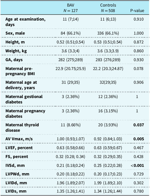
In total, 127 newborns with bicuspid aortic valve and 508 matched controls were included. Demographic and echocardiographic characteristics of the two groups, including maternal characteristics, are shown in Table 1.
Demographic, echocardiographic, and cord blood characteristics

Continuous variables are presented as medians (interquartile ranges) and categorical variable as absolute numbers (percentage). Abbreviations: AV = aortic valve; BAV = bicuspid aortic valve; BMI = body mass index; FS = fractional shortening; GA = gestational age; IVSd = interventricular septum thickness in end-diastole; LVEF = left ventricular ejection fraction; LVIDd = left ventricle internal diameter in end-diastole; LVIDs =left ventricle internal diameter in end-systole; LVPWd= left ventricle posterior wall thickness in end-diastole. Significant values are marked with bold.
The most common subtype of bicuspid aortic valve was type 1 right-left (RL,78.0%), followed by type 1 right-non coronary (RN,11.8%), type 0 anterior posterior (AP,4.7%), type 0 lateral (LAT,3.1%), and type 1 non-coronary-left (NL,2.4%) as the rarest subtype. Among the newborns with bicuspid aortic valve, none had aortic stenosis or coarctation, while 21 (16.5%) had aortic regurgitation (all trivial). The newborns with bicuspid aortic valve had a significantly higher aortic valve outflow velocity (1.00 vs. 0.92 m/s, p = 0.005) compared to matched controls, however, all were within the normal range (see Table 1). No significant differences in left ventricular dimensions or function between newborns with bicuspid aortic valve and matched controls were observed, except for a thinner interventricular septum in diastole (0.21 vs. 0.25 mm, p < 0.001) in newborns with bicuspid aortic valve.
Electrocardiographic findings in newborns with bicuspid aortic valve vs controls
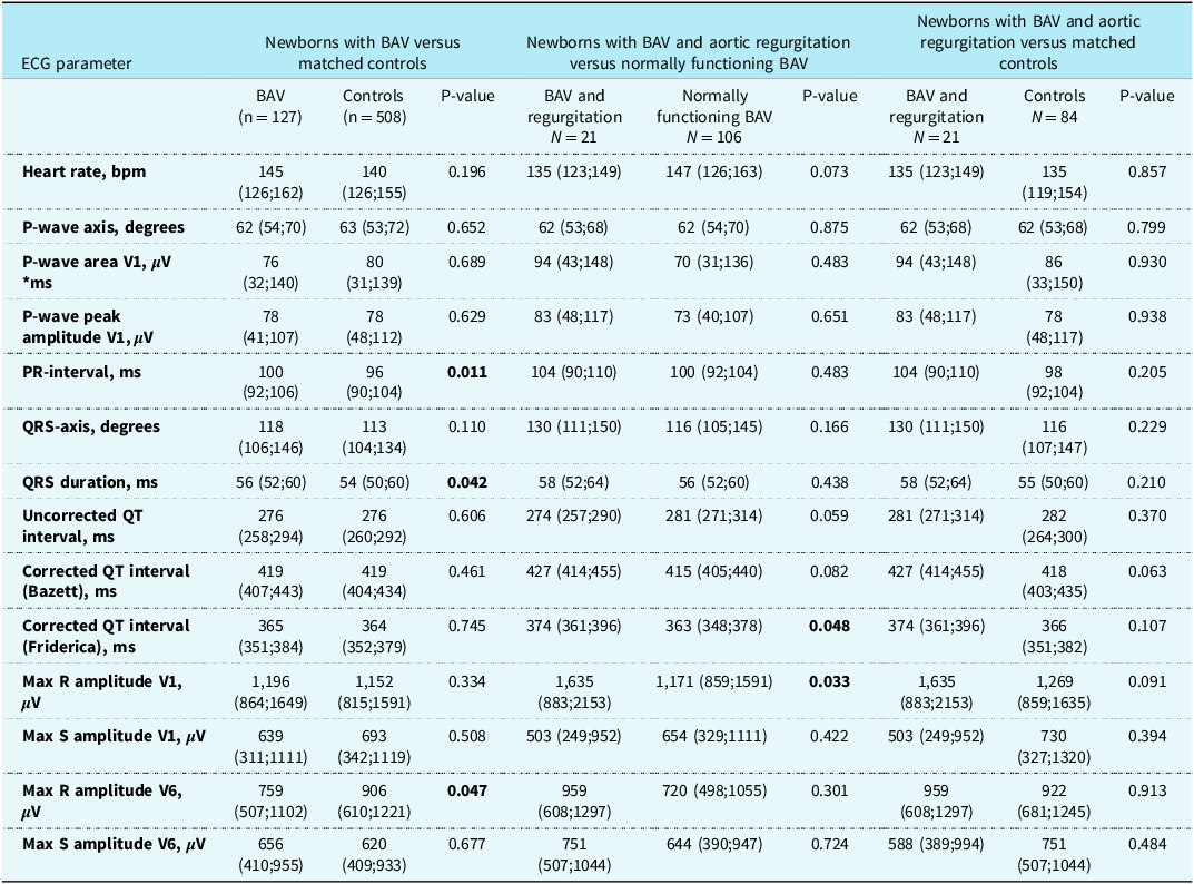
Electrocardiographic parameters for all newborns with bicuspid aortic valve and matched controls are presented in Table 2. Newborns with bicuspid aortic valve had a slightly longer PR-interval (100 vs 96 ms, p = 0.011) and QRS duration (56 vs 54 ms, p = 0.042) and a lower R-wave amplitude in V6 (759 vs 906 µV, p = 0.047) compared to matched controls. We found no other significant differences in electrocardiographic parameters between the two groups. When applying the Bonferroni correction, none of the parameters differed between the groups. Furthermore, no newborns were identified with any degree of atrioventricular block.
ECG parameters of newborns with bicuspid aortic valve and newborn controls

Continuous variables are presented as medians (interquartile ranges). Abbreviations: BAV = bicuspid aortic valve; ECG = electrocardiogram. Significant values are marked with bold.
Electrocardiographic findings in newborns with bicuspid aortic valve with vs without aortic regurgitation
Subgrouping newborns with bicuspid aortic valve according to valve function, we found a significantly shorter QTcFri (363 vs 374 ms, p=0.048) and a significantly lower R-wave amplitude in V1 (1,171 vs 1,635 µV, p = 0.03) in newborns with normally functioning bicuspid aortic valve compared to those with aortic regurgitation. There were no significant differences in PR-intervals or any of the other electrocardiographic parameters studied. When applying the Bonferroni correction, none of the parameters differed between the groups.
When comparing newborns with bicuspid aortic valve and aortic regurgitation to their matched controls, we did not find any differences in the electrocardiographic parameters, as shown in Table 2.
Cord blood electrolytes
Creatinine levels were available for 363 newborns, including 68 (53%) with bicuspid aortic valve and 295 (58%) from the control group. Sodium levels were available in 278 newborns, of whom 69 (54%) had bicuspid aortic valve and 288 (56%) were controls. Newborns with bicuspid aortic valve had significantly lower creatinine levels than matched controls (59 vs 63 µmol/l, p = 0.01), while sodium levels were comparable between the two groups (137 vs 136 mmol/l, p = 0.29). Data on other electrolytes, such as potassium, as well as markers like haemoglobin and glucose, were insufficient for the newborns in this substudy.
Overall, umbilical cord blood samples were collected from 46.9% of all newborns in the Copenhagen Baby Heart Study. Several factors may have influenced this collection rate, including blood coagulation, competing priorities during busy hours in the labour ward, and the need for urgent postpartum care. Reference Vøgg, Basit and Raja16
Discussion
This study describes electrocardiographic findings in a large group of unselected newborns with (near-) normally functioning bicuspid aortic valve compared to matched controls. Newborns with bicuspid aortic valve demonstrated slightly longer PR-intervals and QRS durations along with lower R-wave amplitudes in V6 compared to matched controls. All other ECG parameters were comparable. However, when correcting for multiple testing, none of the parameters significantly differed between the groups.
Adults with bicuspid aortic valve often exhibit electrocardiographic changes reflecting secondary complications such as left ventricular hypertrophy due to aortic valve dysfunction. Reference Suwa, Rahsepar and Geiger18 These electrocardiographic changes include a higher amplitude of the R-wave in V6 combined with a deep S-wave in V1. Reference Suwa, Rahsepar and Geiger18 Suwa et al. investigated 47 adult patients with isolated bicuspid aortic valve, i.e., without aortic stenosis and/or regurgitation, finding no electrocardiographic changes. Reference Suwa, Rahsepar and Geiger18 Our findings align with their results, but we did observe a lower R-wave amplitude in V6, although not reaching statistical significance after Bonferroni correction. Longitudinal studies are necessary to assess whether these initial differences persist and potentially diverge later in life.
In newborns with bicuspid aortic valve and trivial aortic regurgitation (n = 21), we found a longer QTcFri and a higher R-wave amplitude in V1 compared to those with normally functioning bicuspid aortic valve. However, these findings are based on a relatively small subgroup and did not remain significant after Bonferroni correction and may therefore be a result of statistical noise. It should be noted that although QTcFri was prolonged, it did not surpass the published upper cut-off value (QTcFri > 394 ms). Reference Pærregaard, Hvidemose and Pihl17
In this study, we found a slightly longer PR-interval in newborns with bicuspid aortic valve compared to matched controls. In the Copenhagen Baby Heart Study, we have previously shown that unselected newborns with bicuspid aortic valve have higher flow velocities across the valve than newborns with tricuspid aortic valve, Reference Sillesen, Vøgg and Pihl1 indicating a change in dynamics in the bicuspid aortic valve. However, when we divided the bicuspid aortic valve group based on the presence of trivial aortic regurgitation in this study, as an indicator of discrete valvular dysfunction, no PR-interval differences were found. This may be due to the small sample size in subgroups or the regurgitation’s trivial nature not being sufficient to cause a difference. A previous study by Friedman et al. found normal PR-intervals in three adult patients with recently developed high-grade aortic regurgitation, however, follow-up examinations years later revealed prolonged PR-intervals. Reference Friedman, Zaman, Haft and Melendez13 Whether this is because of a remodelling of the aortic valve apparatus and adjacent myocardium is not known.
The slightly longer PR-interval in newborns with (near-) normally functioning bicuspid aortic valve, coupled with previous reports of third-degree atrioventricular block in patients with severe bicuspid aortic valve stenosis, Reference Saha, Bastiaenen, Hayward and McEwan4,Reference Karbasi-Afshar, Jonaidi-Jafari, Saburi and Khosravi14 raises the possibility of a potential underlying intrinsic abnormality rather than being solely attributed to secondary changes to significant valve dysfunction. A longitudinal follow-up study of the newborns with bicuspid aortic valve will provide insights into whether these electrocardiographic findings persist, progress, or resolve over time.
We examined different maternal factors that could potentially influence the findings of this study. We found no significant difference in the prevalence of maternal diabetes in the two groups, and none of the included newborns were exposed to maternal connective tissue disease.
However, our study identified a significantly higher prevalence of maternal thyroid disease in the group of newborns with bicuspid aortic valve compared to the control group. This observation is consistent with prior research suggesting an increased risk of CHDs among offspring of mothers with thyroid dysfunction, including an increased risk of septal defects and outflow tract abnormalities. Reference Mirshekar, Mehran and Faraji Shahrivar21,Reference Vohra and Koren22 Furthermore, a study by Mohammad Ali Mirshekar et al. showed that newborn rats from hypothyroid mothers exhibited electrocardiographic differences including longer PR and QT intervals. Reference Mirshekar, Mehran and Faraji Shahrivar21 Although based on a small sample, and not the primary aim of this study, our findings support previous research and suggests that the increased risk of CHDs may also extend to bicuspid aortic valve. The underlying mechanisms remain incompletely understood but may involve altered maternal–fetal thyroid hormone transfer, changes in placental function, or epigenetic modifications during embryogenesis.
Strengths and limitations
The electrocardiograms of newborns changes as they mature, and their hearts develop. For instance, the heart rate decreases, the PR-interval lengthens, and the left side of the heart enlarges relatively faster than the right side due to increased systemic pressure and decreased pulmonary pressure following birth, reflected in the electrocardiogram as a leftward shift of the QRS axis. Reference Hvidemose, Pærregaard and Pihl19,Reference Lue, Wu and Wang20 As a result, comparing electrocardiograms between newborns can be challenging. However, because of the comprehensive cohort size and systematic assessment in the Copenhagen Baby Heart Study, allowing for the assessment of a wide range of parameters in unselected newborns, we were able to efficiently match cases with bicuspid aortic valve to controls, therefore eliminating potential confounding factors, such as age, which could otherwise account for differences in electrocardiographic findings.
Further, the study design allows for investigation and potentially elimination of a wide variety of factors, such as blood electrolyte levels and maternal diseases that might influence the newborns’ ECG measurements. Multiple studies have established that abnormal potassium levels, whether elevated or reduced, can lead to significant ECG changes, and while this is a relevant factor in ECG interpretation, we were unable to include potassium measurements in the analysis, likely due to sample haemolysis, which can interfere with accurate potassium assessment. Reference Krogager, Kragholm and Skals23
There are other limitations to the study that should be acknowledged. First, the study predominantly included newborns at or near term, which may limit the generalisability of the findings to preterm or sick newborns. Reference Vøgg, Basit and Raja16,Reference Pærregaard, Hvidemose and Pihl17 Second, the study relied on a single electrocardiogram for each newborn, precluding the assessment of changes over time. Reference Pærregaard, Hvidemose and Pihl17 Third, an eight-lead electrocardiogram was used instead of a twelve-lead electrocardiogram, which may restrict the ability to detect subtle changes in the electrocardiographic parameters. Reference Pærregaard, Hvidemose and Pihl17 Fourthly, there may have been a selection bias because of the age at which the newborns where examined (median 11 days), where newborns with severe CHD or other comorbidities had already entered clinical management because of the widespread routine prenatal and postnatal strategies in Denmark and may have declined to undergo further testing in the study although encouraged to participate. Reference Sillesen, Vøgg and Pihl1
Despite certain limitations, this remains to the authors knowledge the most extensive study examining newborns systematically for the presence of a bicuspid aortic valve and its correlation with electrocardiographic findings, allowing for the assessment of these parameters prior to secondary changes.
Conclusion
Newborns from the general population with bicuspid aortic valve demonstrated longer PR-interval and QRS duration and a lower R-wave amplitude in V6 compared to matched controls. Nevertheless, after correcting for multiple testing, no statistically significant difference was found. Further research, including follow-up studies on these newborns, will contribute to determining whether these observed changes represent an early indicator of conductions abnormalities.
Acknowledgements
A huge thanks to all the families who participated in the project.
Financial support
This work was supported by the Danish Heart Association, the Danish Childrens’s Heart Foundation, Candy’s Foundation, the Toyota Foundation, and the Herlev-Gentofte Hospital Research Foundation.
Competing interests
The authors declare none.
Ethical standard
The authors assert that all procedures contributing to this work comply with the ethical standards of the relevant national guidelines on human experimentation and with the Helsinki Declaration of 1975, as revised in 2008, and has been approved by the regional committee of the Capital City Region of Denmark (H-16001518) and the Danish Data Protection Agency (I-Suite no.: 04546, ID-no. HGH-2016-53). The clinical trial registration for the Copenhagen Baby Heart Study is clinicaltrials.gov identifier NCT02753348. Reference Sillesen, Raja and Pihl15