Congenital heart disease is the most common birth defect with reported prevalence of 2.1–9.1 per 1000. Reference Alsaied, McCrindle and Wernovsky1 Most congenital heart disease causing significant haemodynamic alterations require surgical treatments. Medical personnel who are involved in the care of congenital heart disease patients need to understand the anatomy, pathophysiology, and surgery of each defect to effectively care for the patients. Among these, anatomy is the most fundamental and important element that all medical personnel should learn about. Conventional education tools for anatomy of congenital heart disease include textbooks, echocardiography, cardiac angiography, computed tomography, magnetic resonance imaging, direct observation during surgery, and autopsy specimens. However, many of these tools are not always effective in understanding complex three-dimensional structure of the complex congenital heart disease and some are not always readily available.
Modern three-dimensional printing technology has made it possible to create the physical replicas of the complex congenital heart disease. Three-dimensional printed heart models of complex congenital heart disease obviate the need for mentally reconstructing two-dimensional images obtained from conventional diagnostic tools into a three-dimensional structure, and thus may be a very effective education tool for learning anatomy of the complex congenital heart disease. Recently, a few studies reported the utility of three-dimensional printed heart models of congenital heart disease in medical education. Reference Costello, Olivieri and Krieger2–Reference Smerling, Marboe and Lefkowitch8 However, most of these studies utilised models of simple or mildly complex congenital heart disease, such as ventricular septal defect or tetralogy of Fallot, and the education only focussed on medical students or residents. The objective of this study was to evaluate the feasibility and effects of medical personnel education on various kinds of complex congenital heart disease using patient-specific three-dimensional printed heart models.
Materials and methods
Three-dimensional printed heart models
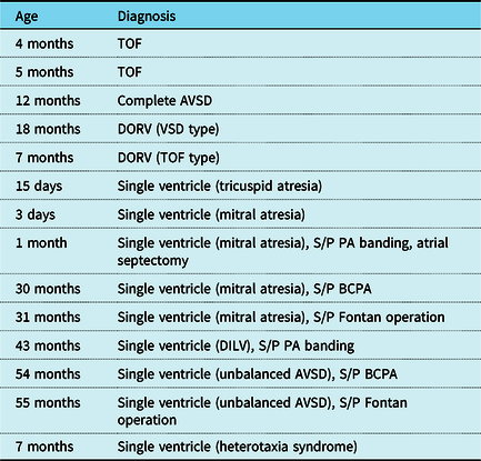
De-identified computed tomography digital imaging and communications in medicine data of the 11 patients with complex congenital heart disease were obtained from a radiology archive. Using these data, 14 kinds of heart models, encompassing 9 kinds of complex congenital heart disease were printed (Table 1).
Summary of the 3D printed heart models

3D = three-dimensional; AVSD = atrioventricular septal defect; BCPA = bidirectional cavopulmonary anastomosis; DILV = double inlet left ventricle; DORV = double outlet right ventricle; PA = pulmonary artery; S/P = status post; TOF = tetralogy of Fallot; VSD = ventricular septal defect.
Three-dimensional printing technique
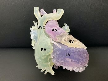
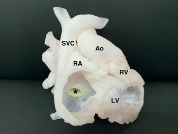
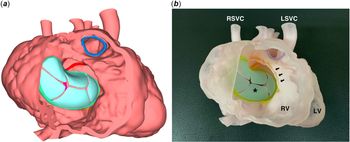
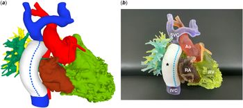
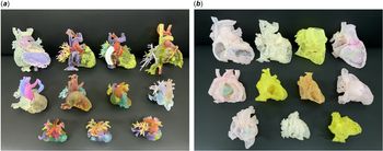
A three-dimensional printing company (MEDICAL IP, Seoul, Republic of Korea) was requested to manufacture the heart models. A commercially available software (MEDIP; MEDICAL IP, Seoul, Republic of Korea) was used for segmentation. Because segmentation requires thorough understanding of the anatomy of various congenital heart disease, an experienced paediatric cardiac surgeon examined, corrected, and confirmed every three-dimensional modelling result via a web-based system provided by the company. Computer-aided design was performed using a commercially available software (Meshmixer; Autodesk, San Rafael, California, United States of America). Blood pool and/or hollow models were printed as needed. The blood pool model is a simple cast of the volume rendered angiogram and is useful for general understanding of the three-dimensional anatomy (Fig 1). The hollow model is best suited for understanding the intracardiac structures, because the inner surface of it represents the precise endocardial surface anatomy (Fig 2). Hollow models were printed with a wall thickness of 1.4–1.8 mm. Because heart valves are not able to be imaged adequately for three-dimensional printing, only the valve annuli were marked with distinct colors using computer-aided design and printed. If necessary, fake valves were created using computer-aided design and printed with a thickness of 1.0 mm (Fig 3). For hollow models, parts of the free wall of the atria and/or ventricles were removed in such ways to best visualise the internal anatomy. For one kind of blood pool model after Fontan operation, the extracardiac conduit was printed as similar to the actual graft used to increase its “realness” (Fig 4). Three-dimensional printing was performed on a commercially available three-dimensional printer (J750; Stratasys, Eden Prairie, Minnesota, United States of America). Blood pool models were printed using a solid material (Vero family) and hollow models were printed using a flexible material (Agilus30 family). A total of 44 replicas were printed and the average cost per replica was 503,150 Korean won (Fig 5).
A 3D printed blood pool model of DILV which was created using CT data obtained from a 43-month-old patient. 3D = three-dimensional; Ao = aorta; CT = computed tomography; DILV = double inlet left ventricle; LV = left ventricle; RA = right atrium; RV = right ventricle; SVC = superior caval vein.

A 3D printed hollow model of the case shown in Figure 1. Parts of the free wall of the RA and LV were removed to visualise the intracardiac structures. The asterisk represents an ASD. 3D = three-dimensional; Ao = aorta; ASD = atrial septal defect; LV = left ventricle; RA = right atrium; RV = right ventricle; SVC = superior caval vein.

An artificially created fake valve in complete AVSD. ( a ) A CAD image showing a fake common AV valve through cut RV free wall. A blue ring represents pulmonary valve annulus. ( b ) A 3D printed hollow model showing a fake common AV valve (an asterisk) through cut RV free wall. Scooped-out ventricular septal crest is observed (arrows). 3D = three-dimensional; AV = atrioventricular; AVSD = atrioventricular septal defect; CAD = computer-aided design; LSVC = left superior caval vein; LV = left ventricle; RSVC = right superior caval vein; RV = right ventricle.

A 3D printed model of mitral atresia which was created using CT data obtained from a 31-month-old patient who underwent extracardiac conduit Fontan operation. ( a ) A CAD image. The extracardiac conduit was artificially expressed as similar to the actual graft used to increase its ‘realness’. ( b ) A 3D printed blood pool model. The extracardiac conduit (an asterisk) looks similar to the commercially available graft used for Fontan operation. 3D = three-dimensional; Ao = aorta; CAD = computer-aided design; CT = computed tomography; IVC = inferior caval vein; RA = right atrium; RV = right ventricle; SVC = superior caval vein.

3D printed models of various kinds of complex CHD. ( a ) Blood pool models. ( b ) Hollow models. 3D = three-dimensional; CHD = congenital heart diseases.

Hands-on seminars
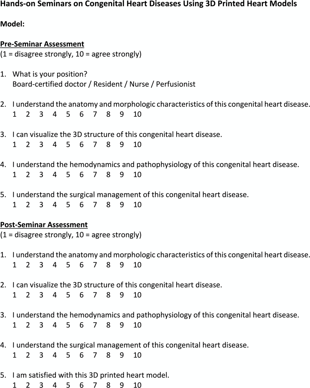
Using the three-dimensional printed heart models, a series of educational hands-on seminars, led by an experienced paediatric cardiac surgeon and a paediatric cardiologist, were conducted for medical personnel who were involved in the care of congenital heart disease patients. Board-certified physicians, residents, nurses, and perfusionists were encouraged to voluntarily participate in the seminars. Each seminar focussed on only one kind of congenital heart disease and contents of each seminar included anatomy, three-dimensional structure, pathophysiology, and surgery for the congenital heart disease . Each seminar was comprised of two components: (1) brief lectures on previously mentioned contents; and (2) an interactive hands-on session utilising the three-dimensional printed heart models. Each seminar was around 60 minutes in duration and included active interaction with the attending instructors throughout the seminar. Likert-type (10-point scale) questionnaires were used before and after each seminar to subjectively evaluate the effects of education (Fig 6).
Likert-type questionnaire which was used to evaluate the effects of education. 3D = three-dimensional.

Statistical analysis
Data were presented as frequencies with percentages, means with standard deviations, or medians with interquartile ranges as appropriate. Comparisons between paired groups were performed using paired t-test or Wilcoxon signed rank test as appropriate. All statistical analyses were performed using SPSS version 24 (IBM Corporation, Armonk, New York, United States of America).
Results
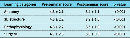
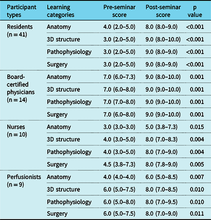
Between November 2019 and June 2020, a total of 16 sessions of hands-on seminar were conducted. The total number of questionnaire responses was 75. Participant types were residents in 41 (54.7%), board-certified physicians in 14 (18.7%), nurses in 10 (13.3%), perfusionists in 9 (12.0%), and unknown in 1 (1.3%). Participants’ specialties were cardiothoracic surgery, paediatric cardiology, neonatology, anesthesiology, radiology, etc. Overall, participants reported significant subjective improvement in understanding anatomy, three-dimensional structure, pathophysiology, and surgery of the congenital heart disease investigated (Table 2). When analysed by participant type, all four groups reported significant subjective improvement in all areas of learning, although it seemed that there were some differences in the level of basal knowledge among the groups (Table 3). Overall satisfaction about the three-dimensional printed heart models was excellent [10 (9–10)].
Overall effects of education (n = 75)

3D = three-dimensional.
Effects of education by participant type

3D = three-dimensional.
Discussion
In the present study, we have shown that the utilisation of patient-specific three-dimensional printed heart models for education on complex congenital heart disease was feasible and improved medical personnel’s understanding of complex congenital heart disease. This improvement was observed in all participant groups, irrespective of their level of basal knowledge regarding the congenital heart disease investigated.
During the past decade, three-dimensional printing technology has been increasingly used in the field of congenital heart disease. Current applications of three-dimensional printing in medicine of congenital heart disease include: (1) planning and simulation for surgical and interventional procedures; (2) hands-on surgical training; and (3) morphology teaching. Reference Yoo, Thabit and Kim9 Surgical planning utilising patient-specific three-dimensional printed heart models has been reported to be of enormous value in repairing various kinds of complex congenital heart disease, including complex forms of double outlet right ventricle. Reference Valverde, Gomez-Ciriza and Hussain10–Reference Vettukattil, Mohammad Nijres, Gosnell, Samuel and Haw14 Hands-on surgical training is changing the paradigm for surgical training in that the learning curve for complex congenital heart surgery may be overcome without adverse effects on patient outcomes. Reference Yoo, Spray, Austin, Yun and van Arsdell15–Reference Van Arsdell, Hussein and Yoo18
Understanding the anatomy of various kinds of congenital heart disease is the most fundamental element that is required for all medical personnel who are involved in the care of congenital heart disease patients. Conventional education tools for anatomy that are readily available in clinical practice include textbooks, echocardiography, and computed tomography. Understanding the anatomy and three-dimensional structure of a specific congenital heart disease with the aid of these tools is not always a straightforward task, especially for the non-experts. For example, although it is not an easy task to visualise the three-dimensional structure of the {S,L,L} type double inlet left ventricle, this can be accomplished promptly with the aid of three-dimensional printed models (Figs 1 and 2). Therefore, three-dimensional printed heart models may be a very effective tool for learning anatomy of congenital heart disease, as evidenced by recent studies. Reference Costello, Olivieri and Krieger2–Reference Smerling, Marboe and Lefkowitch8 However, most of these studies only utilised models of relatively simple congenital heart disease, such as ventricular septal defect or tetralogy of Fallot, and the education only focussed on medical students or residents. In a study by Olivieri and colleagues, 10 kinds of heart models, some of which are complex congenital heart disease, were used to enhance congenital cardiac critical care via simulation training of multidisciplinary intensive care teams. Reference Olivieri, Su and Hynes19 They found that the education was more effective as the case complexity increased.
In the present study, we have created 14 kinds of heart models representing 9 kinds of complex congenital heart disease. Notably, our models included nearly all types of single ventricle and covered all three surgical stages of the “Fontan pathway” (Table 1). Therefore, the participants were able to learn the concept of “three-stage surgical management of single ventricle” as well as the anatomy of various types of single ventricle. We created fake valves in three patients with a common atrioventricular valve (one patient with complete atrioventricular septal defect and two patients with single ventricle and a common atrioventricular valve), because the common atrioventricular valve is one of the key morphologic features of the atrioventricular septal defect (Fig 3). Our models also included tetralogy of Fallot and double outlet right ventricle. Therefore, most of the complex congenital heart diseases were covered by our collection. We believe that this is one of the strengths of our study. As expected, it seemed that the board-certified physicians had higher level of basal knowledge in all areas of learning, compared with other participant groups (Table 3). Interestingly, it seemed that the perfusionists had higher level of basal knowledge in three-dimensional structure, pathophysiology, and surgery, compared with residents and nurses. This may be explained by the fact that the perfusionists are highly experienced personnel with substantial amount of knowledge about congenital heart disease.
Utility of three-dimensional printed heart models is not confined to education of medical professionals. These can be effectively utilised for communication with patients and parents. Reference Biglino, Capelli and Wray20 Currently, we are using the created models for this purpose. In spite of many benefits of three-dimensional printing technology in medicine of congenital heart disease, this has not yet been widely incorporated in clinical practice. Barriers to adoption of this technology include: (1) high costs; (2) lack of expertise in medical image segmentation and three-dimensional modeling; and (3) lack of incentives for hospital administrators to introduce a three-dimensional printing programme. Reference Bramlet, Olivieri, Farooqi, Ripley and Coakley21
The present study was limited by the small sample size. Evaluation of the effects of education was performed in a subjective manner using questionnaires. For further studies in the future, objective tests before and after each seminar will be valuable to evaluate the effects of education. Evaluation of the effects of education by disease type was not performed due to the small sample size. Because brief lectures preceded hands-on sessions, it was not possible to determine the exact contributions of the three-dimensional printed models to the improvements observed.
In conclusion, the utilisation of three-dimensional printed heart models for education on complex congenital heart disease was feasible and improved medical personnel’s understanding of complex congenital heart disease. This education tool may be an effective alternative to conventional education tools for complex congenital heart disease.
Acknowledgements
We gratefully acknowledge the contributions of Yunseon Bang (a pediatric cardiac nurse of the Seoul St. Mary’s Hospital) to this study.
Financial support
This work was supported by the National Research Foundation of Korea (NRF) grant funded by the Korea government (Ministry of Science and ICT) (No. 2019R1F1A1047792).
Conflicts of interest
None.
Ethical standards
The Institutional Review Board of the Seoul St. Mary’s Hospital approved the present study and waived the need for individual patient consent.