Introduction
Childhood maltreatment can have a profound and enduring effect on an individual’s mental health and wellbeing (Tanaka et al., Reference Tanaka, Wekerle, Schmuck and Paglia-Boak2011). Both prospective and retrospective studies have reported significant associations between child maltreatment and internalizing and externalizing problems across the lifespan (Gilbert et al., Reference Gilbert, Widom, Browne, Fergusson, Webb and Janson2009). Despite this well-established association, we know relatively little about the neurocognitive mechanisms through which increased psychiatric vulnerability becomes instantiated after childhood maltreatment. Moreover, there are sparce neurocognitive longitudinal data, which limits our understanding of neurocognitive developmental plasticity. Autobiographical memory (ABM) is a key neurocognitive domain implicated in depression (e.g. Hitchcock et al., Reference Hitchcock, Nixon and Weber2014; Young et al., Reference Young, Bellgowa, Bodurka and Drevets2013) and altered in children who have experienced abuse and neglect (McCrory et al., Reference McCrory, Gerin and Viding2017a and Reference McCrory, Puetz, Maguire, Mechelli, Palmer, Gerin, Kelly, Koutoufa and Viding2017b; Valentino, Toth & Cicchetti, Reference Valentino, Toth and Cicchetti2009).
ABM refers to our memory of personally experienced events. Retrieving autobiographical memories successfully and accurately is thought to play a critical role in scaffolding our sense of self (Conway & Pleydell-Pearce, Reference Conway and Pleydell-Pearce2000), in the development and maintenance of social bonds (Alea & Bluck, Reference Alea and Bluck2003), and in planning of present and prospective actions (Pillemer, Reference Pillemer2003). At the neural level, typical adults engage a network of prefrontal, parietal and temporal regions during ABM processing including the ventromedial prefrontal cortex (vmPFC), middle temporal gyrus (MTG), hippocampus, retrosplenial cortex (rSC) and temporo-parietal junction (Martinelli et al., Reference Martinelli, Sperduti and Piolino2013) and the richness of ABM recall is associated with hippocampal activation (Gilboa & Marlatte, Reference Gilboa and Marlatte2017). Clinical studies of adult patients with depression have reported that while these individuals engage core components of the ABM network, they differentially weight regions within this network during specific ABM recall, depending on the task (i.e. fewer specific, more categorical, and fewer positive ABMs; see Young et al., Reference Young, Bellgowa, Bodurka and Drevets2013).
Individuals with depression also show differences at the behavioral level during ABM recall. The Autobiographical Memory Test (AMT) is a common behavioral paradigm that has been used to index ABM using cue words (Williams & Broadbent, Reference Williams and Broadbent1986). In this task participants recall memories following the presentation of positive and negative cue words; responses are scored in order to index the degree to which the ABMs are specific or ‘overgeneral’ (i.e. underspecified in terms of detail; see Dalgleish et al. (Reference Dalgleish, Williams, Golden, Perkins, Barrett, Barnard and Watkins2007) for a helpful critique of this methodology). Overgeneral ABM has been associated with depression and PTSD (including in child samples) as well as with the risk of developing these disorders in the future (Hitchcock et al., Reference Hitchcock, Nixon and Weber2014; Rawal & Rice, Reference Rawal and Rice2012; Thome et al., Reference Thome, Terpou, McKinnon and Lanius2020). In a previous study investigating ABM, we found that maltreatment experience was associated both with a pattern of overgeneral memory on the AMT, as well as with a pattern of reduced hippocampal activation during an imaging task where fully specified positive and negative memories were recalled (McCrory et al., Reference McCrory, Puetz, Maguire, Mechelli, Palmer, Gerin, Kelly, Koutoufa and Viding2017b).
We have postulated that reduced engagement of the hippocampus during positive memory recall in children who have experienced maltreatment may reflect a neurocognitive vulnerability to depression (McCrory et al., Reference McCrory, Puetz, Maguire, Mechelli, Palmer, Gerin, Kelly, Koutoufa and Viding2017b). Specifically, we have suggested that decreased specificity and salience of positive relative to negative memories in children who have experienced maltreatment may increase the likelihood of a negative inferential style, negative-self schemata, and a ruminative response style, all cognitive vulnerabilities associated with maltreatment and central to cognitive theories of depression and other psychiatric disorders (McCrory et al., Reference McCrory, Gerin and Viding2017a; McCrory et al., Reference McCrory, Puetz, Maguire, Mechelli, Palmer, Gerin, Kelly, Koutoufa and Viding2017b). Such a hypothesis is consistent with recent behavioural findings using an established mind-wandering task that indicated reduced spontaneous generation of positive thoughts in adolescents with maltreatment histories (Hoffmann et al., Reference Hoffmann, Viding, Puetz, Gerin, Sethi, Rankin and McCrory2018).
It should be noted that other neurocognitive systems, in addition to ABM processing, have also been implicated in mental health vulnerability following childhood maltreatment, including the threat, emotion regulation and reward systems (McCrory et al., Reference McCrory, Puetz, Maguire, Mechelli, Palmer, Gerin, Kelly, Koutoufa and Viding2017b). Indeed, the theory of latent vulnerability postulates that an array of neurocognitive systems are likely to adapt to early adverse or neglectful environments in line with the notion of experiential canalization (Blair & Raver, Reference Blair and Raver2012; McCrory & Viding, Reference McCrory and Viding2015). Such adaptations are thought to confer short-term functional advantages in atypical early environments but shape how an individual experiences and negotiates more normative contexts in ways that can increase mental health vulnerability over time. Functional neuroimaging studies of these candidate neurocognitive systems implicated in mental health vulnerability following childhood maltreatment have been predominantly cross sectional in nature (McCrory et al., Reference McCrory, De Brito, Sebastian, Mechelli, Bird, Kelly and Viding2011, Reference McCrory, De Brito, Kelly, Bird, Sebastian, Mechelli, Samuel and Viding2013; McCrory et al., Reference McCrory, Puetz, Maguire, Mechelli, Palmer, Gerin, Kelly, Koutoufa and Viding2017b; McLaughlin et al., Reference McLaughlin, Peverill, Gold, Alves and Sheridan2015; Puetz et al., Reference Puetz, Kohn, Dahmen, Zvyagintsev, Schüppen, Schultz, Heim, Fink, Herpertz-Dahlmann and Konrad2014, Reference Puetz, Viding, Palmer, Kelly, Lickley, Koutoufa, Sebastian and McCrory2016). To date these fMRI studies have generally not tracked changes in neurocognitive functioning. In some instances, a longitudinal follow-up indexing psychological functioning has been included (e.g. Dennison et al., Reference Dennison, Sheridan, Busso, Jenness, Peverill, Rosen and McLaughlin2016; Hanson, Hariri & Williamson, Reference Hanson, Hariri and Williamson2015; Weissman et al., Reference Weissman, Jenness, Colich, Miller, Sambrook, Sheridan and McLaughlin2020). For example, in a large community sample of adolescents with varying degrees of childhood maltreatment, Hanson, Hariri, and Williamson found that the severity of emotional neglect was associated with reduced development of striatal neural response to the receipt of monetary rewards (Reference Hanson, Hariri and Williamson2015). Reduced striatal response was found to partially mediate the association between a history of neglect and depression symptomology two years later. Using social-reward cues, Dennison and colleagues reported that higher striatal responses to social rewards at baseline predicted lower depression symptoms two years later in those with maltreatment histories (Dennison et al., Reference Dennison, Sheridan, Busso, Jenness, Peverill, Rosen and McLaughlin2016). These findings highlight how neural activation differences associated with maltreatment may be related to future mental health functioning.
However, these studies did not examine change over time in functional brain activity. In a recent paper that did consider developmental change at the neural level, Jenness and colleagues (Reference Jenness, Peverill, Miller, Heleniak, Robertson, Sambrook and McLaughlin2021) examined neural circuitry associated with emotion regulation in children who had experienced physical or sexual abuse or witnessed domestic violence versus those who had not had such experiences (Jenness et al., Reference Jenness, Peverill, Miller, Heleniak, Robertson, Sambrook and McLaughlin2021). Elevated activation was reported in the amygdala and the salience network in the maltreated group, who also exhibited increasing recruitment with age in ventrolateral prefrontal cortex during reappraisal in contrast to the control participants who exhibited decreasing recruitment. Differences in the background characteristics of these participants and the control group (e.g. in socioeconomic status and IQ), alongside the lack of matching on pubertal status limits the degree to which the observed neural differences can be confidently attributed to maltreatment experience. However, the study makes an important contribution to the field by investigating, for the first time, functional patterns of neural development in children with maltreated experience using fMRI.
The primary aim of the current study was to investigate developmental neural plasticity in ABM processing by conducting fMRI scans twice over a two-year period in children with documented maltreatment experience. Participants in the maltreated group were recruited from Social Services and carefully matched with control participants who did not have maltreatment experience. At the behavioral level, we expected to observe an overall improvement in the psychological and behavioral functioning of those in the maltreated group, given that the majority were in stable placements. At the neural level, we predicted that both groups of children would engage the core ABM network during recall of positive and negative autobiographical memories (Martinelli et al., Reference Martinelli, Sperduti and Piolino2013) and that this network would be relatively stable over time. In line with the prior work on depression and our own cross-sectional research, we expected that within this network maltreatment experience would be associated with reduced hippocampal activation during positive relative to negative ABM recall (Young et al., Reference Young, Bellgowa, Bodurka and Drevets2013; McCrory et al., Reference McCrory, Puetz, Maguire, Mechelli, Palmer, Gerin, Kelly, Koutoufa and Viding2017b). In view of the extant evidence indicating heightened amygdala response to negative stimuli in maltreated children and in adults at greater risk of depression, we expected greater amygdala activation during negative ABM recall (McCrory et al., Reference McCrory, De Brito, Sebastian, Mechelli, Bird, Kelly and Viding2011, Reference McCrory, De Brito, Kelly, Bird, Sebastian, Mechelli, Samuel and Viding2013; Young et al., Reference Young, Drevets, Bodurka and Preskorn2016; McCrory et al., Reference McCrory, Puetz, Maguire, Mechelli, Palmer, Gerin, Kelly, Koutoufa and Viding2017b). Given improvements in psychological and behavioral functioning, we expected to observe an attenuation of differences in hippocampal or amygdala activation from baseline to follow-up. Finally, we explored whether any regional brain maturational changes within the network of regions engaged by ABM processing differed by group and whether regional maturational changes that differed across group were associated with changes in psychological and behavioral functioning. Any such association would help provide insight into adaptive developmental plasticity.
Method
Participants
Participants had been part of an earlier study on autobiographical memory processing in children with and without maltreatment experiences (McCrory et al., Reference McCrory, Puetz, Maguire, Mechelli, Palmer, Gerin, Kelly, Koutoufa and Viding2017b). At baseline n = 67 children underwent the AMT in MRI (n = 34 Maltreatment group, n = 33 Non-Maltreatment group; see McCrory et al., Reference McCrory, Puetz, Maguire, Mechelli, Palmer, Gerin, Kelly, Koutoufa and Viding2017b for study details). From the initial sample of n = 67, we retained a sample of 37 children (aged 10–14 at baseline), who had both complete and usable data from the baseline assessment and follow-up assessment two years later; 19 with documented experiences of maltreatment (maltreatment group; MT) recruited from social services departments and 18 non-maltreated children with no prior social services contact (non-maltreatment group; Non-MT) recruited via schools and community advertisements. Children who had data at baseline and follow-up (n = 37) and those who did not return for the follow-up (n = 30) did not differ in age (p = .25), gender (p = . 25), IQ (p = .23), ethnicity (p = .24), SES (p = .94), maltreatment severity (p = .26), overall psychological functioning (p = .28) or depressive symptoms T1 (p = .51). MT and Non-MT participants were matched across a range of domains, including gender, SES, IQ and pubertal status and did not differ in these domains (see Table 1). Standard exclusion criteria for all participants included PDD, neurological abnormalities, standard MRI contra-indications and IQ < 70. The average time interval between baseline (2014) and follow-up (2016/2017) was approximately two years, with no significant difference in time between both measurements between the groups (Mean MT group = 794.53, SD = 53.61, t(26) = −1.02, p = .28; Mean Non-MT group = 776.61 days, SD = 25.16). Over the two-year period of follow-up, 84% of the MT participants were in a stable placement. Consent was obtained from the child’s legal guardian and assent was obtained from all children. All procedures were approved by UCL Research Ethics Committee (0895/002).
Background data for the maltreated and non-maltreated groups at baseline and follow-up

Abbreviations: MT = Maltreated group; Non-MT = Non-maltreated group; WASI-IQ = 2-subscale IQ derived from the Wechsler Abbreviated Scales of Intelligence; SDQ-P = Strength and Difficulties Questionnaire Parent report; CASI = Child and Adolescent Symptom Inventory; CASS = Child and Adolescent Social Support Scale.
Measures
Maltreatment history
Children in the maltreatment group had experienced a level of maltreatment requiring Social Services referral. Severity of neglect, emotional, sexual and physical abuse as well as intimate partner violence, were rated by the child’s social worker or adoptive parent on the basis of Social Service’s file information on an amended version of the Kaufman Scales of Child Maltreatment (Kaufman et al., Reference Kaufman, Jones, Stieglitz, Vitulano and Mannarino1994). Ratings from 0 (not present) to 4 (severe) were applied to each category (Kaufman et al., Reference Kaufman, Jones, Stieglitz, Vitulano and Mannarino1994). Maltreatment type was rated as follows: neglect N = 13 (68%); emotional abuse N = 18 (95%) sexual abuse N = 2 (11%); physical abuse N = 2 (11%); intimate partner violence N = 10 (54%). Details regarding maltreatment type and timing of abuse onset, duration and severity at baseline assessment are shown in Table S1 in the supplementary information. In addition, all children completed the CTQ at baseline and at follow up, which indicated that there was no evidence of ongoing maltreatment in the MT sample over the 2-year follow-up period (CTQ, see Table 1). No participant in the non-MT group had a history of referral to Social Services or a rating on the CTQ at a clinical threshold of severity.
Cognitive ability
Cognitive ability was assessed at baseline using two subscales of the Wechsler Abbreviated Scales of Intelligence (Wechsler, Reference Wechsler1999).
Psychiatric symptomatology
The parent-rated Strength and Difficulty Questionnaire total score (SDQ-P) was used to assess overall psychological functioning at both time points (Goodman, Reference Goodman1997). The SDQ-P total score is a composite T-score derived from the subscales conduct problems, hyperactivity-inattention, emotional symptoms, and peer problems, as well as prosocial behaviors. The standardized subscale for MDD of the Child and Adolescent Symptom Inventory (CASI) was completed by the young person’s carer and used to assess symptoms of depression (T-scores) (Lavigne et al., Reference Lavigne, Cromley, Sprafkin and Gadow2009).
Behavioral task and fMRI stimulus generation
Using the Autobiographical Memory Task (AMT; Williams & Broadbent, Reference Williams and Broadbent1986) participants were asked to generate specific memories in response to ten positive (e.g. achieve, caring) and ten negative cue words (e.g. mistake, lonely) for the fMRI task (see supplementary information for a list of cue words used at baseline and follow-up). That is, only specific memories generated were included. A full set of 20 specific memories were generated (10 positive ABMs, 10 negative ABMs) for each participant, in line with prior fMRI studies (Summerfield et al., Reference Summerfield, Hassabis and Maguire2009). Overgeneral memories (i.e. “memories that did not contain at least one specific detail that identifies an event as a distinct episode” with regards to space and time) were excluded (Valentino et al., Reference Valentino, Toth and Cicchetti2009; Williams & Broadbent, Reference Williams and Broadbent1986). The specific memories generated by this approach were rephrased by the participants in 3–4 words to prompt recall during the scanning session. In line with prior studies in adults (Summerfield et al., Reference Summerfield, Hassabis and Maguire2009), a control task was employed, which participants were presented with pictures and brief descriptions of 10 everyday objects. Participants were given 20 seconds to memorize each object. As with the ABMs, key features were captured in brief sentence cues for presentation during fMRI. The cue words chosen at baseline and follow-up were different sets of words that were comparable in terms of frequency in the English language. The memories for both the behavioral task and fMRI stimulus generation were coded for specificity by a trained member of the research team in the lab (blinded) and double rated by a second, independent member of staff on 10% of the memories. Inter-rater agreement was high (average agreement k > .82).
fMRI paradigm
There were three memory conditions: positive ABM recall, negative ABM recall and object recall. The fMRI task consisted of two runs of 15 trials, each comprising 5 positive ABMs, 5 negative ABMs and 5 object memories presented in a pseudo-randomized order. Each trial began with a sentence cue. Participants indicated successful or unsuccessful memory retrieval via a self-paced button press. The text cue then disappeared and the word “Elaborate” was presented during which time the participants recalled the memory or object. Subsequently, participants rated recall difficulty and vividness of the recalled memory on a 5-point Likert scale. There were no group differences or group valence interactions at T1 or T2 in relation to vividness or difficulty (see supplement).
fMRI data acquisition and processing pipeline
Data across both time points were acquired on the same 1.5 tesla Siemens (Siemens Medical Systems, Erlangen, Germany) Avanto MRI scanner with a 32-channel head coil during two runs of approximately 9 minutes each. All data analyses were conducted using the software package SPM12 (www.fil.ion.ucl.ac.uk/spm/software/) implemented in Matlab 2018. Cross-sectional analyses for baseline and follow-up data were carried out using a repeated measures Flexible Factorial Design, while longitudinal data were analysed to test for (group differences in) linear patterns of activation in response to basic and valenced ABM recall with age using the Sandwich Estimator Toolbox for Longitudinal and Repeated Measures Data v2.1.0 (SwE, toolbox for SPM, Guillaume et al., Reference Guillaume, Hua, Thompson, Waldorp and Nichols2014). Precise details of data acquisition parameters can be found in the supporting information.
Analyses
fMRI analyses
Analyses were carried out in the following order: First, we tested the stability of activation in the previously established ABM networks across both groups (i.e., ABM vs. Object recall cross-sectionally analysed at baseline and follow-up), followed by linear changes with age over time (using SwE toolbox, (Guillaume et al., Reference Guillaume, Hua, Thompson, Waldorp and Nichols2014). We then assessed stability of activation in valenced ABM recall (positive vs. negative ABM processing) at baseline at the whole-brain level and using the same ROI’s that showed significant group differences in the wider sample at baseline (i.e. hippocampus for non-MT group vs. MT group, and PHG for MT group vs. Non-MT group in the contrast Positive – Negative ABM as well as the A in the contrast MT group vs. Non-MT group Negative – Positive ABM). We then identified any brain areas that showed an increased engagement over time during positive vs. negative ABM processing with age over time (using SwE toolbox) (Guillaume et al., Reference Guillaume, Hua, Thompson, Waldorp and Nichols2014).
Contrast estimates were extracted from the peak voxels of clusters showing significant group differences in the contrast positive ABM recall vs. negative ABM recall in the follow-up measurement. One regional cluster emerged in the right rSC (extending to the posterior cingulate cortex, pCC) at follow-up and was extracted with MarsBaR Toolbox (Brett et al., Reference Brett, Anton, Valabregue and Poline2002). Whole brain analyses were corrected using cluster-size thresholding according to the most recent method implemented in AFNI’s 3dttest++ of voxel-wise p ≤ .005, ke = 155, which corresponds to p = 0.05, family-wise error (FWE) (AFNI release 2020 20.2.14; Eklund et al., Reference Eklund, Nichols and Knutsson2016; Ward, Reference Ward2000).
Because the size of the effect under investigation was unknown, it was not possible to perform power calculations to determine the optimal sample size. However, based on power calculations as implemented in G-Power software https://stats.idre.ucla.edu/other/gpower/), we were able to determine that our sample size allowed us to detect an effect size of 0.94 with 80% power and alpha equal to 0.05.
Brain-behavior relationships
Correlational analyses were conducted to explore whether any brain regions showing increased engagement between baseline and follow-up were associated with overall psychological functioning (SDQ-P Total Symptom Score) and symptoms of depression (standardized subscale for MDD derived from CASI). The relationships between brain activation and SDQ/CASI data were exploratory and since the two correlational analyses are not independent from each other, we chose to use a standard statistical threshold (p < 0.05) for both comparisons instead of Bonferroni correction which we considered too conservative given the above. Change scores were calculated by subtracting follow-up scores from baseline scores (Follow-Up – Baseline) using SPSS version 24 (3dttest++ IBM Corp. 2012).
Results
Behavioral results
Changes in outcome measures between baseline and follow-up
Psychological functioning
Cross-sectional analyses of the SDQ-P total score at T1 showed that the groups significantly differed on overall psychological functioning at T1 [t(21.84) = −3.65, p = .001] but were no longer statistically significantly different at follow-up [t(31) = −.87, p = .39]. Repeated measures analyses (ANOVA) with group as between-participant factor and SDQ-P score at baseline and follow-up as the within-participant factors showed a significant main effect for group (F(1,31) = 6.70, p = .02) but no main effect of time (F(1,31) = .45, p = .51) or group by time interaction (F(1,31) = 3.10, p = .088). Post-hoc analyses showed that the MT group had significantly elevated SDQ-P scores only at baseline relative to the non-MT group (see Table 1).
fMRI results
Developmental trajectories of neural activity during ABM recall between baseline and follow-up
General ABM processing: ABM vs. object recall
At baseline and follow-up ( cross-sectional analyses)
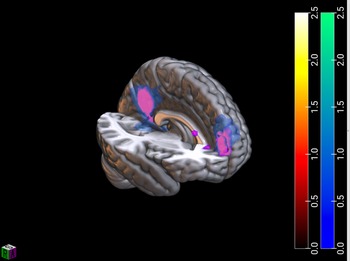
Cross-sectional analyses of the brain regions engaged in ABM vs. Object recall in both groups at follow-up revealed a network consisting of the vmPFC, rSC, pCC, MTG as well as the right hippocampus. This network showed a substantial overlap with the network that we found to be active at baseline (see Figure 1). No group x condition interactions were found.
Cross-sectional analyses of the brain regions engaged in ABM vs. Object recall in both groups at follow-up (T2, depicted in blue-green) revealed a network consisting of the ventromedial prefrontal cortex (vmPFC), retrosplenial cortex (rSC), posterior cingulate cortex (pCC), MTG (middle temporal cortex) as well as hippocampus. This network showed a substantial overlap (depicted in pink) with the network that we found to be active at T1(depicted in red-yellow).

Linear changes with age over time (SwE model)
In order to investigate if any brain areas showed an increase over time in both groups we ran a longitudinal repeated measures model using the SwE (see methods) in the Non-MT-group (Guillaume et al., Reference Guillaume, Hua, Thompson, Waldorp and Nichols2014). No significant increases at the set threshold (p ≤ .005, ke = 155) were observed. However, at a lower cluster extent threshold of ke = 10 increased linear engagement with age was observed in a network of cortical midline structures including the vmPFC and rSC over time. This network resembles the network we found to be active at baseline in both groups and the network that has previously been reported in studies with adults (Summerfield et al., Reference Summerfield, Hassabis and Maguire2009). We found no group differences in linear changes with age in the neural pattern underlying ABM vs. Object processing and no areas that showed a linear decrease with age over time in either group.
Valenced ABM processing: Positive vs. Negative ABM’s
At baseline and follow-up (cross-sectional analyses)
Cross-sectional analyses of regions across both groups more active during positive vs. negative ABM recall at baseline indicated engagement of the right pCC (whole brain) and the left hippocampus (ROI, FWE corrected at p < .05). The opposite contrast revealed no significant differences. This mirrors the findings of the wider sample at baseline (McCrory, Puetz, et al., Reference McCrory, Puetz, Maguire, Mechelli, Palmer, Gerin, Kelly, Koutoufa and Viding2017b). The same analyses at follow-up indicated no valence differences; that is, there was no differential pattern of activation by valence across groups (either whole brain or ROI) at follow-up. Between group comparisons revealed a group x valence interaction for emotion at baseline. This was driven by greater activation for positive vs. negative ABM in the Non-MT group relative to the MT group in the right hippocampus (ROI, FWE corrected at p < .05), again mirroring the pattern observed in the wider sample. However, this group x valence interaction was not evident at follow-up. Other group differences in the parahippocampal gyrus and amygdala reported in the wider study were not observed in this sample.
Linear changes with age over time (SwE model)
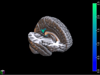
In order to investigate group differences in areas which potentially showed an increase over time during positive vs. negative ABM recall, we ran a repeated measure model using SWE method (Guillaume et al., Reference Guillaume, Hua, Thompson, Waldorp and Nichols2014), which showed significantly increased engagement in the right rSC in the MT group compared to the Non-MT group over time (see Figure 2). We found no areas that showed a greater increase over time in the Non-MT group relative to the MT group, or areas that decreased. In addition, there were no significant group differences for the reverse contrast comparing negative to positive ABMs.
Data analyses with Longitudinal & Repeated Measures Neuroimaging Data (Guillaume et al., Reference Guillaume, Hua, Thompson, Waldorp and Nichols2014), showed significantly increased engagement in the right retrosplenial cortex in the MT group compared to the Non-MT group over time (depicted in blue-green).

Brain-behavior relationships
Overall psychological functioning (SDQ-P) and depression symptoms (CASI)
The right rSC was the only area that showed a differential increase between groups over time during positive versus negative ABM recall. Exploratory post-hoc correlation analysis was conducted to examine whether the observed differential change in rSC between MT and Non-MT was related to psychopathology outcomes for the MT group (overall psychological functioning as measured by SDQ and symptoms of depression as measured by CASI). This analysis indicated that increase in rSC activity over time correlated with an improvement in overall psychological functioning in the MT group as measured by SDQ-P (SDQ-P Total Change Score: r = .53, p = .03). For completeness, we also examined this association in the Non-MT group and did not find a significant relationship (r = −.11, p = .67). The difference between the two correlation coefficients was not significant (Fisher r-to-z transformation one-tailed p = .03; two-tailed p = .07). No significant relationships was detected between rSC activity over time and change in depression scores in the MT group (r = −.11, p = .68), nor in the Non-MT group (Non-MT: r = .27, p = .29).
Discussion
This longitudinal investigation demonstrates functional developmental plasticity in children with documented maltreatment experience compared to a set of control participants carefully matched on socio-economic status, IQ and pubertal status. First, both groups showed relative stability of the ABM brain network, with a pattern of increased engagement of cortical midline structures including the vmPFC, pCC and rSC, reflecting a developmental progression towards network functioning seen in adult samples (Summerfield et al., Reference Summerfield, Hassabis and Maguire2009). Second, the pattern of group differences in relation to hippocampal functioning observed at baseline, was no longer apparent at follow-up. This potentially indicates normalization of hippocampal functioning in the maltreatment group; however, in light of the preliminary nature of this study and sample size, future investigation is required to replicate this finding. Third, we observed significantly increased engagement in the right rSC in the maltreatment group compared to their peers over time, indicating a different developmental maturational pattern in this region of the ABM network. Finally, we note that increased engagement of this region in the maltreatment group was associated with improved behavioral and psychological functioning over the follow-up period that may reflect adaptive developmental plasticity. It will be important for future research to explore whether change in hippocampal activation over time underpins adaptive developmental plasticity associated with reduced risk of depression following maltreatment.
At two-year follow-up we identified a neural network underlying ABM processing comprising cortical midline structures including the vmPFC, rSC, pCC, and MTG as well as the hippocampus. However, this pattern was evident only at a lowered threshold of significance; therefore replication of this finding is required. Notwithstanding this limitation, this network comprised the same regions consistently identified in adult studies of ABM processing (e.g. Summerfield et al., Reference Summerfield, Hassabis and Maguire2009). Increased engagement of the vmPFC, rSC, and pCC between baseline and follow-up assessments suggests a gradual strengthening and maturation of fronto-cingulate networks over adolescence (Summerfield et al., Reference Summerfield, Hassabis and Maguire2009). These changes are an example of the substantive maturational changes that take place during stage of life (Gogtay et al., Reference Gogtay, Giedd, Lusk, Hayashi, Greenstein, Vaituzis, Nugent, Herman, Clasen, Toga, Rapoport and Thompson2004).
Both vmPFC and rSC are key components of the ABM network, thought to serve different roles. Part of the prefrontal executive control network, the vmPFC has been implicated in the activation of appropriate and volitional ABMs and the de-activation of irrelevant ABMs, thereby regulating the recall process and avoiding undesirable intrusions (McCormick et al., Reference McCormick, Ciaramelli, De Luca and Maguire2018; Rawal & Rice, Reference Rawal and Rice2012). The rSC has dense connections with the hippocampus, parahippocampal cortex, and prefrontal cortex, and plays a pivotal role in the construction of coherent and complex ABM scenes (Hassabis & Maguire, Reference Hassabis and Maguire2007; Vann et al., Reference Vann, Aggleton and Maguire2009). In particular, the rSC is hypothesized to be involved in the translational aspect from an allocentric point of view (as indexed by the hippocampus) to a more fine-grained, egocentric representation (Burgess et al., Reference Burgess, Becker, King and O’Keefe2001; Vann et al., Reference Vann, Aggleton and Maguire2009). This hypothesized change in perspective is potentially in line with research implicating both vmPFC and rSC in self-referential processing in adolescents (Bradley et al., Reference Bradley, Colcombe, Henderson, Alonso, Milham and Gabbay2016). We suggest that within a developmental framework, it is conceivable that with increased prefrontal brain maturation in adolescence (Gogtay et al., Reference Gogtay, Giedd, Lusk, Hayashi, Greenstein, Vaituzis, Nugent, Herman, Clasen, Toga, Rapoport and Thompson2004), more fine-grained and accurate constructions of ABMs become possible with an increasing ability to appraise and elaborate on one’s past experiences.
Our analyses of valence-based ABM processing at baseline indicated that children who had been maltreated compared to their peers, showed reduced activation in the right hippocampus when recalling positive versus negative ABMs. Previous research has implicated the hippocampus in processing sensory and perceptual details as well as encoding temporal and spatial context (Burgess et al., Reference Burgess, Becker, King and O’Keefe2001). Differences in the processing of positively-valenced material has been previously observed in maltreated samples and we have speculated that it may contribute to a negative inferential or ruminative style (McCrory et al., Reference McCrory, Gerin and Viding2017a and Reference McCrory, Puetz, Maguire, Mechelli, Palmer, Gerin, Kelly, Koutoufa and Viding2017b). The pattern of attenuated hippocampal activation in children who had experienced maltreatment compared to their non-maltreated peers was no longer observed at follow-up. Such a null finding is difficult to interpret with confidence and should therefore be regarded as provisional. While it may reflect neuronal maturation, it may also have arisen due to statistical noise or reflect differences in developmental timing between the groups (e.g. Gee et al., Reference Gee, Gabard-Durnam, Flannery, Goff, Humphreys, Telzer and Tottenham2013). We would note, however, that the lack of observed differences in hippocampal activation on follow up occurred alongside improvements in psychological and behavioral functioning. While no causal link can be inferred, we would expect such improvements to be reflected in differences in brain function. Further longitudinal research is required to investigate whether specification of positive memories in those who have experienced maltreatment is reliably associated with changes in hippocampal activation and whether such changes are indeed associated with behavioral improvement.
Third, the observed increase in rSC activity over time in those children with maltreatment experience, when recalling positive versus negative ABMs, is of particular interest given the emerging role for this region in current models of ABM processing. This region connects the PFC with the MTL, acting as an interconnected hub for neocortical, hippocampal, parahippocampal, and thalamic regions (Mitchell et al., Reference Mitchell, Czajkowski, Zhang, Jeffery and Nelson2018). In the current study an increase in rSC activity was observed in the MT group from Time 1 to Time 2 during positive as compared to negative ABM recall. This is consistent with a developmental maturational pattern in this region of the ABM network that may support more effective ABM recall in relation to positively valenced memories. Because this developmental maturational pattern was only seen with children with MT histories, it is more likely to relate to resilient functioning following maltreatment, rather than adolescent development more generally. Our post-hoc, exploratory analyses indicated that change in rSC activity was associated with improved psychological functioning in the MT group. However, such a finding should be viewed as preliminary in nature; future longitudinal research in larger samples with multiple assessment periods and detailed assessment of ABM and psychological functioning is required to replicate this finding.
There are a number limitations to the current study. First, as is expected in most longitudinal studies, we lost several participants from baseline to follow-up. It is recognized that retention in longitudinal maltreatment research is highly challenging (Kinard, Reference Kinard1994). Our sample was thus not sufficiently large to robustly examine differences between the sexes, maltreatment types or ethnicity. Our findings, in particular the null finding with respect to hippocampal activation at T2, should be interpreted with caution as the size of the sample impacts on the statistical power with which we were able to detect true differences between the groups, meaning that we were underpowered to detect small effects. The current findings should therefore be regarded as preliminary in nature and they require replication. Previous research indicates that there may be some differential mental health outcomes for boys and girls exposed to early adversity and also that ABM processes can be different between the sexes or maltreatment types (Reese et al., Reference Reese, Haden and Fivush1996), even on the neural level (Puetz et al., Reference Puetz, Viding, Gerin, Pingault, Sethi, Knodt, Radtke, Brigidi, Hariri and McCrory2019). It should also be noted that the developmental window under investigation was relatively narrow (approximately 2 years), and while the longitudinal design permitted data collection at two time points, the study design does not permit inferences about causality. A longer follow-up with multiple assessments would provide a more detailed picture of the development of ABM processing in adolescents who have experienced maltreatment and their typically developing peers. Finally, many different processes are involved in ABM and the present study was unable to disentangle the degree to which encoding, processing, storage or recall differentially contributed to the observed changes in rSC functioning observed in this study. Future studies with more fine-grained ABM paradigms are necessary to shed light on these questions.
Conclusion
This longitudinal study compared two carefully matched groups in order to investigate a neurocognitive mechanism implicated in depression following maltreatment. The current study focussed on the development of ABM functioning and provides preliminary evidence of a pattern of normalization in hippocampal functioning during a two year follow up period. We also report evidence of malleability within a neurocognitive system, reflected in developmental change in rSC activity. This change was associated with improved psychological functioning in children with maltreatment experience, in line with the notion of resilience and recovery. Collectively these findings suggest that neural differences observed in children with maltreatment experience are not fixed. Rather, across development, early differences may normalize or present alongside novel patterns of neural response that may reflect resilient functioning. Future work, that measures neural change over time in children with maltreatment experience, is needed to build on these preliminary findings. Such work can help delineate a mechanistic understanding of latent vulnerability and those neurocognitive processes implicated in resilient functioning.
Supplementary material
To view supplementary material for this article, please visit https://doi.org/10.1017/S0954579421001292
Acknowledgments
The authors thank the children, parents, carers and social workers who generously participated in this research.
Funding statement
This work was funded by a grant to E.MC and E.V from the U.K. Economic and Social Research Council (ES/K005723/1). E.M is supported by a Wellcome Principal Research Fellowship (210567/Z/18/Z).
Conflicts of interest
None to report.