Introduction
Petrous apicitis is defined as osteomyelitis of the petrous apex of the temporal bone. It commonly arises from the contiguous spread of middle-ear disease. It is usually recognised by its association with symptoms of Gradenigo's syndrome, namely ear discharge, neuralgic headache and abducens palsy. However, most cases do not present with the complete triad of symptoms.Reference Chole and Donald1 The often subtle and non-specific symptomatology combined with a reduction in incidence in the post-antibiotic era contributes to its late diagnosis.
The diagnosis of fungal petrous apicitis represents a unique challenge partly due to its rarity as only four previous cases have been reported in the world literature.Reference Kountakis, Kemper, Chang, DiMaio and Stiernberg2 When investigating apparently immunocompetent patients the clinical index of suspicion is lowered and the diagnosis may only be considered following the failure of antibiotic therapy.
We present a case of fungal petrous apicitis in an immunocompetent man complicated by cerebral and nasopharyngeal otogenic abscesses. This represents the first such presentation to our knowledge. We also review the role of surgery both in the diagnosis and treatment of fungal skull base osteomyelitis, and provide a discussion of anti-fungal therapy.
Case report
A 72-year-old man with a history of chronic suppurative otitis media presented with a 4-month history of right-sided otorrhoea and intractable ipsilateral headache. Examination revealed a wet postero-central drum perforation and posterior canal wall oedema. High-resolution computed tomography identified total opacification of the mastoid air cells and medial aspect of the middle ear, but no sign of bone erosion. A tympanotomy revealed milky fluid in the middle-ear cleft with pale but otherwise unremarkable middle-ear mucosa.
The patient was readmitted a week later with nausea and vertigo. A Technetium-99 diphosphonate bone scintigram demonstrated increased osteoblastic activity of the right temporal bone. Oral ciprofloxacin 500 mg two times daily was initiated empirically to treat temporal bone osteomyelitis, but this was poorly tolerated and was therefore changed to Augmentin® 625 mg three times daily.
The patient returned within 72 hours with right-sided facial palsy (House–Brackmann grade IV) and worsening constitutional symptoms. Oral prednisolone 60 mg once daily was initiated in addition to intravenous (IV) Augmentin 1.2 g three times daily. The patient's mastoid was explored; the facial nerve was found to be oedematous and dehiscent at the second genu, and was decompressed from the geniculate ganglion to the stylomastoid foramen.
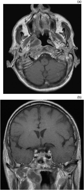
A right hypoglossal palsy and complete facial palsy developed 10 days post-mastoidectomy. A contrast-enhanced magnetic resonance imaging (MRI) scan revealed infiltrative enhancement of the medial temporal bone and an abscess around the eustachian tube orifice (Figure 1). Intravenous ceftriaxone 1.5 g and metronidazole 500 mg were initiated three times daily. The abscess was drained and biopsied endoscopically.
(a) Axial and (b) coronal T1-weighted gadolinium-enhanced magnetic resonance images of the head, demonstrating a low signal intensity collection, with rim enhancement around the right torus tubarius.

A follow-up MRI scan 10 days later demonstrated improvement in the nasopharynx. However, an inferior temporal gyrus abscess had developed, which was managed conservatively under neurosurgical direction with IV ceftriaxone 2 g two times daily, flucloxacillin 1 g four times daily and metronidazole 500 mg three times daily. Magnetic resonance imaging was carried out on a weekly basis.
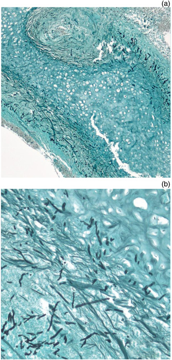
Histological analysis of the nasopharyngeal wall abscess biopsy indicated fungal hyphae on haematoxylin and eosin (H&E) staining, which was confirmed on Grocott's methenamine silver staining (Figure 2). The hyphae exhibited deep tissue penetration, perivascular invasion and acute angle branching of septae, which is in keeping with Aspergillus species. Anti-fungal treatment was initiated under the direction of the regional mycology department, using IV AmBisome® (liposomal amphotericin B) 3 mg/kg once daily for 2 weeks and IV voriconazole 4 mg/kg two times daily for 3 weeks, the latter being continued orally for a further 12 months. Given the possibility of dual fungal and bacterial pathology, co-treatment with antibiotic therapy was continued intravenously for two months and orally with Augmentin 625 mg three times daily for a further month.
Photomicrographs of biopsy specimen (Grocott's methenamine silver stain), showing (a) hyphae penetration and angioinvasion (magnification ×200), and (b) acute septated hyphae typical of Aspergillus species (magnification ×400).

There was symptomatic improvement over the subsequent weeks, with serial MRI scans demonstrating incremental reduction of the intracranial pathology. The facial palsy was still present at three months' follow up and was managed with tarsorrhaphy.
Discussion
Aspergillus is a ubiquitous saprophytic fungus that is inhaled into the upper respiratory tract and a potential commensal of the paranasal sinuses and external auditory canal.Reference Scully and Paes de Almeida3 Factors associated with invasive disease include immunocompromised states such as diabetes mellitus, protracted steroid use, haemopoietic stem cell and solid organ transplantation, critical illness, human immunodeficiency virus infection, and underlying malignancy.Reference Denning4 The early initiation of treatment at the stage of possible infection, prior to microbiological confirmation, has been associated with improved mortality rates.Reference Nivoix, Velten, Letscher-Bru, Moghaddam, Natarajan-Amé and Fohrer5
Fungal middle-ear disease is a condition more commonly seen in the immunocompromised host; only seven cases of Aspergillus mastoiditis have been documented in the immunocompetent (as described by van Tol and van Rijswijk).Reference van Tol and van Rijswijk6 All seven patients were in old age (average age 75 years) and presented with facial palsy and a history of chronic drum perforation. The physical findings were similar to those seen in cases of infection caused by Pseudomonas aeruginosa. All seven cases were managed with aggressive surgical debridement and anti-fungal therapy.
The diagnosis and management of fungal petrous apicitis represents a greater challenge. Only four previous cases of petrous apex aspergillosis have been documented,Reference Eloy, Bederson and Smouha7, Reference Ederies, Chen, Aviv, Pirouzmand, Bilbao and Thompson8 three of which proved fatal with catastrophic intracranial bleeds.Reference Eloy, Bederson and Smouha7 This morbidity reflects the propensity for Aspergillus species to invade blood vessel walls, resulting in a necrotising vasculitis type process. Petrous apex aspergillosis is a category of skull base osteomyelitis. Due to the complications cited above and those of our case, this disease ought to be managed as a form of neuroaspergillosis.
Neuroimaging has an important role in forming the diagnosis of petrous apicitis, as the symptoms and signs of this disease are often non-specific. Computed tomography may demonstrate opacification of air cells at the apex or bone erosion, but MRI features may predate these, with early marrow changes manifesting as T1-weighted hypointensity and T2-weighted hyperintensity in gadolinium-enhanced imaging.Reference Lee, Lee, Kim and Song9 Gallium-67 scanning also has a role in monitoring disease resolution, and in the follow up of petrous apicitis cases.Reference Lee, Lee, Kim and Song9 In this case, the findings of Tc-99m scintigraphy, ophthalmic nerve neuralgia and VIIth cranial nerve palsy led to the diagnosis, which was supported by MRI findings. The XIIth cranial nerve palsy was presumed to reflect a central skull base osteomyelitis.Reference Clark, Pretorius, Byren and Milford10
A recent single institution review highlights the difficulty in differentiating bacterial from fungal skull base osteomyelitis on clinical or radiological grounds.Reference Blyth, Gomes, Sorrell, da Cruz, Sud and Chen11 This emphasises the importance of tissue biopsy if clinical doubt exists or there is a slow response to appropriate antibiotic therapy. In our case, the diagnosis of invasive fungal disease resulted from the fortuitous discovery of an extra temporal ‘peritubular’ otogenic abscess. The tissue samples demonstrated the characteristic acute angle branching of septae and the angioinvasion tendencies of Aspergillus species. Neither culture nor polymerase chain reaction analyses of the tissue specimen were able to isolate a species in our case. Another feature that ought to raise clinical suspicion of invasive Aspergillus disease is that of post-operative clinical deterioration, which in our patient manifested as worsening constitutional symptoms and facial palsy several days following the raising of a tympanomeatal flap. This so called ‘forest fire phenomenon’ has been speculated to result from either the immunoparetic effects of surgery or the local tissue manipulation which stimulates pathogenicity in previously quiescent fungi.Reference Shamim, Siddiqui, Enam, Shah, Jooma and Anwar12
Previous studies have stressed the importance of radical surgery in combination with an anti-fungal agent in treating aspergillus skull base osteomyelitis and neuroaspergillosis.Reference Srinivasan13 The anti-fungal agent of choice has changed over the past decade, with randomised studies demonstrating the greater efficacy of voriconazole over amphotericin B in such cases.Reference Herbrecht, Denning, Patterson, Bennett, Greene and Oestmann14 Voriconazole now represents the recommended first-line treatment for invasive aspergillosis.Reference Walsh, Anaissie, Denning, Herbrecht, Kontoyiannis and Marr15 Although it has excellent oral bioavailability,Reference Amonoo-Kuofi, Tostevin and Knight16 voriconazole is associated with highly variable inter-patient plasma concentrations which requires dosing to be titrated against individual patients' plasma concentrations.Reference Denning, Ribaud, Milpied, Caillot, Herbrecht and Thiel17 Documented side-effects include hypoglycaemia and electrolyte disturbance, and possibly confusion and pneumonitis, all of which may occur in the absence of liver function test abnormality.Reference Boyd, Modi, Howard, Moore, Keevil and Denning18
Parize et al. described a series of patients with Aspergillus necrotising otitis externa who were successfully treated with extensive surgical debridement and voriconazole.Reference Parize, Chandesris, Lanternier, Poirée, Viard and Bienvenu19 Other reports include a case of nasocerebral aspergillosis that was successfully treated with voriconazole and steroids without the need for surgery,Reference Hiraga, Uzawa, Shibuya, Numata, Sunami and Kamitsukasa20 and a case of invasive rhinogenic skull base disease managed with oral voriconazole alone with substantial cost savings.Reference Parsonage, Stafford, Lillie, Moss, Barlow and Thaker21 Although our patient clinically suffered from petrous apicitis, we successfully treated him without recourse to apicectomy, and the cerebral abscess resolved with voriconazole therapy.
• Caution must be taken in empirical treatment of skull base osteomyelitis without microbiological confirmation
• Invasive aspergillosis should be considered early in cases unresponsive to empirical antibiotic therapy, even in the immunocompetent
• Tissue biopsy has an important role in the diagnosis of skull base osteomyelitis
• Aspergillus petrous apicitis and cerebral and nasopharyngeal abscesses may respond to anti-fungal therapy without recourse to radical resection
• Voriconazole is the recommended first-line anti-fungal therapy for invasive aspergillosis
In summary, this paper highlights the limitations of the empirical treatment of temporal bone osteomyelitis without microbiological confirmation. We have stressed the importance of considering a fungal aetiology and early tissue biopsy in cases that do not respond to conventional broad spectrum antibiotic therapy, even in the immunocompetent. We have demonstrated that successful treatment of neuroaspergillosis does not necessarily entail radical surgical resection, and can be achieved by early and sustained anti-fungal treatment under the direction of the regional mycology centre. In addition, to our knowledge, this paper is the first to describe a peritubular otogenic fungal abscess.