Introduction
Culture systems for primordial follicles are important for studying their oocyte development, especially because these follicles are a large potential source of oocytes that could be used in vitro for embryo production. The factors that control primordial follicle activation and further growth of primary follicles are not well understood. Since the cortical region where the primordial follicles are located is poorly vascularized, the development of these follicles is probably regulated by locally produced growth factors.
Endocrine hormones, like FSH, are known to regulate the production of several growth factors that play a critical role in primordial follicle activation and growth. FSH acts by binding to its receptor expressed on granulosa cells (Ulloa-Aguirre et al., Reference Ulloa-Aguirre, Midgley, Beitins and Padmanabhan1995; O'Shaughnessy et al., Reference O'Shaughnessy, Dudley and Rajapaksha1996) and more recent reports indicate its presence in oocytes, suggesting additional sites of action in the ovary (Meduri et al., 2002). Although FSH receptors are expressed from the primary follicles onward (Oktay et al., Reference Oktay, Briggs and Gosden1997), FSH may play an indirect effect on very early follicle development via factors released by larger follicles or ovarian stromal cells. For example, FSH promotes proliferation of granulosa cells via paracrine factors such as IGF-1 and activin (van den Hurk & Zhao, Reference Van Den Hurk and Zhao2005). In addition, FSH regulates expression of kit ligand (KL), growth differentiation factor-9 (GDF-9) and bone morphogenetic protein-15 (BMP-15) in murine follicles (Joyce et al., Reference Joyce, Pendola, Wigglesworth and Eppig1999, Thomas et al., Reference Thomas, Ethier, Shimasaki and Vanderhyden2005). These factors have been implicated in the activation of primordial follicles (van den Hurk & Zhao, Reference Van Den Hurk and Zhao2005).
Studies in vitro have demonstrated that addition of FSH to culture media promotes preantral follicular growth and antrum formation in many species (mouse: Spears et al., Reference Spears, Murray, Alisson, Boland and Gosden1998; murine: McGee et al., Reference McGee, Spears, Minami, Hsu, Chun, Billig and Hsueh1997; human: Wright et al., Reference Wandji, Srsen, Voss, Eppig and Fortune1999; bovine: Gutierrez et al., Reference Gutierrez, Ralph, Telfer, Wilmut and Webb2000; ovine: Cecconi et al., Reference Cecconi, Barboni, Coccia and Mattioli1999; Zhou & Zhang, Reference Zhou and Zhang2005; swine: Mao et al., Reference Mao, Wu, Smith, McCauley, Cantley, Prather, Didion and Day2002). Moreover, it is known that FSH inhibits apoptosis in preantral follicles cultured in vitro (mouse: Baker et al., Reference Baker and Spears1997; rat: McGee et al., Reference McGee, Spears, Minami, Hsu, Chun, Billig and Hsueh1997; human: Roy & Treacy, Reference Roy and Treacy1993). In goats, Silva et al. (Reference Silva, Báo, Lucci, Carvalho, Andrade, Ferreira and Figueiredo2004) demonstrated that FSH, at concentration of 100 ng/ml, increased follicle and oocyte diameters after 5 days of culture, but no effect of FSH on both primordial to primary follicle transition and viability was observed. Clearly, FSH is known to be the main regulator of follicle development in vivo and in vitro, but a specific role or the possibility of dose-dependent action of FSH in promoting primordial to primary follicle transition and growth have not been tested, being essential to investigate whether FSH play a role in this step of folliculogenesis. In addition, most of the studies investigating primordial follicle activation are based on histological evaluation, being very important to use ultrastructural analysis to confirm follicular viability.
Studies with goats are important to improve our knowledge about the factors that control early folliculogenesis in mammals and to explore possible physiological differences among species. Goats are present on all continents and are commercially seen as highly attractive livestock, since they constitute an important source of products such as meat, milk, fibre and skin. Thus, the aim of the present study was to investigate whether FSH has a beneficial role in the survival, activation and further growth of in vitro cultured goat primordial follicles enclosed in ovarian cortex. To this end, both histological and ultrastructural studies were performed to investigate and compare the morphology of follicles before and after culture for 1 or 7 days in the absence or presence of FSH at different concentrations (0, 10, 50 or 100 ng/ml).
Materials and Methods
Source of ovaries
Ovaries (n = 10) from five adult non-pregnant mixed-breed goats (1–3 years of age) were collected at a local slaughterhouse. The animals were cyclic and in good body condition. Then, the ovaries were washed and transported in 0.9% saline solution to the laboratory in thermo flasks with water at 32 °C.
Experimental protocol
At the laboratory, both ovaries from each animal were stripped of surrounded fat tissue and ligaments and cut in half, where after the medulla, large antral follicles and corpora lutea were removed. Following this, the ovarian cortex was divided into 11 fragments of approximately 3 × 3 mm (1 mm thick). One fragment was immediately fixed for classic histological studies (non-cultured controls) while a smaller fragment (1 mm3) was randomly collected and subsequently fixed for ultrastructural examination. The other fragments of ovarian cortex were individually in vitro cultured in 1 ml of culture medium for 1 or 7 days at 39 °C with 5% CO2 in air using a 24-well culture dish. The control medium was minimum essential medium (Cultilab, Brazil) supplemented with ITS (insulin 6.25 μg/ml, transferrin 6.25 μg/ml and selenium 6.25 ng/ml), 0.23 mM pyruvate; 2 mM glutamine; 2 mM hypoxanthine; 1.25 mg/ml BSA, 100 μg/ml penicillin, 100 μg/ml streptomycin (Vetec, Brazil) and 0.25 μg/ml fungizone (MEM+). This control medium (MEM+) was supplemented with different concentrations of porcine FSH (10, 50 or 100 ng/ml – provided by Dr J.F. Beckers, Liége, Belgium). All chemicals used in the present study were purchased from Sigma Chemical Co. unless otherwise indicated. Every 2 days, the culture medium was replaced by fresh medium. Each treatment was repeated five times, thus using the ovaries of five different animals.
Histological analysis and assessment of in vitro follicle growth
To evaluate the morphology of caprine follicles after 1 or 7 days of culture, a small part (1 mm3) from each fragment was randomly removed for TEM studies, while the remainder was fixed in Carnoy for 12 h for histological studies. After fixation, the tissue fragments were dehydrated in a graded series of ethanol, clarified with xylene and embedded in paraffin wax. For each piece of ovarian cortex, 7 μm sections were mounted on slides, stained with periodic acid Schiff and hematoxylin (PAS staining system, Sigma) and examined by light microscopy (Zeiss) at ×100 and ×400 magnification.
The follicles were classified as described by Hulshof et al. (Reference Hulshof, Figueiredo, Beckers, Bevers and Van Den Hurk1994) in primordial (one layer of flattened granulosa cells around the oocyte) or growing follicles i.e. intermediate (one layer of flattened to cuboidal granulosa cells around the oocyte), primary (a single layer of cuboidal granulosa cells around the oocyte) or secondary (oocyte surrounded by two or more layers of cuboidal granulosa cells). These follicles were classified individually as histologically normal when an intact oocyte was present, i.e. an oocyte without a pyknotic nucleus, surrounded by granulosa cells which are well organized in one or more layers and that have no pyknotic nucleus. Degenerated follicles were defined as those with a retracted oocyte, which have a pyknotic nucleus and/or are surrounded by disorganized granulosa cells, which are detached from the basement membrane. From each medium and each culture period, approximately 150 follicles were randomly evaluated.
To evaluate follicular activation and growth, only intact follicles with a visible oocyte nucleus were recorded and the proportion of primordial and growing follicles were calculated at day 0 (control) and after 1 or 7 days of culture in the various media tested. Major and minor axes of each oocyte and follicle were measured with the aid of an ocular micrometer. The averages of the minor and major axes were reported as oocyte and follicle diameters, respectively. These values were used to assess the effect of the hormonal treatment on follicular growth.
Ultrastructural analysis
For ultrastructural analysis, small pieces of ovarian cortex were fixed in 2% paraformaldehyde, 2.5% glutaraldehyde and 0.1 M sodium cacodylate buffer, pH 7.2. After washing the ovarian pieces with sodium cacodylate buffer, they were postfixed in 1% osmium tetroxide, 0.8% potassium ferricyanide and 5 mM CaCl2 in 0.1 M sodium cacodylate buffer. Subsequently, samples were dehydrated in a graded series of acetone and embedded in Spurr's epoxy resin. Firstly, semi-thin sections (3 μm) were cut on an ultramicrotome (Reichert Supernova, German) for light microscopy studies and stained with toluidine blue. Subsequently, follicles classified as histologically normal in semi-thin toluidin blue-stained sections were submitted to ultrastructural analysis. For that purpose, thin sections (70 nm) were cut and then contrasted with uranyl acetate and lead citrate and examined using a Jeol 1011 (Jeol, Tokyo, Japan) transmission electron microscope, operating at 80 kV.
Statistical analysis
Data are expressed as mean ± SEM. The percentages of surviving follicles at all stages, primordial and growing follicles obtained after 1 or 7 days in the various treatments were subjected to arc–sin transformation before analysis of variance (ANOVA). The data as well as the diameter of oocytes and follicles were analysed by ANOVA followed by Fisher's protected least significant difference test (PLSD test) (StatView for Windows, SAS Institute Inc.). Values were considered statistically significant when p < 0.05.
Results
Effect of FSH and culture periods on follicle survival
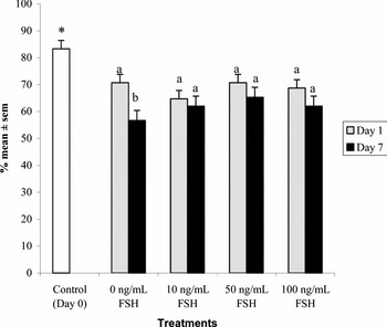
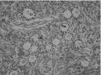
Figure 1 shows the effect of different concentrations of FSH on follicle survival, i.e. the percentage of histologically normal follicles in ovarian tissues after 1 and 7 days of culture. When compared to non-cultured follicles, a significant decrease (p < 0.05) in the percentages of histologically normal follicles (Fig. 2) was observed after 1 and 7 days culture, but no significant effect of FSH on follicle survival was observed. With the increase of the culture period from 1 to 7 days, a significant (p < 0.05) decrease in the percentage of normal follicles was observed in control medium (MEM+), but not in medium with FSH (Fig. 1).
Percentages (means ± SEM) of histologically normal preantral follicles in non-cultured tissue (control) and in tissue cultured for 1 and 7 days in MEM+ and MEM+ supplemented with 10, 50 and 100 ng/ml FSH. (30 follicles evaluated in each one of five replicates per treatment). *p < 0.05, significantly different from non-cultured ovarian cortex tissue (control/D0). (a, b) Different letters denote significant differences between culture periods within the same medium (p < 0.05).

Histological section of non-cultured tissue after staining with periodic acid Schiff–hematoxylin, showing normal primordial and primary follicles. O: oocyte; NU: oocyte nucleus; GC: granulosa cells (×400).

Goat primordial follicle activation and growth during in vitro culture
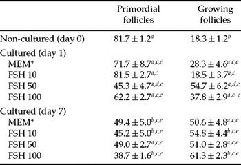
The percentages of primordial and growing follicles in ovarian cortical tissue before and after 1 and 7 days of culture are shown in Table 1. The percentages of primordial and growing follicles in non-cultured cortex were 81.7 and 18.3%, respectively. After 1 day of culture, when compared to non-cultured follicles, a reduction (p < 0.05) of primordial follicles concomitant with a significant increase of growing follicles was observed in all treatments, except when 10 ng/ml FSH was added to the culture medium. Compared to day 1, after 7 days culture, a strong reduction of primordial follicles (p < 0.05) was observed, except in tissues cultured in MEM plus 50 ng/ml FSH. Cortical tissues cultured with 10 or 100 ng/ml of FSH had a significant increase (p < 0.05) in the percentage of growing follicles after 7 days of culture when compared to day 1. At day 1 of culture, addition of 50 ng/ml of FSH to control medium significantly decreased (p < 0.05) the percentage of primordial follicles when compared to non-cultured tissue and other treatments. Furthermore, addition of 50 ng/ml FSH increased significantly (p < 0.05) the percentage of growing follicles when compared to other treatments, except when 100 ng/ml was used.
Percentages (mean ± SEM) of primordial and growing follicles (intermediate, primary and secondary) in non-cultured tissues and in tissues cultured for 1 or 7 days in MEM+ (control medium) and MEM+ supplemented with various concentrations of FSH

Per treatment, 150 follicles were evaluated
a ,b Different letters in the same column denote significant differences between culture periods within the same medium (p < 0.05).
c ,d Different letters in the same column denote significant differences among treatments in the same period (p < 0.05).
ep < 0.05, significantly different from non-cultured ovarian cortex tissue (control/day 0).
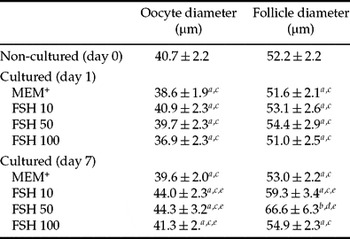
After 7 days of culture in medium containing 10, 50 or 100 ng/ml of FSH, a significant increase in oocyte diameter was seen in unilaminar follicles, i.e., primordial, intermediate and primary follicles together, when compared to non-cultured tissue (Table 2; p < 0.05). In addition, the presence of 10 or 50 ng/ml of FSH promoted a significant (p < 0.05) increase in follicular diameter after 7 days of culture when compared to control. A significant increase in follicle diameter was observed with the increase of culture period from day 1 to 7 only in tissues cultured with 50 ng/ml of FSH. At day 7, tissue cultured in presence of 50 ng/ml FSH had the highest follicle diameter (p < 0.05). Cultured and non-cultured tissue contained variable and relatively low (often zero) numbers of secondary follicles, which were not amenable to statistical comparison.
Oocyte and follicle diameters (mean ± SEM) in non-cultured tissues and in tissues cultured for 1 or 7 days in MEM+ (control medium) and MEM+ supplemented with various concentrations of FSH. Per treatment 150 follicles were evaluated

a,b Different letters in the same column denote significant differences between culture periods within the same medium (p < 0.05).
c,d Different letters in the same column denote significant differences among treatments in the same period (p < 0.05).
e p < 0.05, significantly different from non-cultured ovarian cortical tissues (control/Day 0).
Ultrastructural analysis of goat preantral follicles
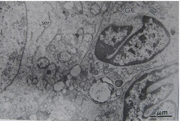
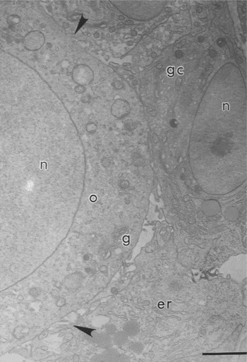
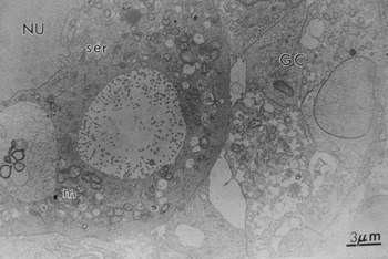
Based on histological results, TEM studies were performed in non-cultured follicles (control) and in follicles cultured for 1 and 7 days in MEM+ (control medium) or MEM+ plus 50 ng/ml FSH. The ultrastructural characteristics of follicles from non-cultured tissue and those cultured in medium without or with 50 ng/ml FSH for 1 day appeared similar, but after 7 days only tissues cultured with FSH had normal follicles. These follicles exhibited sparse vesicles spread throughout the ooplasm. The homogeneous cytoplasm furthermore contained numerous rounded mitochondria with peripheral cristae and continuous mitochondrial membranes, although there were occasional elongated forms with parallel cristae (Fig. 3). Golgi complexes were rarely observed. Both smooth and rough endoplasmic reticulum were present, either as isolated aggregations or as complex associations with mitochondria and vesicles (Fig. 4). The oocyte nucleus had uncondensed chromatin and the nucleolus could generally be observed. In all developmental stages, granulosa cells were small, with a greater nuclear-to-cytoplasm ratio as compared with typical cell structures. The nuclei were irregularly shaped, with loose chromatin in the inner part and small peripheral aggregates of condensed chromatin. Well-developed rough endoplasmic reticulum and mitochondria with well-developed lamellar cristae were the most evident organelles observed in granulosa cells. Gap junctions were abundantly observed between granulosa cells as well as between granulosa cells and the oocyte (Fig. 5).
Electron micrograph of a preantral follicle from a non-cultured control ovarian fragment. Note the homogeneous cytoplasm with numerous rounded mitochondria. GC: granulosa cells; m: mitochondria; ser: smooth endoplasmic reticulum (×6600).

Electron micrograph of a preantral follicle cultured for 1 day in MEM+ (cultured control). Note the great nuclear-to-cytoplasm ratio in granulosa cells. n: nucleus; o; oocyte; g: Golgi complex; gc: granulosa cells; er: endoplasmic reticulum; arrowhead: microvillus (×4000). Bar: 3 μm.

Electron micrograph of a preantral follicle cultured in FSH (50 ng/ml) for 7 days. Note the smooth endoplasmic reticulum and mitochondria, which were the most evident organelles observed in ooplasm. NU: oocyte nucleus; GC: granulosa cells; m: mitochondria; ser: smooth endoplasmic reticulum; v: vesicle (×3300).

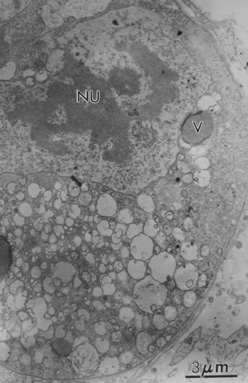
Cortical tissues cultured in control medium for 7 days had histologically normal follicles, but TEM studies revealed some changes in their ultrastructure, which are indicative of degeneration. Such follicles showed an oocyte extremely vacuolated, with the vacuoles often being fused and producing a larger vacuolated area. Organelles were more randomly distributed throughout the cytoplasm and signs of endoplasmic reticulum proliferation and damage to mitochondrial membranes and cristae were observed. The oocyte nucleus appeared misshapen and retracted and had a wavy membrane. Granulosa cells look swollen, while the density of cytoplasmic organelles was low. Frequently, the connection between the oocyte and granulosa cells had disappeared, while organelles were no longer identifiable. Furthermore, granulosa cells showed less contact with each other and exhibited obvious fewer gap junctions (Fig. 6).
Electron micrograph of a preantral follicle cultured for 7 days in MEM+ (cultured control) that were scored as histologically normal at the LM level. Note the extreme vacuolization and the great holes present in the cytoplasm, indicative of degeneration. NU: oocyte nucleus; v: vesicles (×3900).

Discussion
The present study demonstrated the importance of FSH on in vitro activation and growth of caprine primordial follicles in a 7 days culture system. Although very little is known about the regulation of primordial follicle development, FSH seems to be a very effective factor in maintaining follicle viability (Ralph et al., Reference Ralph, Wilmut and Telfer1996; Wandji et al., Reference Van den Hurk, Spek, Hage, Fair, Ralph and Schotanus1996; Saha et al., Reference Saha, Shimizu, Geshi and Izaike2000). However, Silva et al. (Reference Silva, Báo, Lucci, Carvalho, Andrade, Ferreira and Figueiredo2004) did not observed a significant effect of FSH on follicle survival after 5 days culture, probably because a high concentration of FSH (100 ng/ml) was used. In our study, addition of 50 ng/ml of FSH to culture medium was very important to maintain the percentage of normal follicles after 7 days culture. Hsueh et al. (Reference Hsueh, Billig and Tsafriri1994) and Chun et al. (Reference Chun, Billig, Tilly, Furuta, Tsafriri and Hsueh1994) suggested that the diffusion of several essential chemical and physical factors through the basal membrane could be compromised in the absence of FSH. In addition, cultures without FSH more frequently resulted in extrusion of the oocyte from its original follicular structure (Cortvrindt et al., Reference Cortvrindt, Smitz and Van Steirteghem1997), which may be caused either by damage or reduction in number of gap junctions (Amsterdam & Rotmensch, Reference Amsterdam and Rotmensch1987; Hsueh et al.; 1994). In addition, FSH inhibited apoptosis in preantral follicles cultured in vitro (mouse: Baker & Spears, Reference Baker and Spears1997; rat: McGee et al., Reference McGee, Spears, Minami, Hsu, Chun, Billig and Hsueh1997; human: Roy & Treacy, Reference Roy and Treacy1993; Wright et al., Reference Wandji, Srsen, Voss, Eppig and Fortune1999; swine: Mao et al., Reference Mao, Wu, Smith, McCauley, Cantley, Prather, Didion and Day2002).
Addition of 50 ng/ml of FSH to the medium increased the activation rate of primordial follicles as early as day 1 of culture when compared to other treatments. However, after 7 days of culture, follicular activation was similar among all treatments. Ovarian follicular development is known to proceed to primordial and primary stages independently of the action of FSH. This has been observed in mice carrying invalidations of the FSHβ and FSHR genes (Kumar et al., Reference Kumar, Wang, Lu and Matzuk1997; Dierich et al., Reference Dierich, Sairam, Monaco, Fimia, Gansmuller, LeMeur and Sassone-Corsi1998) and also in patients with mutations suppressing the function of the FSHR (Beau et al., Reference Beau, Touraine, Meduri, Gougeon, Desroches, Matuchansky, Milgrom, Kuttenn and Misrahi1998; Touraine et al., Reference Touraine, Beau, Gougeon, Meduri, Desroches, Pichard, Detoeuf, Paniel, Prieur, Zorn, Milgrom, Kuttenn and Misrahi1999). FSH binds to its receptor expressed on granulosa cells (Ulloa-Aguirre et al., Reference Ulloa-Aguirre, Midgley, Beitins and Padmanabhan1995; O'Shaughnessy et al., Reference O'Shaughnessy, Dudley and Rajapaksha1996) and oocytes (Meduri et al., 2002) from the primary follicles onward (Oktay et al., Reference Oktay, Briggs and Gosden1997). Recently, after culture of caprine ovarian cortical tissue for 5 days, Silva et al. (Reference Silva, Báo, Lucci, Carvalho, Andrade, Ferreira and Figueiredo2004) showed that FSH (100 ng/ml) did not promote activation of caprine primordial follicles. Previous studies have confirmed that FSH at this concentration did not influence bovine follicular activation (Fortune et al., Reference Fortune, Kito, Wandji and Srsen1998; Braw-Tal & Yossefi, Reference Braw-Tal and Yossefi1997; Derrar et al., Reference Derrar, Price and Sirard2000). Conversely, we demonstrated that a lower concentration of FSH (50 ng/ml) can improve follicular activation and survival. In addition, Joyce et al. (Reference Joyce, Pendola, Wigglesworth and Eppig1999) reported that FSH stimulates Kit ligand mRNA expression in granulosa cells of preantral follicles. Kit ligand (KL) has been shown to be essential for oocyte growth (Eppig, Reference Eppig2001; Nilsson & Skinner, Reference Nilsson and Skinner2001) and primordial follicle activation (Parrot & Skinne, Reference Parrot and Skinner1999). FSH can also modulate the levels of BMP-15 and GDF-9 in growing follicles (Thomas et al., Reference Thomas, Ethier, Shimasaki and Vanderhyden2005) and these growth factors are essential for primordial and primary follicle development in mice (Dong et al., Reference Dong, Albertine, Nishimori, Kumar, Lu and Matzuk1996) and sheep (Galloway et al., Reference Galloway, McNatty, Cambridge, Laitinen, Juengel, Jokiranta, McLaren, Luiro, Dodds, Montgomery, Beattie, Davis and Ritvos2000).
In the current study, follicular diameter had the highest increase when cultured in presence of 50 ng/ml for 7 days. Itoh et al. (Reference Itoh, Kacchi, Abe, Sendai and Hoshi2002) also demonstrated that 50 ng/ml FSH increased both oocyte and follicular diameters in 13 days cultured bovine follicles. FSH receptor expression has been reported to develop progressively during the transition from primordial to primary to secondary follicle (Oktay et al., Reference Oktay, Briggs and Gosden1997). The presence of FSH receptors in these early follicles presumably explains the effect of FSH on oocyte growth in preantral follicles. Since there are FSH receptors in oocytes, it is possible that FSH must act in both cell types to promote follicular growth and development (Méduri et al., Reference Méduri, Charnaux, Driancourt, Combettes, Granet, Vannier, Loosfelt and Migrom2002). Furthermore, a two-way exchange may occur between the oocyte and granulosa cells and a direct action of FSH on oocytes produces compounds whose diffusion into the granulosa cells is necessary for their proliferation and maturation (Méduri et al., Reference Méduri, Charnaux, Driancourt, Combettes, Granet, Vannier, Loosfelt and Migrom2002). Other authors observed that FSH promotes an increase in follicular diameter and proliferation of granulosa cells (rats: McGee et al., Reference McGee, Spears, Minami, Hsu, Chun, Billig and Hsueh1997; mouse: Nayudu & Osborn, Reference Nayudu and Osborn1992; Cortvrindt et al., Reference Cortvrindt, Smitz and Van Steirteghem1996, Reference Cortvrindt, Smitz and Van Steirteghem1997, Reference Cortvrindt, Hu and Smitz1998; bovine: Wandji et al., Reference Van den Hurk, Spek, Hage, Fair, Ralph and Schotanus1996; Saha et al., Reference Saha, Shimizu, Geshi and Izaike2000; caprine: Silva et al., Reference Silva, Van Den Hurk, Matos, Santos, Pessoa, Moraed and Figueiredo2004; ovine: Cecconi et al., Reference Cecconi, Barboni, Coccia and Mattioli1999; human: Roy & Treacy, Reference Roy and Treacy1993; Wright et al., Reference Wandji, Srsen, Voss, Eppig and Fortune1999). In addition, FSH was found to stimulate antrum formation and steroidogenesis in granulosa cells (Nayudu & Osborn, Reference Nayudu and Osborn1992; Boland et al., Reference Boland, Humpherson, Leese and Gosden1993; Ralph et al., Reference Ralph, Wilmut and Telfer1995, Reference Ralph, Wilmut and Telfer1996; Wandji et al., Reference Van den Hurk, Spek, Hage, Fair, Ralph and Schotanus1996; Abir et al., Reference Abir, Franks, Mobberley, Moore, Margara and Winston1997; Gutierrez et al., Reference Gutierrez, Ralph, Telfer, Wilmut and Webb2000; Wu et al., Reference Wright, Hovatta, Margara, Trew, Winston, Franks and Hardy2000; Mao et al., Reference Mao, Wu, Smith, McCauley, Cantley, Prather, Didion and Day2002; Mitchell et al., Reference Mitchell, Kennedy and Hartshorne2002; Kreeger et al., Reference Kreeger, Fernandes, Woodruff and Shea2005). Adriaens et al. (Reference Adriaens, Cortvrindt and Smitz2004) reported that omission of FSH during the early preantral stage tends to compromise a maximal oocyte developmental competence.
Several authors have emphasized the importance of TEM after in vitro culture of preantral follicles, since it gives close insight into the ultrastructural characteristics of follicles, allowing a better evaluation of their quality (Van den Hurk et al., Reference Van Den Hurk and Zhao1998; Zhao et al., Reference Wu, Nayudu, Kiesel and Michelmann2000; Salehnia et al., Reference Salehnia, Moghadam and Velojerdi2002). In the present study, preantral follicles cultured for 1 day in MEM without or with FSH (50 ng/ml) respectively, appeared ultrastructurally normal, which confirmed the results obtained in the histological studies. However, after 7 days of culture, TEM studies revealed differences in ultrastructural quality of follicles cultured without or with FSH (50 ng/ml). Although they had comparable histological morphology, follicles cultured in the presence of FSH maintained their ultrastructural integrity, while those cultured without FSH showed various signs of initial degeneration after 7 days culture and exhibited more clear degenerative features, like ooplasm vacuolization. Cytoplasmic vacuoles are characteristic signs of degeneration in oocytes (Silva et al., Reference Silva, Lucci, Carvalho, Báo, Costa, Santos and Figueiredo2000), granulosa (Hay et al., Reference Hay, Cran and Moor1976) and cumulus cells (Assey et al., Reference Assey, Hyttel, Roche and Boland1994) and may represent endoplasmic reticulum swelling (Tassel & Kennedy, Reference Tassel and Kennedy1980) or altered mitochondrial structure (Fuku et al., Reference Fuku, Xia and Downey1995). In goat preantral follicles, mitochondria showing extensive swelling and disappearance of their cristae and endoplasmic reticulum that have increased in volume, were previously indicated as the first signs of degeneration (Silva et al., Reference Silva, Báo, Lucci, Carvalho, Andrade, Ferreira and Figueiredo2001).
In conclusion, this study with caprine follicles showed that a concentration of 50 ng/ml FSH is able to promote the activation of primordial follicles and growth of activated preantral follicles. Furthermore, these data support the vital role of FSH in maintaining healthy oocyte growth and follicular ultrastructure after 7 days culture. This culture system should be useful for studying the regulation of early follicular growth and development, especially because these follicles represent a large source of oocytes that could be used in vitro for embryo production.
Acknowledgements
This work was supported by CNPq, FINEP and Fundação Cearense de Apoio à Pesquisa (FUNCAP). Maria Helena Tavares de Matos is a recipient of a grant from CNPq (Brazil). The authors thank Dr Vicente José de Figueiredo Freitas, PhD (LFCR) for the logistical support.