Introduction
Abnormalities in brain morphology have been studied extensively in schizophrenia within the past few decades but less is known about how such brain changes occur in bipolar disorder, especially early in the course of the illness, and also the differences in brain structural changes between bipolar disorder and schizophrenia. Thus far, studies have shown that both disorders share the same genetic determinants (Lichtenstein et al. Reference Lichtenstein, Yip, Björk, Pawitan, Cannon, Sullivan and Hultman2009; Wang et al. Reference Wang, Liu and Aragam2010) and pathogenetic mechanisms (Murray et al. Reference Murray, Sham, van Os, Zanelli, Cannon and McDonald2004), and it has been suggested that schizophrenia and bipolar disorder should be conceptualized as disorders along a psychosis continuum, instead of belonging to separate disease categories (Möller, Reference Möller2003). At present, there is some evidence from neuroimaging studies that brain abnormalities in patients with bipolar disorder and schizophrenia may be distinguishable. A recent meta-analysis revealed that although gray matter (GM) loss in bipolar disorder overlapped substantially with regions of GM reductions in schizophrenia, a volume reduction of the anterior cingulate was specific only to bipolar disorder and has been implicated in emotional processing pathways (Ellison-Wright & Bullmore, Reference Ellison-Wright and Bullmore2010). Smaller cortical GM volume has also been found in schizophrenia but not in bipolar patients (Zipursky et al. Reference Zipursky, Seeman, Bury, Langevin, Wortzman and Katz1997). The bilateral entorhinal cortex, left anterior superior temporal gyrus and right amygdala were reportedly smaller in patients with schizophrenia compared with patients with bipolar disorder, whereas the left amygdala and right anterior superior temporal gyrus were larger in patients with bipolar disorder compared with patients with schizophrenia (Pearlson et al. Reference Pearlson, Barta, Powers, Menon, Richards, Aylward, Federman, Chase, Petty and Tien1997).
Reports of reduced cortical thickness in schizophrenia and bipolar disorder have also surfaced, although few studies compared such brain structural abnormalities between the two patient groups. In a study of cortical thickness in lithium-free bipolar I patients, researchers found that the bilateral prefrontal cortex and the left anterior cingulate cortex were significantly thinner in patients than in healthy individuals, and thinning was more severe in those with a history of psychosis (Foland-Ross et al. Reference Foland-Ross, Thompson, Sugar, Madsen, Shen, Penfold, Ahlf, Rasser, Fischer, Yang, Townsend, Bookheimer and Altshuler2011). This was consistent with the results from an earlier whole-brain cortical thickness study on bipolar patients, where researchers observed cortical thinning in the prefrontal and occipital cortex, in addition to the anterior cingulate cortex (Lyoo et al. Reference Lyoo, Sung, Dager, Friedman, Lee, Kim, Kim, Dunner and Renshaw2006). In schizophrenia, cortical thinning is pronounced and widespread in the frontal, temporal, parietal, occipital and limbic regions (Goldman et al. Reference Goldman, Pezawas, Doz, Mattay, Fischl, Verchinski, Chen, Weinberger and Meyer-Lindenberg2009). Similar findings have been reported in younger schizophrenia patients with a shorter duration of illness (Schultz et al. Reference Schultz, Koch, Wagner, Roebel, Nenadic, Schachtzabel, Reichenbach, Sauer and Schlösser2010). Cortical development has also been shown to differ significantly between childhood-onset schizophrenia patients and healthy controls in a longitudinal study, with thinner cortices reported in patients throughout the 19-year follow-up (Greenstein et al. Reference Greenstein, Lerch, Shaw, Clasen, Giedd, Gochman, Rapoport and Gogtay2006). The only whole-brain study of cortical thickness comparing bipolar disorder patients directly with schizophrenia patients found significant cortical thinning in patients with schizophrenia but not in bipolar disorder, when compared to controls (Rimol et al. Reference Rimol, Hartberg, Nesvåg, Fennema-Notestine, Hagler, Pung, Jennings, Haukvik, Lange, Nakstad, Melle, Andreassen, Dale and Agartz2010).
Neuroimaging studies have suggested that hippocampus and amygdala volume reductions might be magnified over the course of schizophrenia (Adriano et al. Reference Adriano, Caltagirone and Spalletta2012; Shepherd et al. Reference Shepherd, Laurens, Matheson, Carr and Green2012). The volume reduction was only found in the amygdala but not in the hippocampus in adults who were at ultra-high risk of developing psychosis and biopolar I or II disorder (Bechdolf et al. Reference Bechdolf, Wood, Nelson, Velakoulis, Yucel, Takahashi, Yung, Berk, Wong, Pantelis and McGorry2012). Even though there are substantial studies on identifying amygdala and hippocampal volume abnormalities in bipolar and schizophrenia relative to healthy subjects (Strasser et al. Reference Strasser, Lilyestrom, Ashby, Honeycutt, Schretlen, Pulver, Hopkins, Depaulo, Potash, Schweizer, Yates, Kurian, Barta and Pearlson2005), few studies have examined shape deformations in these structures. Subtle loss of volume may be reflected in shape deformations and not in overall volumetric comparisons, allowing better discrimination between schizophrenia and bipolar patients, and healthy controls (Csernansky et al. Reference Csernansky, Joshi, Wang, Haller, Gado, Miller, Grenander and Miller1998). One of the few studies that looked at shape deformations in subcortical structures suggested a lack of overall shape differences in the amygdala and hippocampus in schizophrenia (Shenton et al. Reference Shenton, Gerig, McCarley, Székely and Kikinis2002), whereas another study reported significant deformation in the tail of the hippocampus in schizophrenia patients (Styner et al. Reference Styner, Lieberman, Pantazis and Gerig2004).
To our knowledge, no study has specifically examined cortical thickness and shape abnormalities of the amygdala–hippocampal complex in first-episode schizophrenia (FES) and mania (FEM). Examining the presence of brain structural abnormalities early in the course of illness may help to determine whether these deficits exist and worsen with illness progression, eventually aiding in the identification of structural endophenotypes that precede illness onset. Based on extant findings, we hypothesized that cortical thinning would be evident in both FES and FEM patients, and would be more widespread in FES. In view of previous volumetric magnetic resonance imaging (MRI) findings, we also hypothesized that the hippocampus and amygdala deformities would be found in both FEM and FES.
Method
Subjects
Fifty-six right-handed patients (28 FEM and 28 FES) were recruited from the Institute of Mental Health, Singapore, for the study. Twenty-eight age-, sex- and handedness-matched healthy subjects were recruited from the community through advertisements. Written, informed consent was obtained from all participants after a thorough explanation of the study procedures. The Institutional Review Boards of the Institute of Mental Health and the National Neuroscience Institute approved the study protocol.
All participants were free from a history of neurological illness or a diagnosis of alcohol or drug abuse in the past 3 months prior to the study based on DSM-IV criteria. For healthy controls, there was no family history of psychosis or bipolar disorder and the SCID – Non-Patient version (SCID-NP) was administered to rule out the presence of any other Axis I psychiatric disorder. For patients, a psychiatrist confirmed the diagnosis of mania or schizophrenia using information obtained from the clinical history, past medical records, mental status examination, and administration of the SCID – Patient version (SCID-P; First et al. Reference First, Spitzer, Gibbon and Williams1994). The Positive and Negative Syndrome Scale (PANSS; Kay et al. Reference Kay, Fiszbein and Opler1987) and the Global Assessment of Functioning (GAF; Hall, Reference Hall1995) were administered to all patients with FES and FEM to assess symptom severity and psychosocial functioning. Additionally, the Young Mania Rating Scale (YMRS; Young et al. Reference Young, Biggs, Ziegler and Meyer1978) was administered only to patients with FEM. Handedness was determined following the administration of the modified Edinburgh Questionnaire.
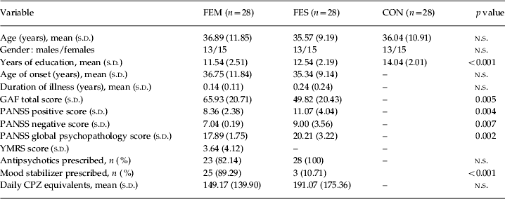
FES and FEM patients receiving psychotropic medications were on stable doses of their medications for at least 2 weeks prior to recruitment, and none had their medications withdrawn for the purposes of the study. Antipsychotics were prescribed in 82.1% (n = 23) of patients with FEM and all patients with FES. Mood stabilizers (mainly lithium or valproate) were administered in 89.3% (n = 25) of patients with FEM and 10.7% (n = 3) of patients with FES. A combination of antipsychotics and mood stabilizers were prescribed for 67.9% (n = 19) of patients with FEM and 10.7% (n = 3) of patients with FES. The antipsychotic dose, defined in mean daily chlorpromazine (CPZ) equivalents, was calculated using standard guidelines (Scott, 2000; APA, 2004). Detailed demographic information and clinical characteristics for the three groups of participants are shown in Table 1.
Demographic and clinical characteristics of the sample (n = 84)

FEM, First-episode mania; FES, first-episode schizophrenia; CON, controls; GAF, Global Assessment of Functioning; PANSS, Positive and Negative Syndrome Scale; YMRS, Young Mania Rating Scale; CPZ, chlorpromazine; s.d., standard deviation; n.s., no significance at a level of 0.05.
Image acquisition
T1-weighted magnetization-prepared rapid gradient-recalled echo [MPRAGE; repetition time (TR) = 7.2 s, echo time (TE) = 3.3 ms, flip angle = 8°] images were acquired using a 3-T whole-body scanner (Philips Achieva) with a SENSE head coil at the National Neuroscience Institute, Singapore. Each T1-weighted volume consisted of 180 axial slices of thickness 0.9 mm without gap [field of view (FOV) 230 mm × 230 mm; matrix: 256 × 256]. The stability of a high signal-to-noise ratio was ensured through a regular automated quality control procedure.
Hippocampus and amygdala delineation and shape analysis
Our delineation and shape analysis procedures of the hippocampus and amygdala have been described in detail elsewhere (Qiu et al. Reference Qiu, Tuan, Woon, Abdul-Rahman, Graham and Sim2010b). In brief, the Markov random field model was first applied to label each voxel in the image volume as GM, white matter (WM) or cerebrospinal fluid (CSF), or subcortical structure (Fischl et al. Reference Fischl, Salat, Busa, Albert, Dieterich, Haselgrove, van der Kouwe, Killiany, Kennedy, Klaveness, Montillo, Makris, Rosen and Dale2002). Because of lack of constraint on structural shapes, this process introduced irregularities and topological errors (e.g. holes) at the structural boundary. This may increase shape variation and thus reduce statistical power to detect group differences in shape. To address this potential pitfall, we generated the hippocampal and amygdala shapes of each individual subject with properties of smoothness and correct topology by injecting an atlas shape into them using an advanced brain mapping tool, the large deformation diffeomorphic metric mapping (LDDMM) algorithm for images (Qiu & Miller, Reference Qiu and Miller2008). The hippocampal and amygdala atlas shapes were obtained by averaging the hippocampal and amygdala shapes of 41 subjects using the LDDMM atlas generation algorithm (Qiu et al. Reference Qiu, Brown, Fischl, Ma and Miller2010a). Each hippocampal and amygdala volume was approximated by the transformed atlas through the LDDMM map. The surface representation of the hippocampal and amygdala shapes was created by composing the diffeomorphic map on the atlas surface.
We applied LDDMM (Vaillant et al. Reference Vaillant, Qiu, Glaunès and Miller2007) to map the atlas surface to the subjects' hippocampal and amygdala surfaces. The Jacobian determinant of the deformation in the logarithmic scale was computed in the local coordinates of the atlas for statistical shape comparison across the clinical population. The logarithmic scale of the Jacobian determinant, termed the ‘deformation map’, represents the ratio of each subject's hippocampal (or amygdala) volume to the template volume in the logarithmic scale; that is, positive values correspond to expansion and negative values to compression of the subject's hippocampus (or amygdala) relative to the atlas at a particular location.
Cortical thickness analysis
Based on the tissue segmentation obtained from FreeSurfer (Fischl et al. Reference Fischl, Salat, Busa, Albert, Dieterich, Haselgrove, van der Kouwe, Killiany, Kennedy, Klaveness, Montillo, Makris, Rosen and Dale2002), an inner surface was constructed at the boundary between WM and GM and then propagated to the outer surface at the boundary between GM and CSF. The cortical thickness was measured as the distance between the corresponding points on the inner and outer surfaces (Fischl & Dale, Reference Fischl and Dale2000) and represented on the fiducial surface where the center of the inner and outer surfaces was located.
For group comparison of the cortical thickness, we used a multi-manifold (MM)-LDDMM algorithm to align individual fiducial surfaces to an atlas that was generated based on the cortical anatomy of 40 subjects (Zhong & Qiu, Reference Zhong and Qiu2010) and transferred the thickness of each individual subject to the atlas. The MM-LDDMM algorithm uses different cortical representations (fiducial surface and sulcal curves) and seeks an optimal diffeomorphic transformation (one-to-one, reversible, smooth, topological preservation) to map one fiducial surface to the other. The algorithm improves the quality of regional alignment by incorporating 14 reliable sulcal curves and controls the quality of global and regional alignment by incorporating the geometry of the fiducial surface. The 14 sulcal curves, including the superior frontal sulcus, inferior frontal sulcus, precentral sulcus, central sulcus, postcentral sulcus, sylvian fissure, anterior segment of the superior temporal sulcus, inferior temporal sulcus, intraparietal sulcus, calcarine sulcus, parieto-occipital fissure, collateral sulcus, superior callosal sulcus and olfactory sulcus, were chosen because they are present consistently and are readily identifiable on the cortex. We extracted them semi-automatically from the fiducial surface using dynamic programming and the protocol described previously (Zhong & Qiu, Reference Zhong and Qiu2010). The accuracy of the MM-LDDMM algorithm for aligning the fiducial surfaces was evaluated by comparing it with the LDDMM curve and LDDMM surface algorithms (Zhong et al. Reference Zhong, Phua and Qiu2010).
Statistical analysis
Demographic variables among the three groups were compared using one-way ANOVAs for continuous variables and the χ2 test for categorical variables.
We examined group differences in amygdala and hippocampal volumes using an ANCOVA with a main factor of diagnosis while controlling for years of education and duration of illness. Post-hoc analysis with Bonferroni correction was performed to examine pairwise group differences in the volumes of these two structures. The group comparisons of the amygdala and hippocampal volumes between patients with FEM and FES were further examined when adjusting for the psychotropic medications, which were measured using mean daily CPZ equivalents (for antipsychotic exposure), or the prescription of mood stabilizers (lithium or valproate).
We examined pairwise group differences in amygdala–hippocampal shapes. For this analysis the shape deformation maps on the atlas surface were smoothed using a 30-mm full-width at half-maximum (FWHM) Gaussian filter (Chung et al. Reference Chung, Robbins, Dalton, Davidson, Alexander and Evans2005). Group comparisons were performed at each point on their atlas surfaces to reveal shape abnormalities in FEM and FES using linear regression with a main effect of diagnosis and years of education and also duration of illness as covariates. Age and gender were not entered into the linear regression because our controls and FEM and FES subjects were age and gender matched. The results at each surface vertex were thresholded at the level of significance (p < 0.001) and then corrected for multiple comparisons at the cluster level of significance (p < 0.05). Each cluster size must be greater than 358 mm2, which was determined based on random field theory (Chung et al. Reference Chung, Worsley, Nacewicz, Dalton and Davidson2010). The same analysis was performed to identify cortical thickness abnormalities in FEM and FES. We replicated the statistical analyses when the psychotropic medications or the prescription of mood stabilizers were considered as additional covariates.
Pearson's correlation analysis was performed to investigate the relationships of the amygdala, hippocampal shape deformation and cortical thickness with PANSS and GAF scores as both clinical measures were collected from all patients. Brain measures were computed as average cortical thickness or shape deformation of the amygdala and hippocampus in the regions with significant pairwise group differences obtained from the above analyses.
Results
Demographics and clinical characteristics
As shown in Table 1, FEM patients received less years of education than healthy controls whereas education level was comparable between the two patient groups. Patients with FES and FEM received comparable antipsychotic doses in terms of mean daily CPZ equivalents and frequencies of antipsychotics prescribed but differed in frequencies of mood stabilizers given. Compared to patients with FEM, patients with FES had a lower GAF total score and higher PANSS positive, negative and global psychopathology scores. Age did not differ significantly among the three groups. The duration of illness and age of onset did not differ significantly among the two patient groups.
Hippocampus and amygdala volume and shape abnormalities
Table 2 lists the means and standard deviations of the amygdala and hippocampal volumes in each group. After adjusting for duration of illness and years of education, group differences in bilateral amygdala volumes were found among the healthy controls and FES and FEM patients (left: p = 0.013; right: p = 0.022). Post-hoc analysis revealed that smaller volumes of the bilateral amygdala were observed in patients with FES, compared with healthy controls (left: p = 0.010; right: p = 0.018). No group differences in bilateral amygdala volumes were found between FEM patients and healthy controls (left: p = 0.241; right: p = 0.598) and between FEM and FES patients (left: p = 0.584; right: p = 0.351). No group differences were found in bilateral hippocampal volumes among the three groups (left: p = 0.141; right: p = 0.165). Further analyses with the antipsychotic dose (mean daily CPZ equivalents) as a covariate revealed no group differences in bilateral amygdala volumes (left: p = 0.768; right: p = 0.517) and bilateral hippocampal volumes (left: p = 0.286; right: p = 0.337) between FES and FEM patients. The inclusion of administration of mood stabilizers as a covariate also revealed no group differences in bilateral amygdala volumes (left: p = 0.969; right: p = 0.783) and bilateral hippocampal volumes (left: p = 0.237; right: p = 0.399) between FES and FEM patients.
Amygdala and hippocampal volumes in healthy controls (CON), patients with first-episode schizophrenia (FES) and patients with first-episode mania (FEM)

Standard deviations are given in parentheses.
a Adjusted for duration of illness and years of education.
Bold values indicate statistical significance.
Fig. 1 illustrates statistical maps for the pairwise group differences in amygdala–hippocampal shape deformation and online Supplementary Fig. S1 shows the pairwise group differences in amygdala–hippocampal shape deformation. Our results reveal that there was a significant surface-inward deformation of the left amygdala in patients with FEM, compared with healthy controls. In patients with FES, the surface-inward deformation extended to the right amygdala and the tail of the left hippocampus. A direct comparison between the two patient groups revealed significant inward deformation of the left hippocampal tail, the right hippocampal body and a small region on the right amygdala in FES compared with FEM. The left hippocampal tail findings for the comparisons between patients with FES and FEM remained unchanged after adjusting for antipsychotic dose or prescription of mood stabilizers (online Supplementary Fig. S2).
Statistical maps for pairwise group comparisons in amygdala–hippocampal shape deformation. The first horizontal row (A and B) shows the difference between controls (CON) and first-episode mania (FEM) patients, the second (C and D) shows the difference between CON and first-episode schizophrenia (FES) patients, and the third (E and F) shows the difference between FEM and FES patients. The larger the t value, the more significant the shape deformation of the latter group compared to that of the former group.
Pearson's correlation analysis revealed that left hippocampal tail deformity (Fig. 1E, colored region) was significantly correlated with a higher PANSS total score (r = 0.269, p = 0.023) in patients with FEM and FES. The amygdala shape deformity or the right hippocampal shape deformity was not significantly correlated with PANSS or GAF scores in patients with FEM and FES.
Cortical thickness abnormalities
Fig. 2 shows that significant cortical thinning in the left lingual gyrus and cortical thickening in the right middle temporal gyrus were revealed in patients with FEM when compared with healthy controls. In FES subjects, cortical thinning was more extensive, and affected regions include the bilateral superior temporal sulcus and fusiform gyrus, the left superior temporal gyrus, inferior temporal gyrus, inferior frontal gyrus, cuneus, parahippocampal gyrus, and the right middle temporal gyrus and occipital-temporal gyrus. The final comparison between FEM and FES patients revealed greater cortical thinning in the left inferior frontal gyrus, left cuneus, right middle temporal gyrus and right superior temporal sulcus in FES compared with FEM (see differences in thickness in online Supplementary Fig. S3). The findings in the temporal lobe remained largely unchanged after adjusting for antipsychotic dose or prescription of mood stabilizers (online Supplementary Fig. S4).
Statistical maps for pairwise group comparisons in cortical thickness. A and B illustrate the difference in cortical thickness in first-episode mania (FEM) patients compared with controls (CON). C and D illustrate the difference in cortical thickness in first-episode schizophrenia (FES) compared with CON. E and F illustrate the difference in cortical thickness in patients with FES compared with patients with FEM. Regions shown in yellow and orange indicate significant cortical thinning in the latter group whereas regions in cooler colors indicate significant cortical thickening.
Pearson's correlation analysis revealed that the thinner cortex in the middle temporal gyrus (Fig. 2B, colored region) is significantly associated with the lower GAF score in patients with FEM or FES (r = 0.238, p = 0.039). No significant associations were found between cortical thickness in the other brain regions and GAF score in patients with FEM or FES.
Discussion
There are several significant findings from this study that are consistent with our hypotheses. First, shape deformation of the hippocampus was exclusive to the FES group. Second, however, shape deformation of the amygdala was not specific to the FEM or FES group. Third, cortical thinning was more widespread in patients with FES compared with FEM, with significant differences between the two patient groups being found within the temporal lobes.
Our observations of shape abnormalities of the amygdala in both FES and FEM compared with controls add to earlier findings of amygdala structural abnormalities in schizophrenia and bipolar disorder. Volumetric reductions of the amygdala have been found in most (Rosso et al. Reference Rosso, Killgore, Cintron, Gruber, Tohen and Yurgelun-Todd2007), but not all, studies (Velakoulis et al. Reference Velakoulis, Wood, Wong, McGorry, Yung, Phillips, Smith, Brewer, Proffitt, Desmond and Pantelis2006) of adolescents and adults with bipolar disorder and also in studies of patients with first-episode bipolar disorder. It is likely that the combined effects of medications, duration of illness and the severity of symptoms, and not just diagnosis per se, play a role in determining the shape and size of the amygdala (Hajek et al. Reference Hajek, Kopecek, Kozeny, Gunde, Alda and Höschl2009). It seems that volume reductions in the amygdala are not exclusive to bipolar patients, as abnormalities have also been reported in both chronic (Suzuki et al. Reference Suzuki, Zhou, Hagino, Niu, Takahashi, Kawasaki, Matsui, Seto, Ono and Kurachi2005) and first-episode, neuroleptive-naïve patients with schizophrenia (Witthaus et al. Reference Witthaus, Kaufmann, Bohner, Özgürdal, Gudlowski, Gallinat, Ruhrmann, Brüne, Heinz, Klingebiel and Juckel2009). The amygdala mediates decision making (Bechara et al. Reference Bechara, Damasio and Damasio2000), allowing humans to recognize emotions in facial expressions (Adolphs et al. Reference Adolphs, Tranel, Damasio and Damasio1994), and may also influence personality (Depue & Collins, Reference Depue and Collins1999). One study suggested that the amygdala also facilitates memory processes involving the hippocampus, such that the amygdala volume correlates positively with verbal memory function in patients with bipolar disorder but negatively in patients with schizophrenia (Killgore et al. Reference Killgore, Rosso, Gruber and Yurgelun-Todd2009), suggesting that the amygdala is implicated in both psychotic conditions.
Unlike the amygdala shape abnormalities, shape deformities in the hippocampal tail were restricted to FES patients alone. As the posterior hippocampus supports memory retrieval in humans and allows for the conscious recall of information (Eichenbaum, Reference Eichenbaum2000), the observed shape abnormalities in the hippocampal tail, together with known hippocampal reductions in schizophrenia, may underlie memory impairments in schizophrenia (Tracy et al. Reference Tracy, Mattson, King, Bundick, Celenza and Glosser2001). This may explain the greater impairments in verbal memory (Altshuler et al. Reference Altshuler, Ventura, Van Gorp, Green, Theberge and Mintz2004) and facial memory (Hill et al. Reference Hill, Reilly, Harris, Rosen, Marvin, DeLeon and Sweeney2009) tasks in schizophrenia compared with bipolar disorder in addition to reported deficits in hippocampal activation during a memory task in patients with schizophrenia compared with bipolar disorder (Hall et al. Reference Hall, Whalley, Marwick, McKirdy, Sussmann, Romaniuk, Johnstone, Wan, McIntosh and Lawrie2010). In a comparison study of remitted and non-remitted FES patients, it was noted that the volume of the left hippocampal tail was reduced significantly in FES patients who did not achieve remission after 6 months of treatment (Bodnar et al. Reference Bodnar, Malla, Czechowska, Benoit, Fathalli, Joober, Pruessner, Pruessner and Lepage2010). Our findings of correlation of shape deformity of the left hippocampal tail with greater severity of psychopathology based on PANSS total scores are in line with these previous findings and clarify further hippocampal structural correlates of significant psychotic psychopathology in FES. Significantly smaller hippocampal volumes, which corresponded to inward shape displacements, have also been observed in non-psychotic, first-degree relatives of schizophrenia patients when compared to controls (Ho & Magnotta, Reference Ho and Magnotta2010), suggesting that hippocampal changes are trait features of schizophrenia, can be observed early in the course of illness and may be a marker of non-remission of the illness.
Cortical thinning was more prominent in FES patients than in FEM patients, particularly in the left cuneus and the right middle temporal gyrus. In patients with FES, brain regions with significant cortical thinning were centered predominantly in the temporal lobe. Our findings are largely consistent with the literature, which found widespread abnormalities in the temporal cortex that can underlie hallucinatory experiences seen in schizophrenia (Chan et al. Reference Chan, Anderson, Pijnenburg, Whitwell, Barnes, Scahill, Stevens, Barkhof, Scheltens, Rossor and Fox2009). Our findings of cortical thinning early in the course of schizophrenia are also consistent with previous reports of worsening cortical thinning with illness progression. A longitudinal study of schizophrenia patients has shown that cortical thinning worsens in the bilateral temporal cortex and left frontal cortex over a 5-year period (van Haren et al. Reference van Haren, Schnack, Cahn, van den Heuvel, Lepage, Collins, Evans, Hulshoff Pol and Kahn2011). In addition, a study comparing ultra-high-risk subjects who later developed schizophrenia with those who did not, also showed reduced GM volume in the temporal lobe in the former group at baseline, prior to the onset of psychosis (Pantelis et al. Reference Pantelis, Velakoulis, McGorry, Wood, Suckling, Phillips, Yung, Bullmore, Brewer, Soulsby, Desmond and McGuire2003), although an increase in the volume of the right cuneus in schizophrenia subjects was found. GM volume reductions involving more widespread brain regions have been observed in more chronic schizophrenia patients sampled from the Northern Finland 1966 Birth Cohort (Tanskanen et al. Reference Tanskanen, Ridler, Graham, Haapea, Veijola, Jääskeläinen, Miettunen, Jones, Bullmore and Isohanni2010) and those with diagnosed with deficit schizophrenia (Cascella et al. Reference Cascella, Fieldstone, Rao, Pearlson, Sawa and Schretlen2010), which may suggest further cortical thinning changes over time and that cortical thickness of the temporal cortex may be a good biomarker of illness progression for schizophrenia.
Even after controlling for psychotropic medications or mood stabilizers, our study did not observe any increases in amygdala or hippocampal volumes or cortical thickness in patients with FEM compared with FES or controls. In more chronic cases, bipolar patients treated with lithium have been shown to have significantly greater GM volumes compared with unmedicated patients and even healthy individuals (Sassi et al. Reference Sassi, Nicoletti, Brambilla, Mallinger, Frank, Kupfer, Keshavan and Soares2002; Hallahan et al. Reference Hallahan, Newell, Soares, Brambilla, Strakowski, Fleck, Kieseppa, Altshuler, Fornito, Malhi, McIntosh, Yurgelun-Todd, Labar, Sharma, MacQueen, Murray and McDonald2011). Long-term lithium use has also been associated with improved verbal memory and increased bilateral hippocampal volumes (4–5%) in bipolar patients (Yucel et al. Reference Yucel, McKinnon, Taylor, Macdonald, Alda, Young and MacQueen2007). Indeed, valproate, the other commonly prescribed mood stabilizer for bipolar disorder, also seems to have neuroprotective effects (Atmaca et al. Reference Atmaca, Ozdemir, Cetinkaya, Parmaksiz, Belli, Kursad Poyraz, Tezcan and Ogur2007), with data suggesting that it enhances the extracellular signal-regulated kinase pathway that regulates neurogenesis (Hao et al. Reference Hao, Creson, Zhang, Li, Du, Yuan, Gould, Manji and Chen2004). Accumulating evidence shows that both lithium (Einat et al. Reference Einat, Yuan, Gould, Li, Du, Zhang, Manji and Chen2003) and valproate (Chen et al. Reference Chen, Zeng, Yuan, Huang, Jiang, Zhao and Manji1999) target neurotropic signaling dysfunction in bipolar disorder (Hao et al. Reference Hao, Creson, Zhang, Li, Du, Yuan, Gould, Manji and Chen2004), leading possibly to improvements in morphometric deficits. Our findings are in contrast with the aforementioned findings, probably because of the brief periods of treatment in early-onset cases. A longitudinal study of these cortical and subcortical changes would be needed to shed light on the neuroplastic effects of specific psychotropic medications in bipolar disorder.
This study has some limitations. First, the numbers of subjects are modest and the findings need to be replicated in larger samples. Second, the study is cross-sectional in nature and longitudinal studies are warranted to better understand the trajectory of these cortical and subcortical changes over time, and also in response to treatment in these patients. Third, correlation with other indices such as WM integrity would confer a better understanding of how the brain structural changes affect functional measures in these conditions.
Conclusions
We found discernible differences in temporal cortical thickness and hippocampal shape between patients with schizophrenia and bipolar disorder in the early stages of the illness. This highlights the need to better understand these biological changes over time with the potential to identify cerebral markers of illness onset, progression, and also treatment targets.
Supplementary material
For supplementary material accompanying this paper visit http://dx.doi.org/10.1017/S0033291712002218.
Acknowledgments
This study was supported by a Young Investigator Award at the National University of Singapore (NUSYIA FY10 P07, A.Q.), a National University of Singapore MOE AcRF Tier 1 (A.Q.), a National Healthcare Group SIG Grant NHG-SIG/05004 (K.S.) and an ASTAR/Singapore Biomedical Imaging Consortium Grant SBIC RP C-009/2006 (K.S.).
Declaration of Interest
None.