Summations
Calcium, as a secondary messenger, is responsible for various cellular bioenergetics and any changes in the MCU can affect the levels of calcium in the cell.
Knockout studies of the MCU complex has shown to cause detrimental effects.
MCU has neuroprotective features and can be studied as a potential therapeutic strategy for AD.
Considerations
MCU complex is poorly studied in the field of AD and not much information is available making it possible for new discoveries.
Effects of ageing and stress on MCU is still unknown.
As a potential therapeutic strategy, its treatment scope is yet to be analysed.
Mitochondria, calcium and reactive oxygen species – The triad in Alzheimer’s disease
Decades of research spent in understanding the aetiology behind Alzheimer’s disease (AD) has nevertheless rendered it evasive. AD is still considered the most common, progressive, fatal neurodegenerative disease and the leading cause of dementia around the world. The characteristic features of AD are slow neuronal loss and gradual cognitive impairment. Early stages of AD are difficult to diagnose and usually postulated as a normal ageing process. It is only as the disease progresses and the individual has difficulties completing his daily duties that the disease gets diagnosed (Piaceri et al., Reference Piaceri, Rinnoci, Bagnoli, Failli and Sorbi2012). AD remains defined by two hallmarks – amyloid plaque formation and tangled tau. Amyloid precursor protein (APP) is a transmembrane protein found in the neuronal cell membrane and tau protein has an important role in the transport of nutrients and other metabolic components throughout the cell. Abnormal cleavage of APP, in cases of AD, gives rise to fragments of different sizes which are insoluble and lead to the formation of amyloid plaques. Simultaneously, hyperphosphorylation of tau protein occurs, which modifies the protein making it lose its transport function resulting in neuronal death (Hawking, Reference Hawking2016) which contributes to neuronal dysfunction and cell death.
The role of mitochondria in ageing and neurodegeneration was first proposed in 1972 by Harman. Calcium was also known to play a central role in AD (Gibson & Thakkar, Reference Gibson and Thakkar2017). By acting as an intracellular messenger, calcium can bind to proteins leading to the activation and inhibition of cellular processes and cellular bioenergetics like energy production. Under normal circumstances during adenosine triphosphate (ATP) production, calcium ions stimulate oxidative phosphorylation (OXPHOS) and Krebs cycle to produce more reactive oxygen species (ROS). An overload of calcium in the matrix will result in the overproduction of ROS. ROS increase in mitochondria due to stress or ageing can cause serious damage to nervous tissues of the brain which leads to AD. Any dysfunction in mitochondria will lead to decreased ATP production, decreased mitochondrial membrane potential and increased oxidative stress causing various diseases – both age-related and not (Coppotelli & Ross, Reference Coppotelli and Ross2016). The underlying connection between ageing, mitochondria, calcium, ROS and AD are poorly understood, though there are several articles individually linking ROS, calcium and mitochondria to AD. In this article, we attempt to describe the importance of mitochondrial calcium uniporter (MCU) which is present in the inner mitochondrial membrane, and the influence of this transporter on calcium and ROS leading to the disruption of mitochondrial function ultimately affecting neurotransmitters like acetylcholine and glutamate, amyloid plaque and tau tangle formation. We reveal with available supporting evidence the effect of MCU on the function of mitochondria and how it may be responsible for the onset of AD.
The brain and oxidative stress
The brain has billions of neurons and they require immeasurable energy for its function. Numerous mitochondria span across the brain and the energy required by them are met through OXPHOS. Being a major source of ROS, the OXPHOS produces hydrogen peroxide, superoxide anion, hydroxyl radical and peroxynitrite (Trifunovic & Larsson, Reference Trifunovic and Larsson2008). Due to its extreme reactivity, ROS is one of the reasons for a high mutation rate in mitochondrial DNA along with other reasons as stated by Payne and Chinnery (Payne & Chinnery, Reference Payne and Chinnery2015). Since neurons absorb the energy produced by mitochondria, together, they have a strategic position in determining cell survival and death by regulating apoptosis making the central nervous system (CNS) vulnerable to ROS.
Numerous studies have claimed that the overproduction of ROS leads to cellular damage, mitochondrial dysfunction, neuronal death and ultimately neurodegenerative diseases (Trifunovic & Larsson, Reference Trifunovic and Larsson2008; Yao et al., Reference Yao, Irwin, Zhao, Nilsen, Hamilton and Brinton2009; Moreira et al., Reference Moreira, Carvalho, Zhu, Smith and Perry2010; Picone et al., Reference Picone, Nuzzo, Caruana, Scafidi and Di Carlo2014; Hawking, Reference Hawking2016; Coppotelli & Ross, Reference Coppotelli and Ross2016; ). Staining the brain of an individual with AD for ROS revealed lots of free radical damage (Praticò et al., Reference Praticò, Lee, Trojanowski, Rokach and Fitzgerald1998; Calingasan et al., Reference Calingasan, Uchida and Gibson1999; Connolly et al., Reference Connolly, Siderowf, Clark, Mu and Pratico2008). Studies have been replicated in mouse models (Praticò et al., Reference Praticò, Lee, Trojanowski, Rokach and Fitzgerald1998) and all these evidence points to the fact that oxidative damage occurs first in the brain, following which Aβ (beta-amyloid) accumulation and other hallmark features of AD occurs. There were reports on changes in mitochondrial distribution and morphology as well (Wang et al., Reference Wang, Su, Lee, Li, Perry, Smith and Zhu2009). With regard to oxidative stress, several studies reported that mitochondrial dysfunction plays a major role in the AD pathogenesis by modifying the Aβ protein and tau protein (Lovell et al., Reference Lovell, Xiong, Xie, Davies and Markesbery2004; Tamagno et al., Reference Tamagno, Parola, Bardini, Piccini, Borghi, Guglielmotto, Santoro, Davit, Danni, Smith, Perry and Tabaton2005).
Based on all the research so far, it was hypothesised that mitochondrial dysfunction could be a major source for ROS in the brains but there is no evidence supporting this claim. Due to ageing, the capacity of the mitochondria slowly gets exhausted making ageing a major contributor to mitochondrial dysfunction. Mitochondria inevitably are one of the prominent players in the development of AD but its involvement in the onset of AD is still shrouded in mystery.
Possible role of calcium in the onset of AD
The wide interest in calcium association in AD pathogenesis perhaps began when excess calcium uptake by the permeability transition pore was shown to result in a downstream process which led to cellular death (Rasola & Bernardi, Reference Rasola and Bernardi2007). Calcium has a pivotal role in the tricarboxylic acid cycle. An increase in calcium increases the metabolic rate by activating the α-ketoglutarate dehydrogenase complex and the pyruvate dehydrogenase complex which generates excess ROS and NADH (Shuttleworth et al., Reference Shuttleworth, Brennan and Connor2003; Pan et al., Reference Pan, Liu, Nguyen, Liu, Sun, Teng, Fergusson, Rovira, Allen, Springer, Aponte, Gucek, Balaban, Murphy and Finkel2013). NADH overload again leads to electron leakage in the electron transport chain (ETC) and the generation of more ROS. ROS and calcium are known to have a symbiotic relationship wherein calcium is necessary for ROS generation and ROS regulates calcium signalling (Gibson & Thakkar, Reference Gibson and Thakkar2017).
The role of calcium in neuronal functions is well studied and ranges from membrane excitability to cell death. At normal conditions, cytosolic calcium is at low concentrations, but studies revealed variations in the level of calcium in individuals with AD (Etcheberrigaray et al., Reference Etcheberrigaray, Hirashima, Nee, Prince, Govoni, Racchi, Tanzi and Alkon1998; Supnet & Bezprozvanny, Reference Supnet and Bezprozvanny2010). Studies have revealed that high concentrations of calcium in the cytosol promotes Aβ protein production (Pierrot et al., Reference Pierrot, Ghisdal, Caumont and Octave2004) and mediates phosphorylation of tau protein in AD (Litersky et al., Reference Litersky, Johnson, Jakes, Goedert, Michael and Seubert1996). Predictably, calcium as a secondary messenger, can directly and indirectly, affect mitochondrial dysfunction, oxidative stress, inflammation, Aβ plaques, neurofibrillary tangles, synaptic plasticity, dendrite pruning, degeneration of neurons resulting in neuronal death all of which are the pathological hallmarks of AD (Fig. 1; Alzheimer’s ACHW, 2017).
Calcium involvement in AD – A stress on the MCU causes an influx of calcium ions resulting in a cascade of events leading to AD.

Link to neurotransmitters
Other than the above-mentioned roles, calcium also controls the release of neurotransmitters. Calcium ions trigger the exocytosis of synaptic vesicles containing the neurotransmitters, thus playing a vital role in neurotransmitter release. At normal conditions, mitochondria move towards the synaptic terminal releasing calcium regulating the neurotransmission of acetylcholine and glutamate (Kannurpatti, Reference Kannurpatti2017). But during the pathophysiological condition, mitochondrial dysfunction can occur disrupting the calcium homeostasis. Increase in calcium concentration affects the release of neurotransmitters displaying one of the features of AD pathology such as behavioural changes and severe dementia. To emphasise the impact of neurotransmitters in AD, currently, the treatments available for AD are mainly cholinesterase inhibitors which have shown to stabilise damage caused to brain cells for a limited time.
The calcium and mitochondria partnership –MCU
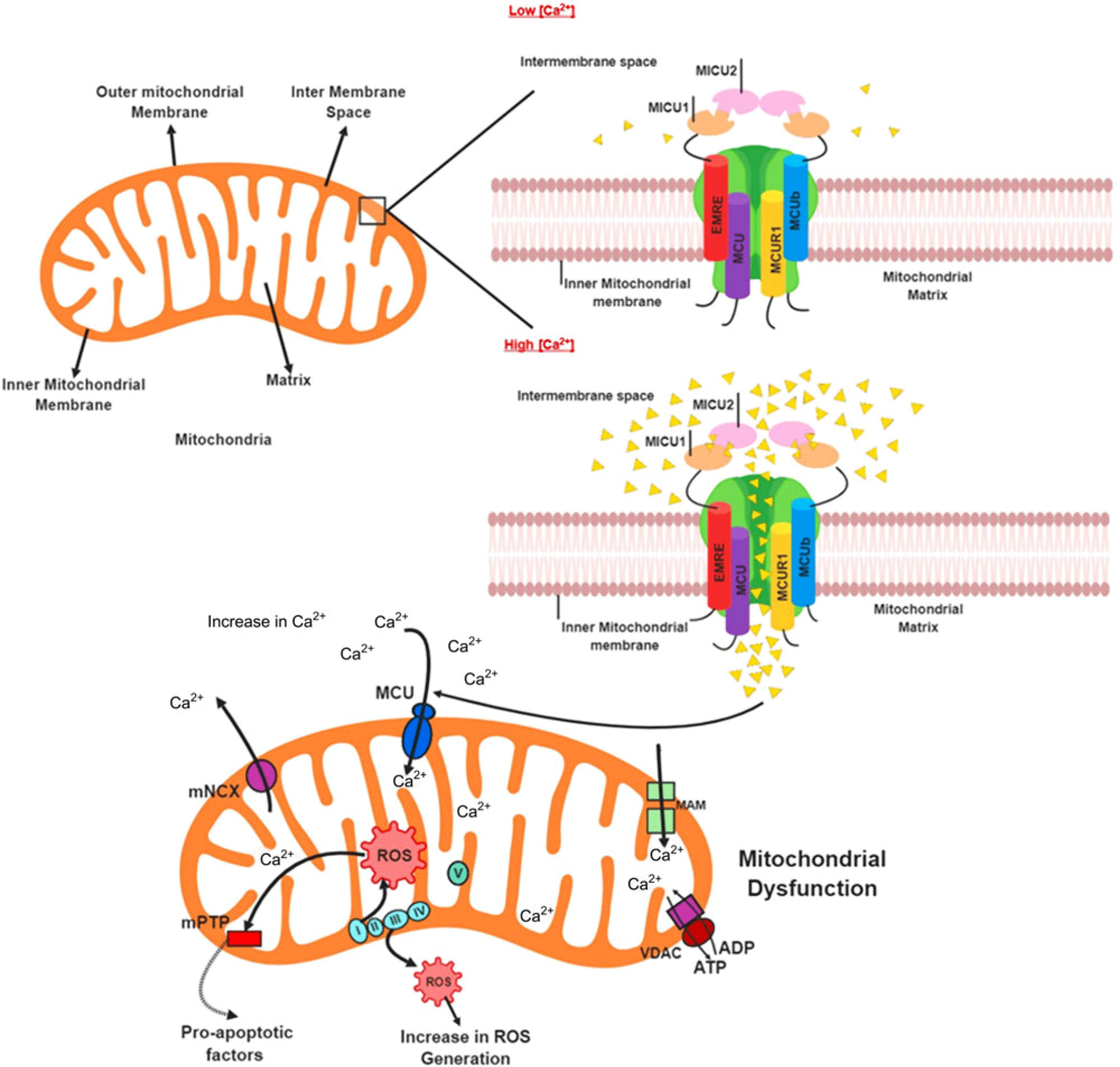
During the ETC, protons shuttle through the mitochondrial membrane creating a negative membrane potential which in turn activates the ATP synthase for the synthesis of ATP. But some of these electrons do not cross the membrane and instead leaks into the inner mitochondrial membrane resulting in the generation of ROS like superoxide radicals (Nakamura et al., Reference Nakamura, Takamura, Matsuzawa-Nagata, Takayama, Misu, Noda, Nabemoto, Kurita, Ota, Ando, Miyamoto and Kaneko2009; Hawking, Reference Hawking2016). ROS cause severe damage to cells but in the normal condition, they behave as normal signalling molecules. The body has defence mechanisms against the overproduction of ROS (both enzymatic and non-enzymatic) and only when these mechanisms are unable to control ROS do the cells get damaged. Another function of the mitochondria is calcium homeostasis and disruption in mitochondrial function that will lead to an imbalance in calcium regulation. Of all the channels involved in transporting calcium, MCU is one of the most selective transporting complexes. Lying in the inner mitochondrial membrane, MCU complex plays a pivotal role in calcium signalling which is critical for the functioning of the CNS. The MCU complex comprises of six subunits – MCU (pore-forming and calcium conducting subunit), MICU1 (mitochondrial calcium uptake protein 1), MICU2 (mitochondrial calcium uptake protein 2), EMRE (mitochondrial essential MCU regulator), MCUR1 (mitochondrial calcium uniporter regulator 1) and MCUb (mitochondrial regulatory subunit).
MCU is the pore-forming homo – oligomer subunit found in the inner – membrane space (Liao et al., Reference Liao, Dong and Cheng2017). Loss of this subunit in its motif results in complete loss of the complex function (Baughman et al., Reference Baughman, Perocchi, Girgis, Plovanich, Belcher-Timme, Sancak, Bao, Strittmatter, Goldberger, Bogorad, Koteliansky and Mootha2011). Both MICU subunits are important gatekeepers of the MCU complex. MICU1 regulates calcium influx by binding to the calcium, forming an oligomer, which in turn activates the MCU subunit. Studies have shown that loss of MICU1 leads to calcium accumulation in mitochondria causing neuronal defects and muscle movement disorders in both mice and humans. So, MICU1 acts as a gatekeeper by controlling the MCU uptake by activating it only when there is high calcium concentration. MICU2 inhibits calcium uptake by MCU when cytosolic calcium is low. MICU3 is claimed to be another member of the MICU family but its function is still unknown. MICU3 subunit is present only in the CNS and believed to have a role restricted only to the CNS. EMRE is a transmembrane protein that helps in assembling the above gatekeepers to maintain calcium homeostasis. The loss of this subunit will result in no calcium uptake. MCUR1 is a positive regulator of the complex required for calcium uptake. MCUb is the negative regulator of the complex. The structure of MCUb is similar to the MCU subunit, but changes in its sequence prevent this subunit from calcium uptake. Contrarily, overexpression of this subunit also results in decreased calcium uptake by MCU (Liao et al., Reference Liao, Dong and Cheng2017).
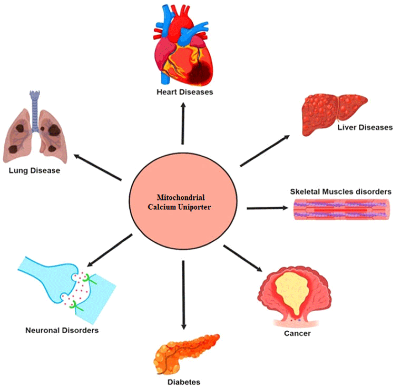
Mitochondrial vitality has been shown to reduce as age progresses. This could be one of the reasons why the natural uptake of calcium by MCU reduces during the later stages of life which can explain why AD is more prone to affect old age. In contrast, during early AD, a mutation or other external factors could occur which affects any of the subunits of MCU complex, causing a change in calcium uptake from the cytosol, resulting in increased concentration of calcium in the mitochondrial matrix. Calcium dysfunction can be the beginning of a vicious cycle wherein imbalance in calcium homeostasis results in elevated calcium concentration in the inner mitochondrial membrane which leads to increased ROS production, mitochondrial dysfunction, decreased ATP synthesis and generation of Aβ plaque and neurofibrillary tangle (Fig. 2; Lin & Beal, Reference Lin and Beal2006; Giorgi et al., Reference Giorgi, Agnoletto, Bononi, Bonora, De Marchi, Marchi, Missiroli, Patergnani, Poletti, Rimessi, Suski, Wieckowski and Pinton2012; Qiu et al., Reference Qiu, Tan, Hagenston, Martel, Kneisel, Skehel, Wyllie, Bading and Hardingham2013). Calcium homeostasis and ROS together are involved in cellular signalling and dysfunction in either one can lead to various disorders like cardiomyopathies, diabetes and neurodegenerative disorders (Fig. 3; DiMauro, Reference DiMauro2004; Görlach et al., Reference Görlach, Bertram, Hudecova and Krizanova2015).
MCU structure and mechanism – when [Ca2+] is less in the intermembrane space, the MCU complex remains closed. As [Ca2+] increases in intermembrane space, MCU opens allowing the entry of Ca2+ to maintain homeostasis. When exposed to an overload of [Ca2+], the intake of Ca2+ into the matrix exceeds its limits resulting in an overproduction of ROS and eventually mitochondrial dysfunction.

Implications of dysfunctional MCU – MCU has been shown to play role in a variety of diseases ranging from neuronal diseases to diabetes.

Evidence supporting the importance of MCU Complex
MCU, previously named CCDC109A, started garnering attention from 2011 (Waldeck-Weiermair et al., Reference Waldeck-Weiermair, Malli, Parichatikanond, Gottschalk, Madreiter-Sokolowski, Klec, Rost and Graier2015). As per its function, the MICU1 subunit rearranges itself to the cytosolic calcium concentration (Waldeck-Weiermair et al., Reference Waldeck-Weiermair, Malli, Parichatikanond, Gottschalk, Madreiter-Sokolowski, Klec, Rost and Graier2015). When there is an increase in concentration, MICU changes its conformation to allow the uptake of calcium into the matrix. Other studies including mouse models also have shown how knockdown of MICU1 causes an increase in calcium uptake (Logan et al., Reference Logan, Szabadkai, Sharpe, Parry, Torelli, Childs, Kriek, Phadke, Johnson, Roberts, Bonthron, Pysden, Whyte, Munteanu, Foley, Wheway, Szymanska, Natarajan, Abdelhamed, Morgan, Roper, Santen, Niks, van der Pol, Lindhout, Raffaello, De Stefani, den Dunnen, Sun, Ginjaar, Sewry, Hurles, Rizzuto, Duchen, Muntoni and Sheridan2014; Waldeck-Weiermair et al., Reference Waldeck-Weiermair, Malli, Parichatikanond, Gottschalk, Madreiter-Sokolowski, Klec, Rost and Graier2015; Liu et al., Reference Liu, Liu, Holmström, Menazza, Parks, Fergusson, Yu, Springer, Halsey, Liu, Murphy and Finkel2016). Apart from MICU1, mutations in the other MCU subunits also restricted the calcium uptake (Baughman et al., Reference Baughman, Perocchi, Girgis, Plovanich, Belcher-Timme, Sancak, Bao, Strittmatter, Goldberger, Bogorad, Koteliansky and Mootha2011; De Stefani et al., Reference De Stefani, Raffaello, Teardo, Szabò and Rizzuto2011). Knockdown studies of MCU and MICU1 have revealed various possible implications due to the loss of this uniporter. Cell lines like HeLa were used to discover increased sensitivity to apoptosis (Mallilankaraman et al., Reference Mallilankaraman, Doonan, Cárdenas, Chandramoorthy, Müller, Miller, Hoffman, Gandhirajan, Molgó, Birnbaum, Rothberg, Mak, Foskett and Madesh2012), increase in mitochondrial ROS (Mallilankaraman et al., Reference Mallilankaraman, Doonan, Cárdenas, Chandramoorthy, Müller, Miller, Hoffman, Gandhirajan, Molgó, Birnbaum, Rothberg, Mak, Foskett and Madesh2012; Csordás et al., Reference Csordás, Golenár, Seifert, Kamer, Sancak, Perocchi, Moffat, Weaver, de la Fuente Perez, Bogorad, Koteliansky, Adijanto, Mootha and Hajnóczky2013), increase in ATP synthesis by decreasing pyruvate dehydrogenase (PDH) phosphorylation (Mallilankaraman et al., Reference Mallilankaraman, Doonan, Cárdenas, Chandramoorthy, Müller, Miller, Hoffman, Gandhirajan, Molgó, Birnbaum, Rothberg, Mak, Foskett and Madesh2012). A decrease in PDH phosphorylation was replicated in mouse skeletal muscles by Pan et al. (Reference Pan, Liu, Nguyen, Liu, Sun, Teng, Fergusson, Rovira, Allen, Springer, Aponte, Gucek, Balaban, Murphy and Finkel2013). A study by Kwon et al. showed that tampered serine/threonine kinase LKB1 disrupted calcium homeostasis in MCU leading to changes in neurotransmitter release (Kwon et al., Reference Kwon, Sando, Lewis, Hirabayashi, Maximov and Polleux2018). In humans, loss of function of MICU1 resulted in neurological defects and movement disorder (Logan et al., Reference Logan, Szabadkai, Sharpe, Parry, Torelli, Childs, Kriek, Phadke, Johnson, Roberts, Bonthron, Pysden, Whyte, Munteanu, Foley, Wheway, Szymanska, Natarajan, Abdelhamed, Morgan, Roper, Santen, Niks, van der Pol, Lindhout, Raffaello, De Stefani, den Dunnen, Sun, Ginjaar, Sewry, Hurles, Rizzuto, Duchen, Muntoni and Sheridan2014). Mouse models also showed similar results (Liu et al., Reference Liu, Liu, Holmström, Menazza, Parks, Fergusson, Yu, Springer, Halsey, Liu, Murphy and Finkel2016; Antony et al., Reference Antony, Paillard, Moffat, Juskeviciute, Correnti, Bolon, Rubin, Csordás, Seifert, Hoek and Hajnóczky2016). Downregulation or knockdown of MICU1 in hepatic cells affects repair mechanisms and decreases calcium accumulation (Antony et al., Reference Antony, Paillard, Moffat, Juskeviciute, Correnti, Bolon, Rubin, Csordás, Seifert, Hoek and Hajnóczky2016). Similarly, in a study using Drosophila, silencing of MICU1 by RNA interference caused a shortening of the lifespan of dopaminergic neurons as well as age-related loss in climbing ability of the flies (M’Angale & Staveley, Reference M’Angale and Staveley2017). Another study using mouse neurons and patient fibroblasts showed that inactivation of MICU1 or MCU increased neuronal injury (Verma et al., Reference Verma, Callio, Otero, Sekler, Wills and Chu2017).
Knockout mice studies
Finkel lab first created the MCU KO mice. In contrast to WT mice, mitochondria from MCU KO mice were unable to uptake calcium (Harrington & Murphy, Reference Harrington and Murphy2015). Mice studies have revealed that there is a suppression in the intake of calcium when the mitochondria in the forebrains of MCU nulls were studied. These mice were also shown to have reduced neuronal cell death in the forebrain mitochondria due to decreased calcium uptake (Nichols et al., Reference Nichols, Elustondo, Warford, Thirumaran, Pavlov and Robertson2017). A study with MICU1 KO mice showed that cellular bioenergetics were impaired and that none of the MICU1 KO pups were able to survive after birth (Antony et al., Reference Antony, Paillard, Moffat, Juskeviciute, Correnti, Bolon, Rubin, Csordás, Seifert, Hoek and Hajnóczky2016). Another study also revealed that knockdown of MCU affected the release of neurotransmitters (Qiu et al., Reference Qiu, Tan, Hagenston, Martel, Kneisel, Skehel, Wyllie, Bading and Hardingham2013). MCU complex has not been considered as a key factor in the development of AD to date. These studies show that MCU can affect neuronal death, ROS and ATP levels and neurotransmitter release through the secondary messenger, calcium making its role in the elusive mechanism of AD vital.
Works on this MCU complex are not limited to neurodegenerative diseases and are quite popular even in other fields like cardiac and cancer studies. The ablation of the MCU complex has shown to lead to complete inhibition of calcium in skeletal muscles. Though the deletion of MCU causes reduced uptake of calcium, the cells cannot escape from hypoxic-ischemic damage (Pan et al., Reference Pan, Liu, Nguyen, Liu, Sun, Teng, Fergusson, Rovira, Allen, Springer, Aponte, Gucek, Balaban, Murphy and Finkel2013; Mammucari et al., Reference Mammucari, Gherardi, Zamparo, Raffaello, Boncompagni, Chemello, Cagnin, Braga, Zanin, Pallafacchina, Zentilin, Sandri, De Stefani, Protasi, Lanfranchi and Rizzuto2015; Gherardi et al., Reference Gherardi, Nogara, Ciciliot, Fadini, Blaauw, Braghetta, Bonaldo, De Stefani, Rizzuto and Mammucari2018).
These studies reveal that the MCU complex plays a major role in maintaining the calcium homeostasis and mitochondrial function. Further research is required to understand its potential use as a therapeutic tool for the treatment of numerous disorders it may be associated with (Qiu et al., Reference Qiu, Tan, Hagenston, Martel, Kneisel, Skehel, Wyllie, Bading and Hardingham2013; Liao et al., Reference Liao, Hao, Chen, He, Yuan and Cheng2015; Soman et al., Reference Soman, Keatinge, Moein, Da Costa, Mortiboys, Skupin, Sugunan, Bazala, Kuznicki and Bandmann2017; Depp et al., Reference Depp, Bas-Orth, Schroeder, Hellwig and Bading2018). Many unanswered questions remain like the existence of the MCU complex as an unrivalled opponent in the development of neurodegenerative diseases and if its neuroprotective properties can be used to develop a cure for these diseases. Knockdown studies of MCU and MICU subunits have shown its role in calcium maintenance but the roles of the other remaining subunits are still unclear. The possibilities are limitless, but it is possible that the other three seemingly unthreatening, not widely studied subunits, may have a major role to play in calcium homeostasis. It is also possible that other undiscovered or unrelated pathways other than the ones mentioned in this article interlink with calcium homeostasis contributing to the onset of AD. In that case, many questions will arise paving the way to an unexplored area of AD.
Conclusion
It has not been long since interest in MCU complex has developed. The scope of research is still at its infant stage and the research possibilities are limitless. Many aspects of this uniporter are still vague, and the roles of the subunits still must be scrutinised. Mouse models have shown interesting results though most of them are related to skeletal muscle pathology. An imbalance in calcium homeostasis can result in a cascade of events that explains many of the AD pathophysiologies. The effects of ageing, external stress and mutations on the MCU complex contributing to neurological conditions like AD remains to be studied. If the role and impact of MCU complex can be proven, it will pave a whole new dimension in the understanding of neurodegenerative diseases and will also envision an imaginary connexion that may connect all neurodegenerative diseases. Having neuroprotective properties, MCU also has the potential to be further studied for the treatment of neurodegenerative diseases.
Acknowledgements
The authors would like to thank Bharathiar University, India for providing the necessary infrastructure facilities required for completing the article. The authors would also like to thank all members of our lab for their continuous support in completing this article.
Author contributions
AV and Dr. VB contributed to concept development, designing of article. AV has drafted the article and MI has made a necessary correction. VB contributed by generating of figures. Dr. VB also contributed by approving the final version of the article.
Financial support
This research did not receive any specific grant from funding agencies in the public, commercial or not-for-profit sector. Figures are created by Biorender.
Conflict of interest
There are no conflicts of interest.