INTRODUCTION
The family Parthenopidae MacLeay, 1838 is currently divided into two subfamilies: Parthenopinae MacLeay, 1838 and Daldorfiinae Ng & Rodriguez, 1986. Daldorfiinae comprises four genera with 17 species and Parthenopinae 32 genera and 123 species (Ng et al., Reference Ng, Guinot and Davie2008).
The adult morphology of the parthenopids has been examined recently and several changes in its systematics were proposed (Tan & Ng, Reference Tan and Ng2007; Tan & Low, Reference Tan and Low2014). However, there is very little information about their larval morphology and most larval descriptions deal only with the first zoeal stages (ZI). Complete larval development is only known for two species, Platylambrus serratus (H. Milne Edwards, 1834) by Yang (Reference Yang1971) and Enoplolambrus validus (De Haan, 1837) by Kurata and Matsuda (Reference Kurata and Matsuda1980) and Terada (Reference Terada1985). For the remaining species, the larval development descriptions are partial or unavailable. The first known description, assigned to Lambrus massena (Roux, 1830), was published by Gourret (Reference Gourret1884), and later Cano (Reference Cano1893) described three zoeal stages as Lambrus sp. Already in the 19th century, Aikawa (Reference Aikawa1937) described the first zoea of Enoplolambrus validus (as Lambrus validus) and Lebour (Reference Lebour1944) identified and illustrated one megalopa from plankton attributed to Parthenopidae. Bourdillon-Casanova (Reference Bourdillon-Casanova1960) and Heegaard (Reference Heegaard1963) reported the first zoeal stage of Parthenopoides massena (as Lambrus massena). Thiriot (Reference Thiriot1973) also reported the ZI of Distolambrus maltzami (Miers, 1881) (as Heterocrypta maltzami) reared in the laboratory and five zoeal stages and one megalopa from plankton of P. massena. Heegaard (Reference Heegaard1963) described the first zoeal stage of Derilambrus angulifrons (Latreille, 1825) (as Lambrus angulifrons) and Kurata (Reference Kurata1970) illustrated and described the first zoea of Heterocrypta granulata (Gibbes, 1850). More recently, Guerao and Abelló (Reference Guerao and Abelló1999) described the first zoeal stage of Spinolambrus macrochelos (Herbst, 1790) (as Parthenope macrochelos), and Ng and Clark (Reference Ng and Clark2000) described the first zoeal stage of Rhinolambrus pelagicus (Rüppell, 1830), both from larvae hatched in the laboratory. Rice and Williamson (Reference Rice and Williamson1977) and Paula (Reference Paula1987) attributed larvae described from plankton samples to parthenopids but did not identify genus or species.
In the present work, we compare and analyse all these data, revise the larvae from plankton attributed to this family, and make a summary of the larval features that characterize parthenopids and which can be used to distinguish them from other brachyuran larvae.
Many brachyurans are clearly distinguishable in adult form but have larval and juvenile forms that are difficult to identify to species level. In some instances, the larvae are distinguishable but not easily matched with the correct adult form. A classic tool for helping to identify larvae collected in the field is to use complete descriptions of larvae obtained in laboratory cultures from clearly identified parental females. Current molecular tools such as DNA barcoding ensure that specimens collected in the field are identified correctly. These specimens collected in the field have clear advantages over specimens which have been reared in the laboratory; for example, González-Gordillo & Rodríguez (Reference Gonzalez-Gordillo and Rodríguez2000) reported morphological differences between larvae collected in the plankton and those reared in the laboratory from ovigerous females, although both inhabit the same locality.
The use of molecular markers has demonstrated to be a powerful tool for accurately identifying plankton specimens (Pan et al., Reference Pan, McBeath, Hay, Pierce and Cunningham2008; Pardo et al., Reference Pardo, Ampuero and Véliz2009; Ampuero et al., Reference Ampuero, Veliz and Pardo2010; Marco-Herrero et al., Reference Marco-Herrero, Torres, Cuesta, Guerao, Palero and Abelló2013). In the present study, we identified the megalopa stages of Derilambrus angulifrons and Parthenopoides massena, collected in the plankton, using partial sequences of the mitochondrial genes 16S and Cox1 as DNA barcodes.
Derilambrus angulifrons is known from the eastern Atlantic: south-western Spain (Cuesta Mariscal & González-Gordillo, Reference Cuesta Mariscal and González-Gordillo1992) and the Mediterranean Sea (d'Udekem d'Acoz, Reference D'Udekem d'Acoz1999) at depths from 2 m (Števčić, 1990) to 40 m (Zariquiey Álvarez, Reference Zariquiey Álvarez1968). In this area this species lives on sandy mud, muddy detritus and coralligenous bottoms (d'Udekem d'Acoz, Reference D'Udekem d'Acoz1999). Parthenopoides massena is distributed in the east Atlantic from northern Europe to Guinea and Mediterranean coasts (d'Udekem d'Acoz, Reference D'Udekem d'Acoz1999) where they inhabit mainly sandy and calcareous algae bottoms at 3–141 m depth (Zariquiey Álvarez, Reference Zariquiey Álvarez1968; Števčić, 1990).
In the present study the megalopa of Derilambrus angulifrons is described and illustrated in detail for the first time and the megalopa of Parthenopoides massena is compared with the previous description by Thiriot (Reference Thiriot1973).
MATERIALS AND METHODS
Collection of the megalopae
Megalopae were collected in the course of three different projects. Three megalopae of Derilambrus angulifrons were captured in July 2007 from the plankton of the Guadalete estuary (Cádiz-SW Spain) (36°35′24.09″N 6°13′46.19″W) in a campaign of plankton sampling in this estuary in the context of the project ‘Transporte y reclutamiento larvario de crustáceos bentónicos litorales: importancia de los agentes forzadores costeros y regimen mareal’ (CTM2005-00024/MAR). Two megalopae of Parthenopoides massena were collected in two different stations in the Mediterranean Sea, one in the Gulf of Naples (40°49′10.51″N 14°14′05.09″E) in September 2009 and another one off the Balearic Islands (39°43.27′N 02°13.07′E) in July 2010.
Morphological descriptions
Drawings and measurements were made using a Wild MZ6 and Zeiss Axioskop compound microscope with Nomarski interference, both equipped with a camera lucida. All measurements were made using an ocular micrometer. Descriptions were based on all collected megalopae. The following measurements were taken for the megalopa: cephalothorax length (CL), measured from the tip of rostrum to posterior margin of cephalothorax; and cephalothorax width (CW), measured as the cephalothorax maximum width (mesobranchial regions). In Figures 3B, C and 4B the plumose setae are drawn truncated.
The larvae are described using the basic malacostracan somite plan from anterior to posterior and appendage segments are described from proximal to distal, endopod then exopod (Clark et al., Reference Clark, Calazans and Pohle1998).
DNA extraction, amplification and sequencing
The identification of larval stages was based on partial sequences of the 16S rDNA and Cox1 genes. Total genomic DNA was extracted from muscle tissue from pereiopods of the megalopae, and incubated for 1–24 h in 300 µl lysis buffer at 65°C. Protein was precipitated by addition of 100 µl of 7.5 M ammonium acetate and subsequent centrifugation, and DNA precipitation was obtained by addition of 300 µl of isopropanol and posterior centrifugation. The resulting pellet was washed with ethanol (70%), dried, and finally resuspended in Milli-Q distilled water.
Target mitochondrial DNA from the 16S rRNA and Cox1 genes was amplified with polymerase chain reaction (PCR) using the following cycling conditions: 2 min at 95°C, 40 cycles of 20 s at 95°C, 20 s at 45–48°C, 45 s (16S) or 47 s (Cox1) at 72°C, and 5 min 72°C. Primers 1472 (5′- AGA TAG AAA CCA ACC TGG -3′) (Crandall & Fitzpatrick, Reference Crandall and Fitzpatrick1996) and 16L2 (5′-TGC CTG TTT ATC AAA AAC AT-3′) (Schubart et al., Reference Schubart, Cuesta and Felder2002) were used to amplify 540 bp of 16S, while primers COH6 (5′- TAD ACT TCD GGR TGD CCA AAR AAY CA -3′) and COL6b (5′- ACA AAT CAT AAA GAT ATY GG -3′) (Schubart & Huber, Reference Schubart and Huber2006) allowed amplification of 670 bp of Cox1. PCR products were sent to New Biotechnic and CISA-INIA companies to be purified and then bidirectionally sequenced.
Sequences were edited using the software Chromas version 2.0. The obtained final DNA sequences were compared with those from adult specimens of several Iberian brachyuran crabs obtained in the context of the MEGALOPADN project. Adult and larval sequences for both genes are deposited in GenBank under accession numbers (KP057806-KP057819).
RESULTS
Barcode identification
In the context of the MEGALOPADN project we have obtained the DNA mitochondrial sequences of 16S and Cox1 genes for almost all the Iberian brachyuran crabs. Therefore we can compare the sequences obtained from the megalopae with those in our alignments and database. For Parthenopidae we have got the sequences of the Iberian representatives of Derilambrus angulifrons, Distolambrus maltzani, Parthenopoides massena and Spinolambrus macrochelos. The sequences of the megalopae from Guadalete estuary perfectly fit those of Derilambrus angulifrons and those of the megalopae from the Balearic Islands and Naples with the sequences of Parthenopoides massena. No differences (100% match) were found between the 16S (546 bp) and Cox1 (667 bp) sequences of D. angulifrons and the Guadalete estuary megalopae. Also the Mediterranean megalopae sequences math 100% with 16S sequence of P. massena. In the case of Cox1, while the Naples megalopa sequence (613 bp) also matches 100% with those of P. massena, the Balearic Island megalopa sequence differs in 4 mutations out of 667 bp from the Cox1 sequence of P. massena.
Megalopa description
Family Parthenopidae MacLeay, 1838
Genus Derilambrus Tan & Ng, Reference Tan and Ng2007
Derilambrus angulifrons (Latreille, 1825)
(Figures 1 & 2)
Size: CL = 1.78 ± 0.08 mm; CW = 0.91 ± 0.06 mm; N = 3
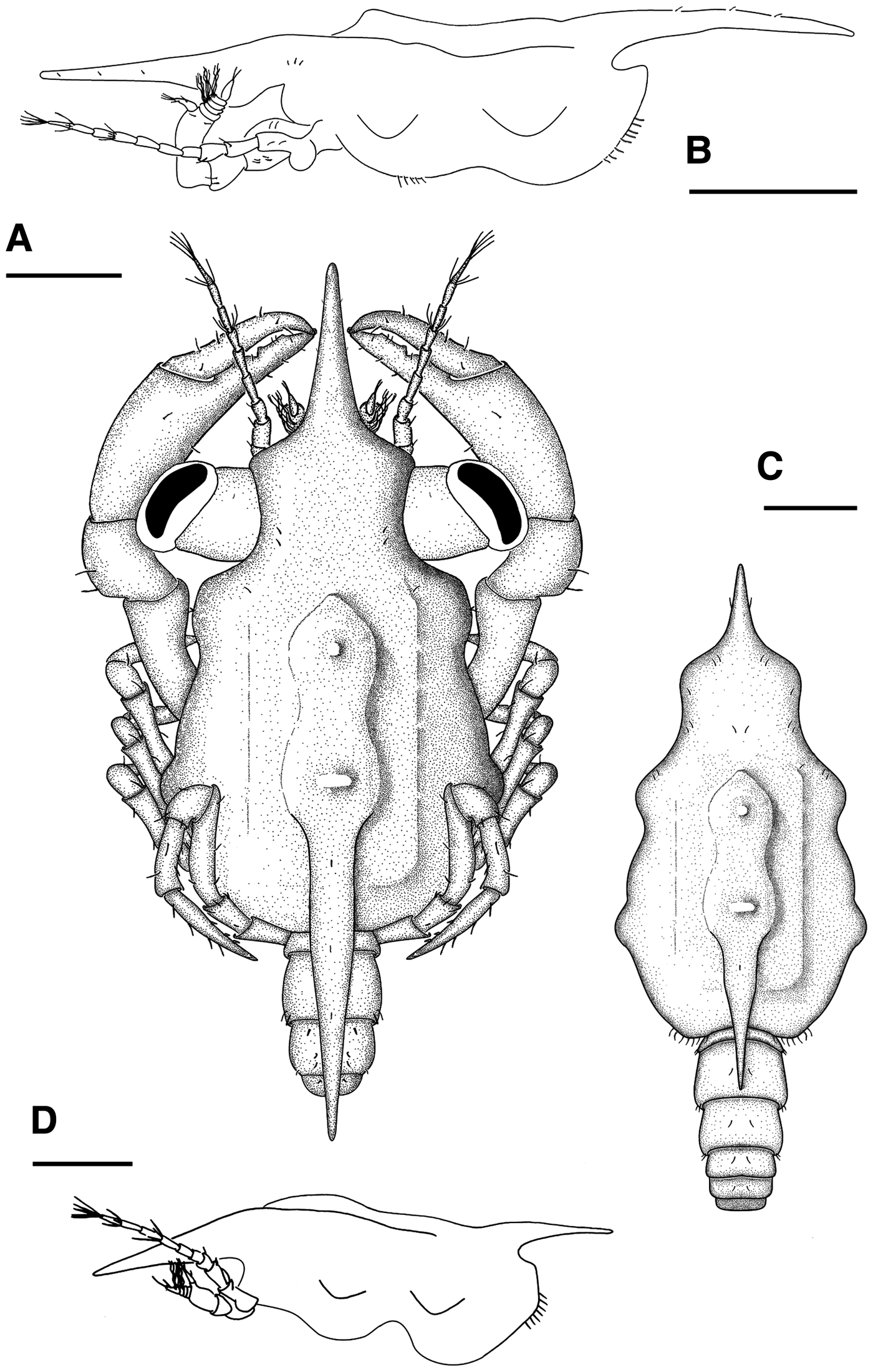
Derilambrus angulifrons (Latreille, 1825) Megalopa, (A) general dorsal view; (B) lateral view of the cephalothorax. Parthenopoides massena (Roux, 1830) Megalopa, (C) dorsal view; (D) lateral view of the cephalothorax. Scale bars= 0.5 mm.

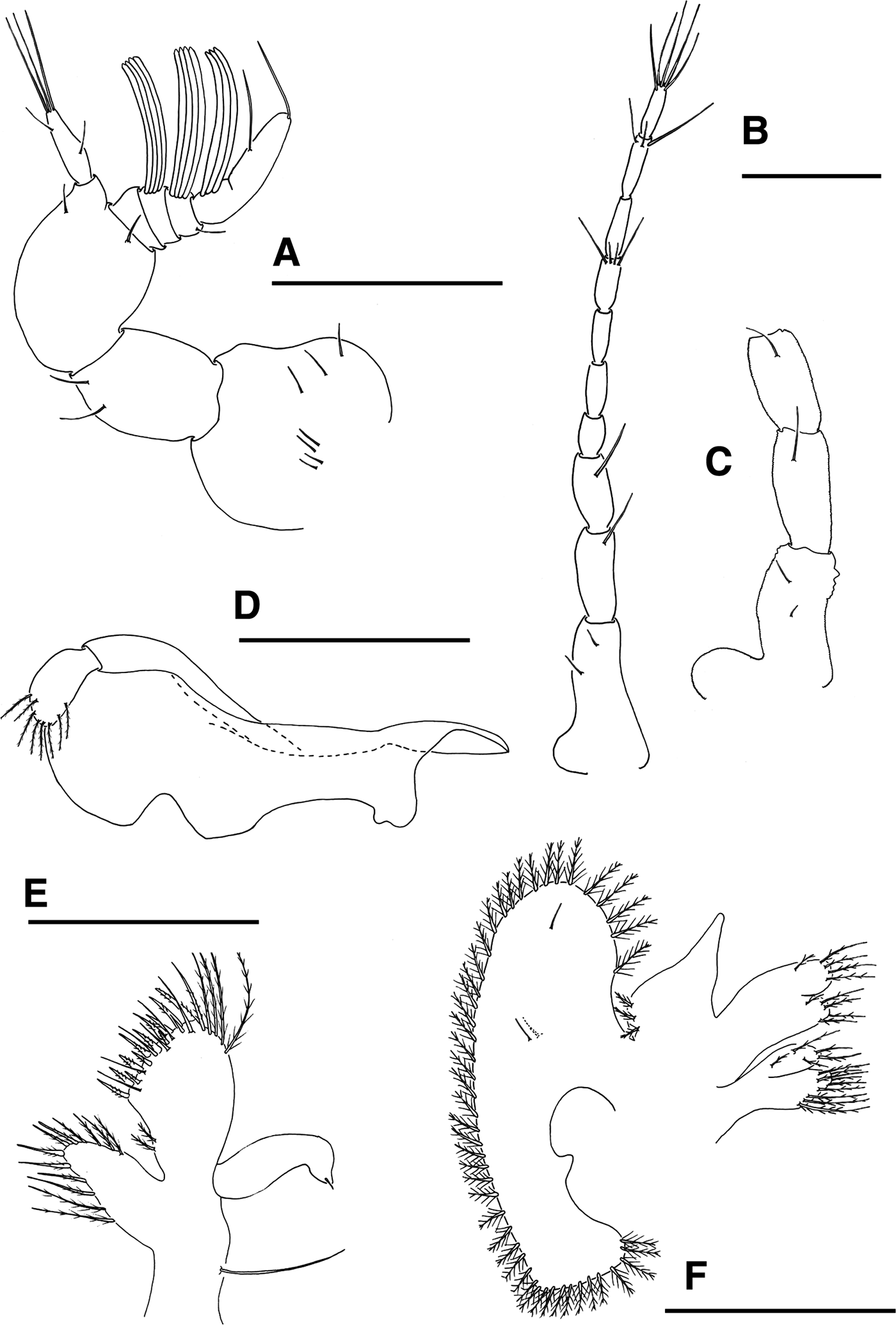
Derilambrus angulifrons (Latreille, 1825) Megalopa, (A) antennule; (B) antenna, (C) detail of the peduncle of antenna; (D) mandible; (E) maxillule; (F) maxilla. Scale bars = 0.2 mm.

Cephalothorax (Figure 1A, B) Longer than broad, with long, thin and straight rostrum with 3 pairs of minute setae; a pair of lobes on the mesobranchial regions with hepatic regions moderately inflated; 2 tubercles, 1 on metagastric region and 1 on urogastric region; prominent long spine present on cardiac region backwards with few minute unpaired setae; setation as drawn; dorsal organ present; eyes stalked.
Antennule (Figure 2A) Peduncle 3-segmented with 7, 2, 2 simple setae; unsegmented endopod with 1 medial, 1 subterminal and 3 terminal simple setae; exopod 4-segmented with 0, 0, 1, 2 simple setae; segments 2–4 with 4, 4 and 3 aesthetascs respectively.
Antenna (Figure 2B, C) Crenulated peduncle 3-segmented with 2, 1, 1 simple setae respectively, proximal segment with stout and ventrally directed process; flagellum 7-segmented with 0, 0, 0, 4, 0, 3, 5 simple setae respectively.
Mandible (Figure 2D) Palp 2-segmented with 8 plumodenticulate terminal setae on distal segment.
Maxillule (Figure 2E) Coxal endite with 8 plumose setae plus 4 plumodenticulate setae on margin; basial endite with 14 marginal cuspidate, 10 subterminal plumodenticulate, and 2 proximal plumose setae; endopod unsegmented with 1 terminal simple setae; long exopodal simple seta present.
Maxilla (Figure 2F) Coxal endite bilobed with 9 + 5 terminal plumose setae; basial endite bilobed with 5 + 5 sparsely plumodenticulate setae; endopod unsegmented with 3 short plumodenticulate setae on base; exopod (scaphognathite) with 47–48 marginal plumose setae plus 3 small simple setae, 2 dorsal and 1 ventral, on lateral surface.
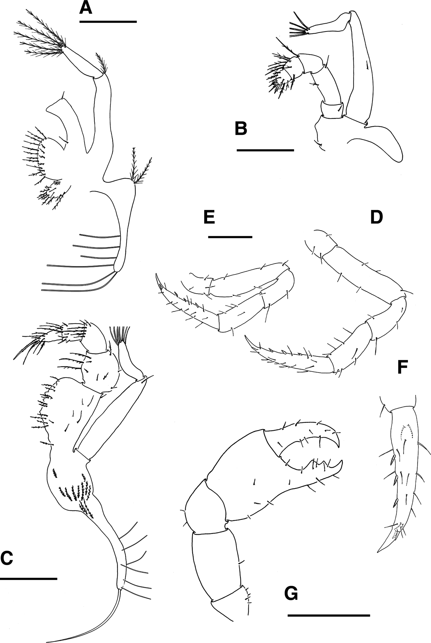
First maxilliped (Figure 3A) Epipod triangular shaped with 8 setae, 2 proximal plumodenticulate and 6 distal long setae; coxal endite with 13 plumose setae; basial endite with 17 sparsely plumodenticulate setae; endopod reduced, unsegmented and with 2 simple setae; exopod 2-segmented with 1 plumodenticulate distal seta on proximal segment and 5 terminal plumose setae on distal segment.
Second maxilliped (Figure 3B) Epipod reduced without setae; protopod with 1 simple seta; endopod 5-segmented with 1 (simple), 2 (simple), 1 (long simple), 7 (plumodenticulate) and 9 (3 cuspidate, 6 plumodenticulate) setae, respectively; exopod 2-segmented with 1 medial simple seta on proximal segment and 5 terminal plumose setae on distal segment.
Derilambrus angulifrons (Latreille, 1825) Megalopa, (A) first maxilliped; (B) second maxilliped; (C) third maxilliped; (D) second pereiopod; (E) fifth pereiopod; (F) detail of the dactylus of pereiopods II–V (G) cheliped. Scale bars = (A–E) 0.2 mm and (F) 0.5 mm.
Third maxilliped (Figure 3C) Epipod with 6 subterminal and 1 terminal long setae; protopod with 12 plumodenticulate setae; endopod 5-segmented, margin of the proximal segment denticulate, and 19, 10, 6, 8, 7 sparsely plumose setae respectively; exopod 2-segmented with 1 distal simple seta on proximal segment and 7 terminal plumose setae on distal segment.
Pereiopods (Figure 3D–G) Cheliped setation as drawn, fixed finger lower margin with 2 prominent teeth; pereiopods II–V thin and setose, inner margin of dactyl with 3 stout ventral spines and 1 pair subterminal shorter spines; setation as illustrated. Long setae (feelers) on dactylus of pereiopod V absent.
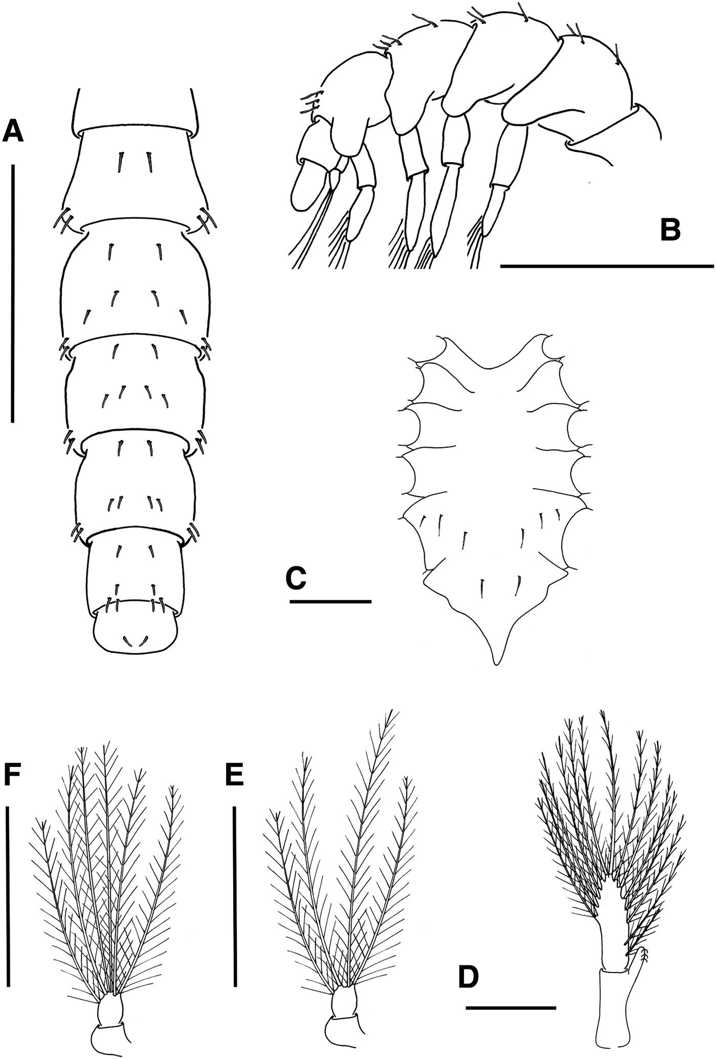
Sternum (Figure 4C) Maxilliped sternites completely fused with 2 simple setae, cheliped sternites with 3 simple setae each, pereiopod sternites 2–5 without setae; sternal sutures are interrupted medially.
Derilambrus angulifrons (Latreille, 1825) Megalopa, (A) pleon, dorsal view; (B) pleon, lateral view; (C) sternum; (D) third pleopod; (E) uropod. Parthenopoides massena (Roux, 1830) Megalopa, (F) uropod. Scale bars = (A, B) 0.5 mm and (C, D) 0.2 mm.
Pleon (Figure 4A, B) Six pleonites; pleonite I without setae; setation of pleonites II–VI as shown; pleonite VI reduced.
Pleopods (Figure 4B, D & E) Present on pleonites II-VI; endopods unsegmented with 3 cincinuli; exopod unsegmented with 11–14 long plumose natatory setae; uropod 2-segmented, proximal segment without setae, distal segment with 4 terminal plumose natatory setae.
Telson (Figure 4A) Reduced, subquadrate, with 1 pair of dorsal setae.
DISCUSSION
The systematic relationships of Parthenopidae have been controversial for a long time. In several works since 1862 to the present, its systematic position has changed from Calappidae (Strahl, Reference Strahl1862) to Brachyryncha (Yang, Reference Yang1971), passing through Cancridae (Lebour, Reference Lebour1928; Aikawa, Reference Aikawa1935) and Oxyryncha (Bouvier, Reference Bouvier1940; Balss, Reference Balss1957). Guinot (Reference Guinot1977, Reference Guinot1978) elevated the Parthenopidae to a superfamily in the section Heterotramata, which was later corroborated with larval morphology (according to Rice, Reference Rice1980), and currently this is the most widely accepted status. Tan (Reference Tan2004) and Tan & Ng (Reference Tan and Ng2007) have carried out the most recent and comprehensive revision of Parthenopoidea, which Ng et al. (Reference Ng, Guinot and Davie2008) follows. According to these authors, Parthenopoidea contains only one family, Parthenopidae, divided into two subfamilies, Daldorfiinae (4 genera and 17 species) and Parthenopinae (32 genera and 123 species). In spite of all these studies, its phylogenetic relationships are still unresolved, and it is only clear that it is not related to Majoidea (Yang, Reference Yang1971; Ahyong et al., Reference Ahyong, Lai, Sharkey, Colgan and Ng2007). However, it has been suggested that based on adult morphology there are relationships with Aethroidea, Calappoidea, Trapezoidea and Plagusiidae, among others (see Tan & Ng, Reference Tan and Ng2007), and based on larval morphology there are relationships with Cancroidea (Lebour, Reference Lebour1928; Aikawa, Reference Aikawa1937) and Cyclometopa in general (Rice, Reference Rice1980).
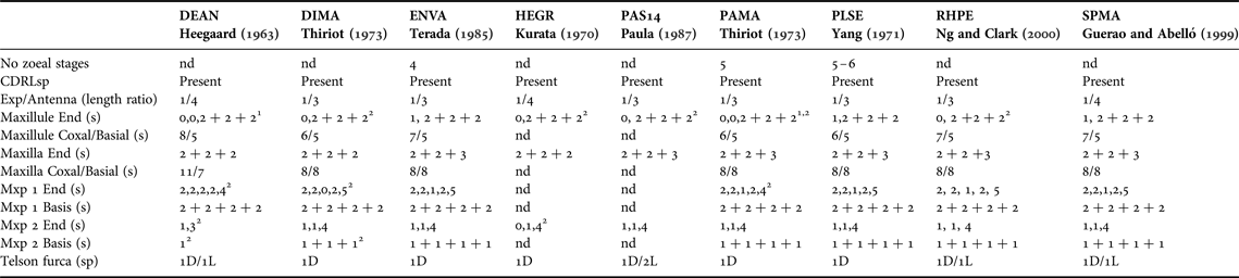
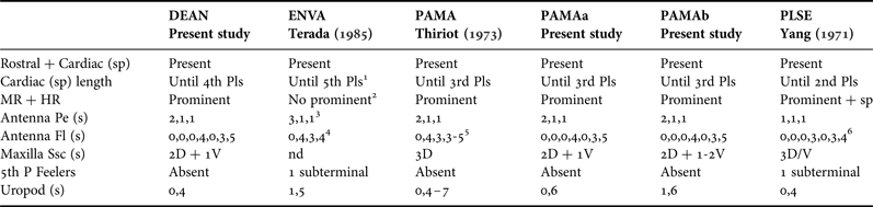
Larval studies have contributed to the resolution of problems in the systematic classification of brachyuran crabs (Rice, Reference Rice1980; Marques & Pohle, Reference Marques and Pohle1998; Clark & Guerao, Reference Clark and Guerao2008; Clark, Reference Clark, Martin, Crandall and Felder2009; Marco-Herrero et al., Reference Marco-Herrero, Torres, Cuesta, Guerao, Palero and Abelló2013) because the morphology of larval stages gives an insight into the relationships between brachyuran taxa. Larval characters may reflect relationships even better than adult morphology (Rice, Reference Rice1980). Nevertheless, there are still few data on larval development for parthenopids and most larval descriptions deal only with the first zoeal stages and partial descriptions of intermediate zoeae from plankton samples. In the present study we compare all known descriptions of the larval stages of parthenopids (see Tables 1 & 2).
Morphological comparison of the known zoea I of Parthenopidae
CDRLsp, cephalothorax dorsal, rotral and lateral spines; D, dorsal; DEAN, Derilambrus angulifrons; DIMA, Distolambrus maltzani; End, endopod; ENVA, Enoplolambrus validus; Exp, exopod; HEGR, Heterocrypta granulate; L, lateral; Mxp, maxilliped; nd, no data; PAMA, Parthenopoides massena; PAS14, unidentified parthenopid larvae collected in the plankton; PLSE, Platylambrus serrata; RHPE, Rhinolambrus pelagicus; s, setation; sp, spines; SPMA, Spinolambrus macrochelos.
1Probably a mistake, it is 2-segmented.
2Probably authors' mistakes, some seta overlooked.
Morphological comparison of the known megalopa of Parthenopidae
Fl, flagellum; HR, hepatic region; MR, mesobranchial region; P, pereiopod; PAMAa, Parthenopoides massena megalopa from Balearic Island plankton; PAMAb, P. massena megalopa from Gulf of Naples plankton; Pe, peduncle; Pls, pleon somite; Ssc, scaphognathite surface; V, ventral; rest of abbreviations as in Table 1.
1Until 3rd Pls according to Kurata & Matsuda (Reference Kurata and Matsuda1980).
2Prominent and with spines, according to Kurata & Matsuda (Reference Kurata and Matsuda1980).
30,1,0, according to Kurata & Matsuda (Reference Kurata and Matsuda1980).
40,0,0,2,0,3,4, according to Kurata & Matsuda (Reference Kurata and Matsuda1980).
5Thiriot (Reference Thiriot1973) overlooked the segmentation of the first three segments.
6Based on the drawing of Fig 8a by Yang (Reference Yang1971).
In parthenopid larvae there is no single character that distinguishes them from the rest of the brachyuran superfamilies (see Yang, Reference Yang1971; Rice, Reference Rice1980) but there is a set of features that can be used to identify them. Summarizing the set of characters proposed by Yang (Reference Yang1971) and Rice (Reference Rice1980), including some modifications and new features, the 9 diagnostic characteristics of the parthenopid zoeal stages are: (i) the cephalothorax has well developed and smooth dorsal, lateral, and rostral spines and the dorsal and rostral spines are longer than cephalothorax length; (ii) the antenna shows a long protopodal process (but never reaching the tip of the rostral spine) with 2 rows of spinules, an exopod about 2/3 of the protopod length with 2 unequal length terminal setae (the longer seta can reach the tip of protopod, and in some cases is described as setulose); (iii) endopod of maxillule and maxilla with 1,2 + 2 + 2 and 2 + 2 + 3 setae respectively; (iv) basis of maxillipeds 1 and 2 with 2 + 2 + 2 + 2 and 1 + 1 + 1 + 1 setae respectively; (v) endopod of maxilliped 2 with 1,1,4 setae; (vi) dorsolateral processes are present on pleonal somites II and III; (vii) usually long acute posterolateral processes on somites III-V; (viii) telson forks bear one pair of well-developed dorsomedial spines and sometimes there are 1 or 2 lateral setae present; (ix) three pairs of posterior processes on telson through development. Moreover, Yang (Reference Yang1971) described another character: a well-developed forehead and posterodorsal protuberances on the cephalothorax that appears in the majority of parthenopids (absent in Rhinolambrus pelagicus by Ng & Clark, Reference Ng and Clark2000), although this is also very common in larvae of other brachyurans.
According to the few previous studies describing the complete larval development of parthenopids the number of zoeal stages is variable. Four were described for Enoplolambrus validus (see Terada, Reference Terada1985) and five for Parthenopoides massena (see Thiriot, Reference Thiriot1973) and Platylambrus serrata (see Yang, Reference Yang1971), although in this last case an extra sixth zoeal stage was also recorded. The common characters related to changes through development are, besides the general increase in the number of setae, the appearance of the sixth somite of the pleon from zoea III on, and the addition of one plumodenticulate seta on the distal segment of the endopod of the first maxilliped also from zoea III on.
The megalopa stage has only been described for three species of parthenopids, P. serrata, P. massena and E. validus, and now in the present study it is also described for D. angulifrons. Although it is early to draw conclusions about the typical morphological characters for megalopa of parthenopids, all known megalopae share the features listed in Table 2. The main distinctive characters are: (i) the presence of well-developed rostral and cardiac spines horizontally directed, (ii) antennal flagellum with seven segments, (iii) 3 simple setae on the scaphognathite surface and (iv) dactylus of fifth pereiopod without feelers (only 1 long seta described in P. serrata) and with 3 ventral spines and 1 pair of subterminal spines.
In the present study the megalopa of Derilambrus angulifrons is described for the first time based on three specimens collected in the plankton and identified by DNA barcode. These megalopae show all common characters described above as typical of parthenopid megalopae. The main distinctive feature that separates them from the only other known megalopae of the family with an overlapping distribution, Parthenopoides massena, is the length of the cardiac spine. In D. angulifrons the cardiac spine is longer, exceeding the third somite of the pleon, while that of P. massena is shorter and never reaches the third pleonal somite. In the present study, two megalopae of P. massena collected in the plankton have also been identified by DNA barcode techniques. Comparing them with the megalopa described by Thiriot (Reference Thiriot1973) from plankton samples confirmed that the assignment of these megalopae to P. massena was correct. Nevertheless, we found one difference between the two megalopae studied: the antennal flagellum is 7-segmented, while Thiriot (Reference Thiriot1973) described only 4 segments. This fact affects the key for the identification of Mediterranean brachyuran megalopae by Pessani et al. (Reference Pessani, Tirelli and Flagella2004) who based the identification of Parthenopoides massena (according to Thiriot, Reference Thiriot1973) on the number of antennal segments. This dichotomy separates P. massena (8–9-segmented) from Cancer pagurus Linnaeus, 1758 and two species of Atelecyclus Leach, 1814 (11-segmented), the numbers for P. massena should be corrected to 10-segmented, which will still make a valid separation possible. A feature not described by Thiriot (Reference Thiriot1973) is the sternal plate, which in the two specimens studied here has the same setation as D. angulifrons (see Figure 4C). In addition, the number of setae of the uropods described by Thiriot (Reference Thiriot1973) was 0, 4–7, but in the two specimens studied here it was 0, 6 and 1, 6.
With respect to the other larval stages collected in the plankton samples and attributed to Parthenopidae, not all the zoeae described by Rice and Williamson (Reference Rice and Williamson1977) as ASM16-ASM19 fit exactly with the features mentioned above for parthenopid zoeae. While ASM16 and ASM17 are clearly zoeae II-V of unidentified parthenopids, ASM18 and ASM19 show remarkable differences, for example they have different types of antennae (exopod very reduced), and the spines of the cephalothorax have spinules. ASM18 also differs in the setation of the endopod of the maxillule and second maxilliped, and in the case of ASM19 (zoeae II–III) the telson has a fourth pair of the distal process. Although Rice & Williamson (Reference Rice and Williamson1977) state that these differences correspond to intergeneric variability and that the specimens definitely belong to the parthenopids we believe that some of the differences, especially those in the mouthpart setation pattern, are not acceptable as intrafamilial variability. Unfortunately there are still a lot of brachyuran families without larval data. Therefore, at this point it is not possible to attribute ASM18 and ASM19 to another family with certainty, although in some aspects they are close to Xanthoidea and Cancroidea.
Paula (Reference Paula1987) described zoea I of unidentified parthenopids as Parthenope S14 and Parthenopidae S15. Parthenope S14 clearly corresponds to a zoea of Parthenopidae, with a setation of the endopod of the maxillule 0, 2 + 2 + 2. The absence of this seta in the proximal segment was also described in the zoeal stages of P. massena, according to Heegaard (Reference Heegaard1963) and Thiriot (Reference Thiriot1973), and D. angulifrons (see Heegaard, Reference Heegaard1963) and Rhinolambrus pelagicus (see Ng & Clark, Reference Ng and Clark2000), although it is present in other species (see Table 1). Normally this is not a setation pattern that shows variability at intrafamilial level; therefore, the significance of this variability is not currently easy to evaluate due to the low number of species studied. Kurata and Matsuda (Reference Kurata and Matsuda1980) describe ‘1 rudimentary seta on proximal segment which may be very difficult to see in early stages’; therefore that this seta was overlooked by some authors cannot be discarded. Paula (Reference Paula1987) states that Parthenopidae S15 resembles ASM19 (Rice & Williamson, Reference Rice and Williamson1977); therefore, according to the issues mentioned above, these larvae must not be attributed to this family.
There is also a megalopa collected in the plankton attributed to Parthenopidae by Lebour (Reference Lebour1944). She gave a brief description and illustration, and based on the elongated cheliped, long rostral and cardiac spines, and lack of feelers on the dactyl of the fifth pereiopods, it was attributed to Parthenopidae. All these characters support this identification, except the general shape of the cephalothorax and the long chelipeds, as they are very different with respect to the rest of the known megalopae of parthenopids. Especially the chelae that clearly resemble those of the adult forms. It is possible that this stage could be an intermediate anomalous specimen between megalopa and first crab.
Cano (Reference Cano1891) described a megalopa that he assigned to Goneplax rhomboides Linnaeus, 1758, but later Ingle & Clark (Reference Ingle and Clark1983) when they described the complete larval development of G. rhomboides showed that Canós megalopa does not belong to this species. However, according to the description, although brief and incomplete, in the figures it is clear that it corresponds to a parthenopid larva because it shares the characters described above for parthenopid megalopa.
Rice (Reference Rice1981) examined the phylogenetic significance of the brachyuran megalopae and commented that this stage was the only phase of the brachyuran life cycle that had not been previously examined for classificatory evidence. Later Martin (Reference Martin1988) studied the phylogenetic significance of the brachyuran megalopa in the case of Xanthidae. It is difficult to apply the megalopa morphology to infer phylogenetic relationships for Parthenopoidea considering that currently there are only known descriptions for five species. The most conspicuous features are the characteristic cephalothorax with long rostral and cardiac spines, and a pair of lobes on the mesobranchial region with hepatic regions moderately inflated. The long rostral and cardiac spines are features shared with Cancridae (see for example the megalopae of Atelecyclus rotundatus by Hong & Ingle (Reference Hong and Ingle1987) and Cancer pagurus by Ingle (Reference Ingle1981)), but it can be distinguished from them by the number of segments of the antennal flagellum and setae of the uropods, as well as by the absence of feelers on the dactylus of the fifth pereiopod.
Relationships between Parthenopidae and Cancridae have been proposed in the past (Lebour, Reference Lebour1928; Aikawa, Reference Aikawa1935) but there have been no new studies on this matter since then. The first molecular phylogeny including data of parthenopids was made in the context of their systematic position with respect to Majoidea (Hultgren & Stachowicz, Reference Hultgren and Stachowicz2008), where it is clear that there are no relationships with majoids, and in a global phylogeny of Podotremata (Ahyong et al., Reference Ahyong, Lai, Sharkey, Colgan and Ng2007) where its systematic relationships was not resolved. In both cases, representatives of Cancridae were not included in the molecular phylogenies. However, in a recent exhaustive phylogeny of brachyuran crabs (Tsang et al., Reference Tsang, Schubart, Ahyong, Lai, Au, Chan, Ng and Chu2014) an important number of taxa have been analysed and on this occasion representatives of Crancridae have been included. The results place Parthenopidae in the same clade as Aethridae, Cancridae and Calappidae, with a closer relationship with Calappa philargius (Linnaeus, 1758), the only representative of Calappidae. While relationships with Cancridae are as expected those with Calappidae are not supported by larval data.
New data on the larval morphology of more genera of Parthenopinae and representatives of the subfamily Daldorfiinae, as well as new molecular phylogenies comprising members of all Heterotramata superfamilies, with a wider representation of Parthenopidae, Cancridae, Aethridae and Calappidae species are needed to determine the phylogenetic position of this taxon.
ACKNOWLEDGEMENTS
We are especially grateful to Carlos Sánchez Nieto for his assistance in the laboratory work, Catherine Stonehouse for her careful language corrections and Asvin P. Torres (Instituto Español de Oceanografía) and. Maria Grazia Mazzocchi (Stazione Zoologica Anton Dohrn) who helped in collecting the megalopae of P. massena.
FINANCIAL SUPPORT
The research was carried out within the framework of the projects MEGALOPADN (CGL2009-11225) and ‘Transporte y reclutamiento larvario de crustáceos bentónicos litorales: importancia de los agentes forzadores costeros y regimen mareal’ (CTM2005- 00024/MAR), both co-financed by the ‘Ministerio de Economía y Competividad (MINECO)’ Spanish Plan R + D + I and FEDER. Elena Marco-Herrero acknowledges the pre-doctoral FPI Fellowships supported by MINECO (BES-2010-033297 and EEBB-I-13-07501).