INTRODUCTION
Many hydroids are symbiotic with other benthic organisms such as algae, sea grasses, sponges, cnidarians, molluscs, polychaetes, bryozoans, echinoderms, tunicates and vertebrates (Boero & Bouillon, Reference Boero, Bouillon and Rhode2005). Some of these epibionts live on the external skeletal structures of the host (e.g. several hydractiniids on the shells of gastropod molluscs, or Proboscidactyla spp. on the rim of polychaete tubes), but others, like those symbiotic with bryozoans or sponges, settle directly on the living epithelium or inside the tissues of the host (Puce et al., Reference Puce, Bavestrello, Di Camillo and Boero2007).
Few hydroid species live in association with other cnidarians: the hydroid epibiosis on the perisarc of other hydroid species (e.g. Hebella spp., Lafoeidae and Campanulariidae) is the most common association. Moreover, several hydroids live as epibionts on dead cnidarians exploiting hard coral skeletons or the gorgonian axis as substrate (Gili et al., Reference Gili, Lopez-González and Bouillon2006).
The symbiosis between hydroids and living anthozoans involves twelve athecate species belonging to six families (Ptilocodiidae, Coryniidae, Tubulariidae, Asyncoryniidae, Cladocoryniidae and Zancleidae): Ptilocodium repens Coward, Reference Coward1909, Hydrichthella epigorgia Stechow, Reference Stechow1909, Sarsia medelae Gili et al., Reference Gili, Lopez-González and Bouillon2006, Ralpharia magnifica Watson, Reference Watson1980, Ralpharia coccinea Watson, Reference Watson1984, Ralpharia neira Petersen, Reference Petersen1990, Ralpharia gorgoniae Petersen, Reference Petersen1990, Ralpharia sanctisebastiani (da Silveira & Migotto, Reference Silveira and Migotto1984), Ralpharia parasitica (Korotneff, Reference Korotneff1887), Asyncoryne philippina (Hargitt, Reference Hargitt1924), Pteroclava krempfi (Billard, Reference Billard1919) and Zanclea gilii Boero et al., Reference Boero, Bouillon and Gravili2000.
Zanclea gilii is the only species living in association with a so far unidentified tabular hard coral (F. Boero, personal communication), while the remaining species are associated with octocorals. Ptilocodium repens was collected on the sea pen Ptilosarcus sinuosus (Coward, Reference Coward1909), Ralpharia magnifica and R. coccinea on the soft coral Parerythropodium membranaceum (Watson, Reference Watson1980, Reference Watson1984), Hydrichthella epigorgia on the gorgonian Anthoplexaura dimorpha and on the soft corals Bellonella rigida and Dendronephthya sp. (Hirohito, 1988), and Pteroclava krempfi on the soft coral Cladiella krempfi (Billard, Reference Billard1919) and on unidentified gorgonians (Hirohito, 1988; Boero et al., Reference Boero, Bouillon and Gravier-Bonnet1995).
Five species have always been observed in association with gorgonians: R. neira on Melitodes ochracea (Petersen, Reference Petersen1990), R. gorgoniae on Gorgonia ventalina (Petersen, Reference Petersen1990), R. sanctisebastiani on Lophogorgia punicea (da Silveira & Migotto, Reference Silveira and Migotto1984), R. parasitica on an unidentified gorgonian (Korotneff, 1887; Petersen, Reference Petersen1990) and S. medelae on three different species, Thouarella sp., Primnoisis sp. and Notisis sp. (Gili et al., Reference Gili, Lopez-González and Bouillon2006).
The aim of this work was to study the hydroids symbiotic with octocorals in Indonesian waters, focusing on the morphological modifications resulting from the symbiosis.
MATERIALS AND METHODS
The samples were gathered by diving in the Bunaken Marine Park and in the Lembeh Strait (North Sulawesi, Indonesia) during the years 2004–2006.
The collected samples were fixed in 4% formaldehyde after examination under a stereomicroscope.
Drawings were made from fixed material, while the colours mentioned in the descriptions are from living material. The nematocysts were measured in fixed material. For scanning electron microscopy (SEM) analysis, some portions of colonies fixed in 4% formaldehyde were rinsed and gradually dehydrated in ascending ethanol concentrations. The samples were then dried in a critical point dryer, coated with gold-palladium in a Balzer Union evaporator, and examined with a Philips XL20 scanning electron microscope.
Histological examinations were made in thin sections of resin-embedded samples. The specimens were dehydrated in a graded ethanol series (one-day steps), embedded in a cold-curing resin (Technovit 8100), and finally mounted on plastic supports. The sections obtained by a microtome were stained with toluidine blue, then analysed with a compound optical microscope.
Type material of the new species was deposited in the Museo di Storia Naturale di Genova, Italy (MSNG).
SYSTEMATICS
Hydrichthella epigorgia Stechow, Reference Stechow1909, Reference Stechow1913, Reference Stechow1923; Yamada, Reference Yamada1959; Bouillon, Reference Bouillon1967; Millard & Bouillon, Reference Millard and Bouillon1973; Bouillon, Reference Bouillon1978; Hirohito, 1988; Bouillon et al., Reference Bouillon, Medel and Peña Cantero1997, Reference Bouillon, Gravili, Pagès, Gili and Boero2006.
Hydrichthella doederleini Stechow, Reference Stechow1926; Yamada, Reference Yamada1959.
Material examined
One fertile colony on the soft coral Siphonogorgia sp., Bualo (Bunaken National Park, North Sulawesi, Indonesia); water depth: 15 m. Collected by C.G. Di Camillo & S. Puce, February 2005.
Description
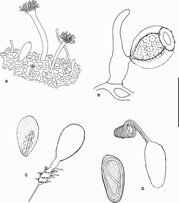
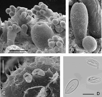
Polymorphic colony, pink-orange colour, growing on the soft coral surface (Figures 1A & 2A). The polyps arise from a dense reticular, encrusting hydrorhiza covered by perisarc. Each polyp has a short perisarc cup at its base. Gastrozooids (up to 1.7 mm high, up to 1 mm wide) sac shaped, with mouth, lacking tentacles (Figures 1A & 2A). Gonozooids (up to 1 mm high) similar to gastrozooids in shape, each bearing one gonophore (Figures 1B & 2B). Two types of dactylozooids: one (up to 4 mm high) with 4–20 capitate tentacles, the other filiform (up to 2 mm high) with a swollen tip (Figures 1A & 2A).
Hydrichthella epigorgia. (A) Colony; (B) gonozooid bearing medusoid; (C) undischarged and discharged microbasic eurytele; and (D) undischarged and discharged desmoneme. Scale: A, 2 mm; B, 500 µm; C, 15 µm; D, 10 µm.

Hydrichthella epigorgia. Scanning electron microscopy photographs of: (A) a colony; (B) a gonozooid; (C) tentacle capitation of a dactylozooid showing discharged microbasic euryteles and desmonemes; and (D) microphotograph of a microbasic eurytele and desmonemes. Scale: A, 200 µm; B, 100 µm; C, D, 10 µm.

The gonophores develop into eumedusoids with eight red tentacular bulbs, four radial canals and velum (Figure 1B). Only female gonophores were observed: numerous pink coloured eggs developed around the manubrium of the attached medusoids. When released, the medusoids swam actively.
Cnidome: microbasic euryteles (12.5–16.5 × 6–8 µm undischarged, 12.5–14 × 6–8 µm discharged) abundant on all types of dactylozooids and on hydrorhiza, rare on gastrozooid mouth (Figures 1C & 2C,D); desmonemes 10–11 × 4.5–5 µm undischarged abundant on tentaculate dactylozooids (Figures 1D & 2C,D). The medusoid cnidome was indeterminable.
Remarks
Bouillon (Reference Bouillon1978) described Hydrichthelloides reticulata from Papua New Guinea living on different types of substrate. The colony had two kinds of dactylozooids, tentaculate and filiform, and possessed a net of stolons surrounded by perisarc. Hirohito (1988) observed colonies of Hydrichthella epigorgia growing on different substrate, such as the gorgonian Anthoplexaura dimorpha, the soft corals Dendronephtya sp. and Bellonella spp., and on rocks. The colonies growing on the gorgonian showed an encrusting hydrorhiza covered by naked coenosarc, while on other substrate the hydrorhiza was reticular and perisarc covered. The observation that the type of the hydrorhiza depends on the substrate lead Hirohito (1988) and Bouillon et al. (Reference Bouillon, Gravili, Pagès, Gili and Boero2006) to define the genus Hydrichthelloides as a junior synonym of Hydrichthella, and we concur with this consideration.
We attributed our Indonesian specimens to the species H. epigorgia owing to the production of eumedusoids with a velum and tentacular bulbs.
Bouillon (Reference Bouillon1967) hypothesized that H. epigorgia could perhaps release its gonophores as free, short-lived medusoids. Our observations confirm this hypothesis, showing that the gonophores are released and are able to swim actively before spawning.
Hydrichthella epigorgia has so far been recorded from Japan (Stechow, Reference Stechow1909; Hirohito, 1988) and the Seychelles (Bouillon, Reference Bouillon1967). Our find represents the first record for the Indonesian area.
Relationship with the host
The hydroid grows on the host surface and the interaction is probably limited by the presence of the perisarc layer. The hydrorhiza net is dense and in preserved specimens detaches spontaneously as a film from the gorgonian surface. Apart from the formation of the perisarc covered hydrorhiza, no other mutual influences on the morphology were observed.
Material examined
One fertile and three sterile polyps on the red gorgonian Ellisella sp., Lembeh Strait (North Sulawesi, Indonesia); water depth: 15 m. Collected by M. Boyer, May 2006.
Description
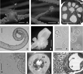
Solitary polyp, or colony with up to two polyps, growing on the gorgonian that completely covers the hydrocaulus up to the neck region with its coenenchyme (Figures 3A & 4A, B). The hydrocaulus is enveloped by a thin layer of perisarc and in the largest specimens reaches 5 cm in height. In specimens still uncovered by the gorgonian coenenchyme, the hydrocaulus is deep orange in colour and reveals longitudinal ridges against the light (Figure 3A). The stem enlarges distally and has up to four transversal constrictions (Figure 3A). The coenosarc is parenchymatic with 8–10 longitudinal gastrovascular canals of variable diameter (Figure 4C). Living hydranths are deep orange in the basal cup, while the portion including the aboral and oral tentacles is milky white. At the base of the hydranth, there is a whorl of 25–26 filiform aboral tentacles, about 5 mm in length, tapering in diameter from about 180 µm to about 40 µm at the tip (Figures 3A & 4D). The hypostome is cone-shaped and the mouth is surrounded by 30–32 oral tentacles arranged in 4–5 irregular whorls (Figure 3A).
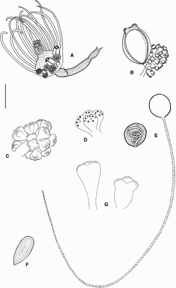
Ralpharia neira. (A) Fertile polyp; (B) medusoid with a nematophore group; (C) magnification of the nematophore group; (D) nematophore; (E) undischarged and discharged heterotrichous anisorhizas; (F) undischarged ?isorhizas; and (G) nematophores of the Ralpharia gorgoniae Petersen, Reference Petersen1990 type material. Scale: A, 3 mm; B, 400 µm; C, 150 µm; D, 70 µm; E, 10 µm; F, 6 µm; G, 300 µm.

Ralpharia neira. Photographs in situ of: (A) a polyp inducing a coenenchyme gall production; (B) a polyp with the hydrocaulus enveloped by the gorgonian coenenchyme; (C) microphotographs of the hydrocaulus coenosarc divided by gastrovascular canals; (D) an aboral tentacle; (E) a nematophore group and medusoids at different stages of development; (F) ?isorhizas and stenoteles from aboral tentacles; (G) undischarged heterotrichous anisorhizas from nematophores; (H) discharged heterotrichous anisorhizas from nematophores; (I) transversal section of the hydrocaulus enveloped by the gorgonian; and (L) histological section of the hydrocaulus enveloped by the gorgonian; a, gorgonian axis; c, gorgonian coenenchyme; ce, coenosarcs; p, perisarc. Scale bars: A, 5 mm; B, 4 mm; C, L, 200 µm; D, 100 µm; E, 300 µm; F, G, 10 µm; H, 30 µm; I, 600 µm.

The large fertile polyp shows 5–6 well-developed blastostyles made up of a branched stalk bearing gonophores proximally and a group of nematophores distally (Figure 3A,B & 4E). In young hydranths, the blastostyles are absent while small groups of nematophores are already visible between the aboral tentacles. Immature gonophores (more than 20 at different stages of maturation) are orange, bell-shaped medusoids (up to 600 µm high and 400 µm wide) with a circular canal, four radial canals and four white tentacle bulbs containing nematocysts (Figure 3B). Each group of nematophores (550–600 µm in diameter) consists of 15–20 milky white elements (80 µm high and 70 µm wide), shaped like a garlic bulb, with four lobes armoured distally with about 20 nematocysts each (Figure 3C,D). Very young gonophores have a shape and dimension intermediate between a medusoid and nematophore (Figures 3B & 4E).
No medusoid release was observed.
Cnidome: oral tentacles: large stenoteles (10–12 × 8–10 µm), small stenoteles (6–7.5 × 4.5–5 µm), heterotrichous anisorhizas (8–10 × 8–10 µm).
Aboral tentacles: large stenoteles (10–12 × 8–10 µm), small stenoteles (6–7.5 × 4.5–5 µm) (Figure 4F), heterotrichous anisorhizas (8–10 × 8–10 µm), ?isorhizas (9–10 × 4–5 µm) (Figures 3F & 4F).
Nematophores and immature medusoids: large stenoteles (10–12 × 8–10 µm), heterotrichous anisorhizas (10–10 × 8–10 µm undischarged, 8–9.5 × 8–9 µm discharged, threat 150–170 µm, 2 µm wide proximally and 0.5 µm distally) (Figures 3E & 4G,H).
Remarks
The collected specimens of Ralpharia share with R. sanctisebastiani, R. neira, R. gorgoniae and R. rosetta the presence of grouped nematophores placed on the distal portion of the blastostyle. Nevertheless, they differ from R. rosetta in the blastostyle shape, which in this species is long, slender and unbranched, and in the host species (Watson, Reference Watson1999). The nematophores of the studied specimens are garlic bulb-shaped with four lobes (Figure 3D), while those of R. sanctisebastiani are button-shaped (da Silveira & Migotto, Reference Silveira and Migotto1984; Petersen, Reference Petersen1990). They also differ from those of R. gorgoniae, whose polyp is very large with tri-lobate to triangular or heart-shaped nematophores (Petersen, Reference Petersen1990) (Figure 3G). The general polyp shape and dimension and the nematophore shape agree with the description of R. neira given by Petersen (Reference Petersen1990). Although Petersen did not mention the capsule here tentatively identified as ?isorhiza, we attributed it to R. neira. Petersen has perhaps overlooked this capsule type. Moreover, our specimens come from the same geographical region as R. neira. A comparison between the type material of R. neira and R. gorgoniae confirmed our initial identification. Petersen (Reference Petersen1990) did not provide a catalogue number for the holotype of R. neira. However, Petersen's material is kept by the Zoological Museum of Copenhagen which provided us with a sample labelled ‘Ectopleura neira Petersen, Th. Mortensen's Key Island Expedition, collected by Th. Mortensen 1 June 1922, off Neira, Banda, Indonesia, 10 m depth’. In the tube there are numerous polyps detached from a branch of Melithodes ochracea (also present in the tube). All these data correspond to Petersen's description and therefore this specimen must be considered as the type material of R. neira (syntypes).
In the same collection there is also a tube labelled ‘Gorgonia ventalina (Linnaeus) (ref. no. 282). Collected by Chantal De Ridder, 19 August 1976, Galeta Reef, Panama (Atlantic side), on the edge of the reef at 1.5–2 m depth’. In the tube there is a portion of Gorgonia ventalina and a separate small tube without a label containing one fertile polyp. The morphology of this polyp corresponds to the description of Ralpharia gorgoniae Petersen, Reference Petersen1990. The data on the label also corresponds to Petersen's description of R. gorgoniae. Therefore, this polyp must be the holotype of Ralpharia gorgoniae Petersen, Reference Petersen1990.
In his description of R. neira, Petersen (Reference Petersen1990, p. 189) observed ‘The whole shape of the nematophore resembles a medusa, and it is tempting to interpret these nematophores as transformed medusa buds’. The study of our specimens reveals the presence of the same nematocysts in the nematophore lobes and in the medusoid tentacular bulbs and of intermediate stages between medusoids and nematophores. These observations confirm Petersen's idea, indicating that nematophores are medusoids that do not reach maturity.
Relationship with the host
The presence of the hydroid induces several modifications in the gorgonian. When the planula settles on the gorgonian surface, the gorgonian tissue reacts by producing a kind of gall that envelops the base of the young polyp (Figure 4A). The coenenchyme subsequently grows along the hydrocaulus covering it up to the hydranth neck region (Figure 4B). The coenenchyme covering the hydroid does not form polyps (Figure 4B). The transversal section of the enveloped hydrocaulus revealed that the gorgonian skeletal axis enveloped the perisarc tube of the hydroid (Figure 4I & L). Progressing from the exterior to the interior, the coenenchyme, the skeletal axis, the hydroid perisarc and the hydroid coenosarc form consecutive layers (Figure 4I & L). The gorgonian skeleton is absent in the association involving R. sanctisebastiani and Lophogorgia punicea as reported by da Silveira & Migotto (Reference Silveira and Migotto1984).
Clava krempfi Billard, Reference Billard1919; Van Praёt, Reference Van Praët1979.
Pteroclava krempfi: Weill, Reference Weill1931, Reference Weill1934, Reference Weill1936, Reference Weill1937; Ranson, Reference Ranson1937; Leloup, Reference Leloup1937; Picard, Reference Picard1957; Kramp, Reference Kramp1961; Vervoort, Reference Vervoort and Rees1966; Bouillon, Reference Bouillon1974; Petersen, Reference Petersen, Larwood and Rosen1979; Bouillon, Reference Bouillon1985; Hirohito, 1988; Petersen, Reference Petersen1990; Boero et al., Reference Boero, Bouillon and Gravier-Bonnet1995; Bouillon et al., Reference Bouillon, Gravili, Pagès, Gili and Boero2006.
? Syncoryne crassa Pictet, Reference Pictet1893; Hartlaub, Reference Hartlaub1905; Mayer, Reference Mayer1910; Prévot, Reference Prévot1959; Schuchert, Reference Schuchert2003.
Material examined
Fertile colony on the red gorgonian Astrogorgia sp., Siladen (Bunaken National Park, North Sulawesi, Indonesia); water depth: 12 m. Collected by C.G. Di Camillo & S. Puce, February 2005. Fertile colony on the red gorgonian Astrogorgia sp., Negeri (Bunaken National Park, North Sulawesi, Indonesia); water depth: 15 m. Collected by C.G. Di Camillo & S. Puce, February 2005.
Description
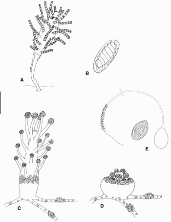
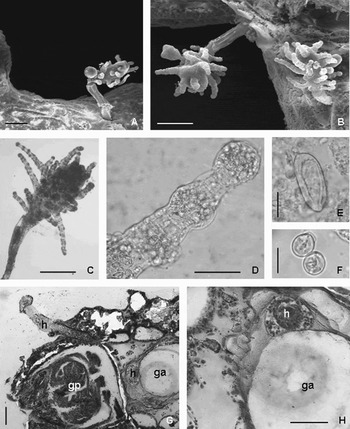
Colony growing on gorgonians, with perisarc-covered hydrorhiza embedded in the host tissues (Figures 5A & 6A–C). Hydranths up to 2 mm high, borne on long pedicels covered by a distally enlarging perisarc tube; hydranth body elongated, with 4–5 moniliform oral tentacles and up to 25 moniliform scattered tentacles (Figures 5A & 6C,D); one to four patches of large nematocysts under the oral tentacles. Medusa buds single or in groups of up to three on short pedicels scattered in the mid-region of the hydranth (Figure 6B).
Pteroclava kremfi. (A) Polyp; (B) macrobasic eurytele. Zanclea timida sp. nov.; (C) extended polyp; (D) contracted polyp; and (E) undischarged and discharged telotrichous macrobasic mastigophore. Scales: A, 200 µm; B, 25 µm; C, D, 100 µm; E, 20 µm.

Pteroclava kremfi. Scanning electron microscopy photographs of: (A) a polyp arising from the gorgonian; (B) magnification of polyps with gonophores; (C) microphotograph of polyp; (D) moniliform tentacle; (E) undischarged macrobasic eurytele; (F) undischarged stenoteles; (G) histological section of the gorgonian hosting the hydroid; and (H) magnification of the histological section showing the hydrorhiza near to the gorgonian axis. ga, gorgonian axis; gp, gorgonian polyp; h, hydrorhiza. Scale bars: A,C, 200 µm; B, 250 µm; D, 50 µm; E, 15 µm; F, 10 µm; G,H, 100 µm.

Medusa stage not observed.
Cnidome: macrobasic euryteles (40–41 × 15–17 µm undischarged, 37–38 × 14–15 µm discharged) in patches under oral tentacles (Figures 5B & 6E), and stenoteles of two sizes (10 × 9–10 µm and 5 × 4–5 µm undischarged) mainly on tentacles (Figure 6F).
Remarks
Boero et al. (Reference Boero, Bouillon and Gravier-Bonnet1995) noted the presence of a long pedicel in specimens inhabiting hosts rich in superficial spicules like gorgonians, while in specimens symbiotic with soft corals the pedicel is less evident. In accordance with these observations, the Indonesian specimens living on gorgonians have a long, perisarc-covered pedicel (Figures 5A & 6A–C).
This species has been recorded from Vietnam (Billard, Reference Billard1919), Japan (Hirohito, 1988), Papua New Guinea (Boero et al., Reference Boero, Bouillon and Gravier-Bonnet1995) and Réunion Island (Boero et al., Reference Boero, Bouillon and Gravier-Bonnet1995). Our specimens represent the first record from Indonesia.
Relationship with the host
Billard (Reference Billard1919) described the species with the hydrorhiza embedded in a soft coral tissue, while Hirohito (1988) observed the perisarc-covered hydrorhiza growing on a gorgonian host. The Indonesian specimens have polyps arising from a gorgonian tissue which completely hides the hydrorhiza. In fact, the longitudinal and transversal sections of a colonized gorgonian show the perisarc-covered hydrorhiza creeping below the gorgonian polyps and running along the host axis (Figure 6G) that sometimes tends to incorporate it (Figure 6H).
Type material
Holotype: sterile colony on the Paratelesto sp., Siladen (Bunaken National Park, North Sulawesi, Indonesia); water depth: 25 m; [MSNG54191]. Collected by G. Bavestrello, July 2004.
Diagnosis
Zanclea living on Paratelesto sp. Perisarc absent, hydrorhiza with frequent clusters of nematocysts. Hydranth with a rigid basal portion, cup-shaped, lacking tentacles. The tentaculate distal portion of the hydranth is able to completely retract inside the basal portion leaving the spherical ends of the tentacles outside. Telotrichous macrobasic, mastigophores in a ring on the distal part of the hydranth basal portion.
Description
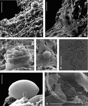
Colony characterized by a reticular hydrorhiza creeping on Paratelesto sp. and filling spaces between sclerite bundles. A perisarc is absent and the hydrorhiza is characterized by several globular cells and frequent clusters of nematocysts (Figures 5C,D & 7A,D). The polyps (up to 500 µm high) arise among the octocoral anthocodia. The hydranths are pear-shaped and show a short hypostome surrounded by 5–6 oral capitate tentacles (diameter of capitula 50 µm); 15–16 aboral capitate tentacles are scattered on the hydranth body (diameter of capitula 27, 5–30 µm) (Figures 5C & 7B). The basal portion of the hydranth is cup-shaped, semi-rigid, lacking tentacles and armoured distally by a large nematocyst ring (Figures 5C,D & 7B,C). The distal, tentaculate portion of the hydranth is able to retract completely inside the basal portion leaving the capitula of the oral tentacles outside (Figures 5D & 7C).
Zanclea timida sp. nov. Scanning electron microscopy (SEM) photographs of: (A) colony showing polyps and hydrorhiza with clusters of nematocysts (arrows); (B) extended polyp; (C) contracted polyp; (D) magnification of nematocyst cluster; microphotograph of (E) undischarged telotrichous macrobasic mastigophore; SEM photographs of (F) capsule of discharged telotrichous macrobasic mastigophore; and (G) shaft end of telotrichous macrobasic mastigophore. Scale bars: A, 200 µm; B, 100 µm; C, 50 µm; D,E, 20 µm; F, 5 µm; G, 10 µm.

Medusa buds not observed.
Cnidome: egg-shaped telotrichous macrobasic mastigophores (undischarged capsule 20–21 × 15–15.5 µm, discharged capsule 18–19 × 15 µm, shaft 300 µm) in a ring on the distal part of the hydranth basal portion, on the hydrorhiza scattered or arranged in clusters (Figures 5E & 7E–G); stenoteles of two sizes (undischarged capsule 10–11 × 9–10 µm and 5–6 × 5–6 µm) on tentacle capitations, on the hydrorhiza both scattered and in clusters.
Etymology
The specific name is derived from the latin word timidus meaning ‘shy’ and refers to the tendency of the polyps to retract frequently into the basal part of the hydranth.
Remarks
Zanclea timida represents the only Zanclea species living in association with an octocoral. Zanclea timida differs from the other described species by the presence of macrobasic mastigophores and by the hydranth differentiated into a rigid, cup-shaped basal portion and a narrow contractile tentaculate distal portion. A nematocyst ring marks the boundary between the basal and distal portion. While several species symbiotic with bryozoans can retract inside the skeleton of the hosts, Z. timida is able to retract inside its own basal ‘cup’. When retracted, the polyp becomes almost spherical and the capitula of the tentacles armed with nematocysts protrude out of the ‘cup’, whose rim is also surrounded by nematocysts (Figure 5D). Moreover, similarly to Z. divergens and Zanclella diabolica Boero et al., Reference Boero, Bouillon and Gravili2000, whose hydrorhiza is also provided with nematocyst clusters emerging from the bryozoan skeleton (Boero et al., Reference Boero, Bouillon and Gravili2000; Puce et al., Reference Puce, Cerrano, Boyer, Ferretti and Bavestrello2002), Zanclea timida presents frequent stolonal nematocyst clusters protecting its exposed stolons.
Relationship with the host
The hydrorhiza grows on the gorgonian surface and appears to be devoid of perisarc. The only protection against living enemies is provided by the stolonal nematocyst clusters. No specific tissue reactions owing to the association are evident either in the hydroid or in the octocoral. Several hydroids arise rather close to the Paratelesto polyps, suggesting a trophic relationship between the two. Boero et al. (Reference Boero, Bouillon and Gravier-Bonnet1995) made a similar observation for the hydroid Pteroclava krempfi that occurred in close proximity to the polyps of the gorgonian host, even touching their tentacles.
DISCUSSION
With the exception of Zanclea gilii, growing in a hard coral, all hydroid species associated with anthozoans are symbiotic with octocorals. Pteroclava krempfi and Hydrichthella epigorgia are able to colonize both soft corals and gorgonians and the hydroid morphology depends on the different hosts. Pteroclava krempfi, inhabiting hosts rich in spicules, such as gorgonians, forms a long pedicel, while the pedicel is less pronounced in specimens living in soft corals (Boero et al., Reference Boero, Bouillon and Gravier-Bonnet1995). The hydrorhizal net of H. epigorgia is naked in colonies associated with the gorgonian Anthoplexaura dimorpha, but covered by perisarc in specimens living on other octocorals (Hirohito, 1988). A similar phenomenon occurs in the symbiosis between hydroids and bryozoans, where the bryozoan reaction to the presence of different hydroid species leads to different hydrorhizal covering patterns (Puce et al., Reference Puce, Bavestrello, Di Camillo and Boero2007).
Most of the hydroid species associated with anthozoans show a perisarc-covered hydrorhiza which separates the coenosarc from the host tissue, although Ptilocodium repens, some H. epigorgia specimens, Z. gilii and Z. timida lack a distinct perisarc. Thus, in several symbiotic genera belonging to different families (e.g. Hydrichthys, Proboscidactyla, Zanclea and Eutima), the perisarc loss appears to be related to the symbiotic life. In several Zanclea species associated with bryozoans the stolons lack a perisarc sheath. The protection provided by the bryozoan skeletons seems to make the perisarc covering superfluous (Puce et al., Reference Puce, Cerrano, Boyer, Ferretti and Bavestrello2002, Reference Puce, Bavestrello, Di Camillo and Boero2007). The tendency of the Zanclea species to suppress perisarc formation is also evident in the anthozoan inhabiting Zanclea. In Z. gilii the hydrorhiza is completely embedded in the coral tissue and naked (Boero et al., Reference Boero, Bouillon and Gravili2000). It can probably exploit the protection offered by its host, while in Z. timida the stolons grow in grooves in the surface of the octocoral. Although being thus quite exposed, the presence of nematocyst clusters on the stolons seems to offer enough protection to eliminate the need for a perisarc covering.
In hydroids inhabiting bryozoans, the polyps are able to retract into the bryozoan skeleton and only the tentacular capitations armed with nematocysts remain exposed on the surface (Boero et al., Reference Boero, Bouillon and Gravili2000; Puce et al., Reference Puce, Cerrano, Boyer, Ferretti and Bavestrello2002, Reference Puce, Bavestrello, Di Camillo and Boero2007). Similarly, the polyps of Z. timida retract into their basal cup whose edge is surrounded by a nematocyst ring and from which only the tentacular nematocyst clusters stick out (Figures 5D & 7C). A similar reaction was described by Schuchert (Reference Schuchert2007) for the filiferan hydroid Garveia grisea.
The presence of the hydroid can modify the octocoral morphology, inducing the production of new branches whose skeleton totally or partially envelops the hydrocaulus. Lophogorgia punicea covers the hydrocaulus of Ralpharia sanctisebastiani only with a coenenchyme layer without a supporting axial skeleton (da Silveira & Migotto, Reference Silveira and Migotto1984). Ellisella sp. produces its axial skeleton around the Ralpharia neira stem (Figure 4I & L). Although the hydrocaulus of Pteroclava kremfi is exposed, the hydrorhiza is deeply embedded in the gorgonian tissue, creeping along the gorgonian axis (Figure 6G,H).
The possible behavioural interactions between hydroids and octocorals are still undescribed, but there are some indications of existing trophic relationships. In fact, the polyps of P. krempfi, Sarsia medelae and Z. timida, appear to be denser in the areas at the base of the octocoral polyps (Boero et al., Reference Boero, Bouillon and Gravier-Bonnet1995, Gili et al., Reference Gili, Lopez-González and Bouillon2006). Moreover, Boero et al. (Reference Boero, Bouillon and Gravier-Bonnet1995) observed preserved polyps in the act of touching the gorgonian tentacles, suggesting a trophic behaviour similar to that reported for numerous other symbiotic hydroids (Boero & Hewitt, Reference Boero and Hewitt1992; Piraino et al., Reference Piraino, Bouillon, Boero and Bouillon1992; Bavestrello et al., Reference Bavestrello, Puce, Cerrano, Balduzzi and Mills2000; Boero et al., Reference Boero, Bouillon and Gravili2000; Puce et al., Reference Puce, Cerrano, Boyer, Ferretti and Bavestrello2002, Reference Puce, Bavestrello, Di Camillo and Boero2007).
The symbiosis with anthozoans evolved independently in six athecate hydroid families. However, while S. medelae and P. krempfi are the only known species of their respective families, Ptilocodiidae and Zancleidae each contain two species associated with anthozoans. Most Zanclea species are in fact closely associated with other marine invertebrates, mainly with bryozoans (Boero et al., Reference Boero, Bouillon and Gravili2000; Puce et al., Reference Puce, Cerrano, Boyer, Ferretti and Bavestrello2002, Reference Puce, Bavestrello, Di Camillo and Boero2007), but also with bivalves (Cerrano et al., Reference Cerrano, Amoretti and Bavestrello1997), hexacorals (Boero et al., Reference Boero, Bouillon and Gravili2000), octocorals (present paper) and sponges (unpublished). Likewise, of seven known Ralpharia species, six are symbiotic with octocorals.
ACKNOWLEDGEMENTS
The authors thank Dr Carlo Cerrano for the octocoral identification and Dr Massimo Boyer for underwater pictures of Ralpharia neira. This work was financially supported by MIUR (Italian Ministry of Education, University and Research). The comments provided by Dr Peter Schuchert and an anonymous referee greatly improved the manuscript.