Introduction
The common opportunistic fungal infections of the nose and paranasal sinuses involve aspergillosis and zygomycosis (mucormycosis) in an immunocompromised host. Entomophthorales are a group of primitive fungi belonging to the phylum zygomycetes, which are known clinically to cause zygomycosis. Conidiobolus is one of the six families belonging to the entomophthorales group, and the species Conidiobolus coronata is one of the 27 species belonging to the conidiobolus genus.Reference Ellis, Merz and Hay1 The conidiobolus species was classified by Rixford and Gilchrist in 1896 (Table I).Reference Ribes, Vanover Sams and Baker2
1862 taxonomical classification of Conidiobolus coronata by Rixford and GilchristReference Ribes, Vanover Sams and Baker2

Fungi of the entomophthorales group are true pathogens, infecting primarily immunocompetent hosts.Reference Ribes, Vanover Sams and Baker2 Among these fungi, C coronata induced fungal infections may cause a more chronic disease of the nasal mucosa, with spread to the subcutaneous tissue.
Case report
A 49-year-old man presented with complaints of bilateral nasal discharge and nasal blockage of three months’ duration. These symptoms were worse on the left side. There was no history of fever, recurrent upper respiratory tract infection, sneezing, headache or weight loss. There was also no history of bronchial asthma, diabetes mellitus, pulmonary tuberculosis, or previous, prolonged use of any oral medications such as steroids or antineoplastic drugs.
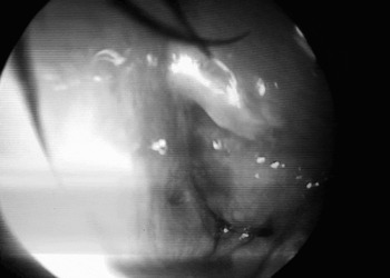
On anterior rhinoscopy, the left inferior turbinate appeared enlarged (Figure 1). The left middle turbinate and middle meatus could not be visualised. The right inferior turbinate, middle turbinate and meati appeared normal. There was no palpable cervical lymphadenopathy.
Endoscopic view of enlarged, congested inferior turbinate in contact with the nasal septum.

Within the left nasal cavity, a 30° nasal endoscope could not be passed along the floor of the nasal cavity beyond the area corresponding to the cartilaginous part of the septum. The inferior turbinate was congested and enlarged, and topical decongestion had no effect. The swelling corresponding to the left inferior turbinate appeared diffuse, and the osteomeatal complex could not be assessed endoscopically. This clinical picture prompted suspicion of a benign tumour arising from the lateral nasal wall.
On general examination, the pulse rate was 84 beats/minute, the respiratory rate 16 breaths/minute and the patient was afebrile.
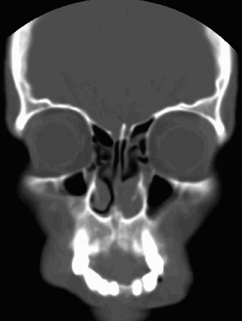
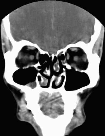
Coronal computed tomography scanning of the paranasal sinuses with contrast showed mild mucosal thickening in the ethmoid sinuses bilaterally and in the left sphenoid sinus. The left inferior turbinate was impinging on the nasal septum, with synechiae formation (Figure 2). There was a deviated nasal septum with a bony spur on the left (Figure 3). There was no evidence of soft tissue enhancement. The chest X-ray was normal.
Coronal computed tomography scan of the paranasal sinuses, showing left inferior turbinate hypertrophy impinging on the nasal septum, with synechiae formation.

Coronal computed tomography scan of the paranasal sinuses, showing nasal septum deviation to the left with a bony spur also on the left.

Laboratory investigations were within normal limits, including complete blood picture, bleeding parameters and erythrocyte sedimentation rate. Biochemical analyses of blood sugar, renal function and serum electrolytes were normal. Testing for human immunodeficiency virus was negative.
A biopsy from the mass was reported as showing acute inflammation with an eosinophilic infiltrate. There was no evidence of malignancy. Special stains for fungi were negative. However, the pathologist requested a deeper, more representative biopsy due to clinical suspicion of malignancy.
The patient was scheduled for excision of the left nasal mass under general anaesthesia. Under 30° endoscopic vision, a small synechia between the inferior turbinate and septum was released. Using turbinectomy scissors, the left inferior turbinate was excised. A sharp, bony, cartilaginous spur was found to be impinging into the inferior turbinate. The mucoperiosteum was elevated over the spur and a spurectomy was performed. The cut edge of the inferior turbinate was coagulated with a KTP 532 laser (Laserscope, San Jose, USA) set at 7 W and 125 Joules. The resected inferior turbinate was sent for histopathological analysis and the mucoidal discharge for fungal culture.
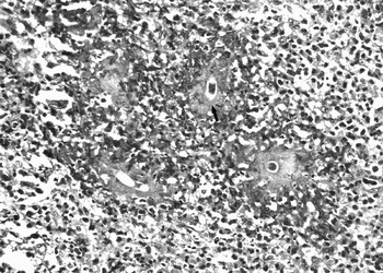
The biopsy specimen showed multiple fragments of tissue lined by focally hyperplastic respiratory epithelium with distinct subepithelial oedema. Some fragments were devoid of lining epithelium and were diffusely infiltrated by sheets of lymphocytes, plasma cells, neutrophils and eosinophils with granulation tissue like appearance. Numerous granulomas composed of epithelioid histiocytes and foreign body giant cells were seen around fragments of hyphae enveloped by Splendore Hoeppli material (Figure 4). These hyphae were broad and thin-walled with occasional septations. No angioinvasion was seen. These were the morphological features of entomophthoromycosis by C coronata. Fungal culture was requested for confirmation; however, this was negative.
Cross-section of fungal hyphae showing Splendore Hoeppli phenomenon (H&E; ×400).

The patient was discharged the following day. He was advised to take cotrimoxazole DS tablets, one tablet twice daily for seven days, and to continue normal saline nasal douches.
After seven days, crust from the excised area of the left inferior turbinate was removed in the out-patient department with a 30° endoscope. The patient was empirically commenced on itraconazole 100 mg twice daily for three weeks.
On review after three weeks, the nasal wound had healed. The patient had no complaints. There was no crusting seen on nasal endoscopy.
The patient was followed up for one year. There was no evidence of recurrent granuloma formation at the same site or in the surrounding tissues.
Discussion
The presence of any fungal infection in humans usually implies that the host defence mechanisms have been compromised by disease or therapeutic intervention. These conditions are associated with leucopenia or impaired white cell function without leucopenia.
Mucormycosis or zygomycosis is caused by members of the order entomophthorales and mucorales. In general, fungi of the order entomophthorales cause a more chronic disease of the nasal mucosa and subcutaneous tissue, whereas those of the order mucorales cause more severe forms of disease. Thus, clinically, manifestations of zygomycosis may vary from a superficial infection to a disseminated infection with high mortality. All such infections are confined to the tropics.
The genus conidiobolus contains 27 species. Of these, C coronatus and C incongruous are the only species known to cause human disease. Entomophthoromycosis caused by conidiobolus species results in a chronic inflammation or granulomatous disease limited to the nasal submucosa and maxillofacial tissue. The patient presents with nasal obstruction and sinus pain. On examination, there may be nasal polyps or palpable, firm, subcutaneous facial nodules in the nasal and perinasal regions. Generalised, progressive facial swelling may occur.Reference Ellis, Merz and Hay1 Disseminated entomophthorales infection affecting immunocompetent patients is rare, as these organisms rarely invade blood vessels.Reference Ribes, Vanover Sams and Baker2 The mode of transmission may be rhinocerebral or pulmonary. A case of disseminated conidiobolus infection with endocarditis has been reported in a cocaine abuser. The dissemination occurred via skin abrasions in the lower extremities.Reference Jaffey, Haque, El-Zaatari, Pasarell and McGinnis3 Another case of disseminated rhinofacial zygomycosis affected by C coronata caused bilateral distortion of facial tissues, leading to disfigurement.Reference Costa, Porto, Pegas, Reis, Pires and Lacaz4
Fungal culture may not be reliable for diagnostic purposes, since the fungal hyphae easily lose viability. Histopathological analysis is the most rapid diagnostic method. The Splendore Hoeppli phenomenon is the characteristic feature, wherein the hyphae are 5–15 μm wide, infrequently septate, thin-walled and surrounded by an eosinophilic sheath.
A combination of surgical resection and antifungal treatment is effective in cases of isolated, early C coronata fungal granuloma. Treatment of submucosal infection with surgical resection is controversial, due to rapid spread of infection and unsuccessful outcomes. The combination of trimethoprim and sulphamethoxazole has been used, although with inconsistent response. Of the antifungal drugs, itraconazole is preferred for subcutaneous and deep-seated tropical mycosis due to its limited side effects. Itraconazole given for a prolonged period of time (ranging from six to 19 months) has a 65 per cent response rate.Reference Restrepo5 Posaconazole is a newer antifungal triazole which has been approved for the treatment of zygomycosis.Reference Chayakulkeeree, Ghannoum and Perfect6
The current case of inferior turbinate fungal granuloma is noteworthy in that a rare fungal species – Conidiobolus coronata – affected an immunocompetent individual. A large bony spur impinging into an enlarged inferior turbinate probably caused minor trauma, allowing implantation of spores and formation of a chronic fungal granuloma limited to the inferior turbinate. Clinically, this posed a diagnostic dilemma as to whether the lesion was benign or malignant. No similar cases have previously been reported. Our patient's lesions had distinguishing histopathological features which were diagnostic. Radical resection and KTP 532 laser coagulation of the excision margin, in order to prevent spread of infection, may represent a successful mode of treatment. Empirical post-operative treatment with itraconazole for six weeks may prevent relapse.