Introduction
Cystic hydatid disease (CHD), a chronic zoonotic disease that results from infection with the larval stage of the dog tapeworm, Echinococcus granulosus, is medically and economically one of the most important zoonoses (Schantz, Reference Schantz, Long and Prober1997; Thompson, Reference Thompson, Gillespie and Pearson2001; Rostami Nejad et al., Reference Rostami Nejad, Hoseinkhan, Nazemalhosseini, Cheraghipour, Abdinia and Zali2007a). The disease is endemic worldwide and is widespread in the Mediterranean region (Euzeby, Reference Euzeby1991). Hydatid cysts are commonly found in sheep, camels, cattle, goats and humans throughout Iran. Sheep are the most common and important intermediate hosts of E. granulosus in Iran, followed by cattle and goats (Mobedi et al., Reference Mobedi, Madadi and Arfaa1970; Moghaddar et al., Reference Moghaddar, Oryan and Hanifpour1992; Oryan et al., Reference Oryan, Moghadar and Gaur1994; Mehrabani et al., Reference Mehrabani, Oryan and Sadjjadi1999; Dalimi et al., Reference Dalimi, Motamedi, Hosseini, Mohammadian, Malaki, Ghamari and Ghaffari-far2002; Fasihi Harandi et al., Reference Fasihi Harandi, Hobbs, Adams, Mobedi, Morgan-Ryan and Thompson2002; Rostami Nejad et al., Reference Rostami Nejad, Abdinia and Cheraghipour2007b).
Based on morphological, biological, biochemical and especially molecular-biological characteristics, E. granulosus has now been divided into eight main genotypes (G1–G8). These include the important sheep strain (G1), two bovid strains (G3, G5), a horse strain (G4), the camelid strain (G6), a pig strain (G7) and a cervid strain (G8) (Bowles et al., Reference Bowles, Blair and McManus1992; Pearson et al., Reference Pearson, Le, Zhang, Blair, Dai, McManus, Craig and Pawlowski2002). A ninth genotype (G9) has recently been described from humans and pigs in Poland (Scott et al., 1996). The G1 sheep strain of E. granulosus is the most widespread and important zoonotic genotype (Craig et al., Reference Craig, Rogan and Allan1996).
A few molecular epidemiological studies have been carried out on Iranian isolates. The first of these was by Zhang et al. (Reference Zhang, Eslami, Hosseini and McManus1998) who analysed sequence variation within the cox1 and nad1 genes based on the polymerase chain reaction-restriction fragment length polymorphism (PCR-RFLP) method for 16 isolates of E. granulosus collected from human cysts and domestic animals, including cattle, sheep, goats and camels at slaughterhouses in Tehran, and in central and southern Iran. Their study clearly indicated that the common sheep strain (G1 genotype) and the camel/dog strain (G6 genotype) both occur in Iran, the latter being uncommon. The G1 genotype was present in all four human isolates examined and was more prevalent in domestic animals than the camel-restricted G6 genotype (Zhang et al., Reference Zhang, Eslami, Hosseini and McManus1998).
In a second study, isolates of E. granulosus were collected from humans and other animals from different geographical areas of Iran and characterized using both DNA (PCR-RFLP of the ITS1 region in the nuclear ribosomal gene cluster) and morphological criteria. The presence of the sheep and camel strains/genotypes was again demonstrated (Fasihi Harandi et al., Reference Fasihi Harandi, Hobbs, Adams, Mobedi, Morgan-Ryan and Thompson2002).
Based on these surveys, we designed new specific primer pairs targeting the mitochondrial 12S rRNA gene to characterize different E. granulosus isolates occurring in Iran. This approach is straightforward and more rapid than the PCR-RFLP method used previously.
Materials and methods
Sample processing
Liver and lung samples were obtained from 30 sheep, cattle and goats. Fifteen isolates were provided from the abattoir of Lorestan Province (Aleshtar) located in western Iran and another 15 isolates were from Mazandaran Province (Noor) located in northern Iran. The viability of protoscoleces was determined by microscopic examination of unstained material to detect flame cell movement, and also by the use of a vital dye (eosin 0.1%). Cysts were processed separately and in this study an isolate represents protoscoleces collected from an individual hydatid cyst. Nine of ten bovine cysts and three of ten ovine cysts were unfertile and material from these was not used. Protoscoleces were aspirated from cysts, rinsed in saline, fixed in 70% (v/v) ethanol, and transported to the Foodborne and Diarrheal Diseases Laboratory for analysis. From each cyst, protoscoleces were removed and stored at − 20°C. The protoscoleces were rinsed several times with phosphate buffered saline (PBS) to remove the ethanol prior to DNA extraction. Reference material of the G6 genotype was provided from a slaughterhouse in the south of Tehran Province.
DNA extraction
DNA extraction methods for E. granulosus were evaluated with four different methods, including the Tissue Extraction Kit (Qiagen, Germany), a modified Cinnagen extraction kit (Cinnagen, Iran), phenol–chloroform (Sambrook et al., Reference Sambrook, Fritsch and Maniatis1989) and modified phenol–chloroform methods. Efficiency of the DNA extraction method was determined by the degree of success in PCR.
Tissue Extraction Kit
In case of the Tissue Extraction Kit (Qiagen, Germany), DNA was extracted according to the manufacturer's guidelines.
Modified Cinnagen extraction kit
Before starting the DNA extraction process, DNG™-Plus solution (Cinnagen, Iran) was warmed at 37°C for 20 min with gentle shaking. Then 300 μl of sample was mixed with 600 μl of DNG™-Plus and vortexed for 20–25 s. The sample should be a completely homogeneous suspension at this step. Then 450 μl of isopropanol was added and mixed by vortexing. For low concentrations of DNA, samples were kept at − 20°C for 20 min. Then samples were centrifuged at 12,000 rpm for 10 min and the supernatant gently poured off. Two millilitres of 75% ethanol were added to the pellet, mixed by vortexing for 3–5 seconds and centrifuged at 12,000 rpm for 5 min. Once again the supernatant was gently poured off and the pellet dried at 65°C. The DNA pellet was dissolved in 50 μl of sterile distilled water by gentle shaking at 65°C for 5 min. A final centrifugation for 30 s at 12,000 rpm pelleted undissolved material, leaving pure DNA in the supernatant.
Phenol–chloroform
DNA was extracted according to the phenol–chloroform method of Sambrook et al. (Reference Sambrook, Fritsch and Maniatis1989).
Modified phenol–chloroform
Protoscoleces were washed three times with PBS. Then 500 μl lysis buffer containing 1% sodium dodecyl sulphate (SDS) was added to 300 μl protoscoleces. At the next step 2 μl of proteinase K was added, vortexed and then incubated for 2 h at 55°C and 15 min at 80°C. Next, 650 μl phenol–chloroform–isoamyl alcohol (25:24:1) was added and spun at 12,000 rpm for 2 min. The supernatant was removed and, after chloroform addition (same volume), mixing and spinning (12,000 rpm for 2 min), 1/10 the volume of sodium acetate (3 m, pH = 5.2) and 500 μl ethanol were added and the tube kept at − 20°C for 30 min. Thereafter, it was spun at 12,000 rpm for 10 min and the pellet was washed using 70% ethanol. Later, the pellet was dissolved in 30 μl sterile super double-distilled water, placed at 37°C for 15 min and subsequently stored at − 20°C until required for PCR.
Sequencing
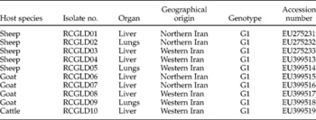
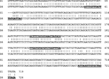
Following consideration of different sequences of E. granulosus in GenBank (AF297617 and AB208063 were used as main GenBank sequence accession numbers for G1 and G6 genotypes, respectively), new and specific primer pairs were designed for the two genotypes known to exist in Iran. G1 and G6 12S rRNA gene sequences were aligned using MegAligne software (DNASTAR Inc., Madison, Wisconsin, USA) and primers were designed that were likely to be strain specific. Relevant portions of the 12S rRNA for G1 (259 bp) and G6 (676 bp) were amplified and sequenced using an ABI 3130X Genetic Analyzer (see table 1). Sequence data were compared with available sequences in GenBank. Figure 1 shows the alignment of G1 and G6 12S rRNA sequences.
Host and geographical origin of Iranian isolates of Echinococcus granulosus genotyped and sequenced in this study.

Alignment of 12S rRNA nucleotide sequences of the G1 (GenBank accession no. AF297617) and G6 (GenBank accession no. AB208063) strains. Regions used for primer design are underlined. The outer pair of primers was designed to amplify only the G6 genotype and the inner pair the G1 genotype. Numbering is for the 12S gene according to the original annotations in the GenBank entries. According to the annotation in AF297617, the 12S rRNA gene ends at position 700 in the G1 genotype.

Amplification of G1 and G6 genotypes
G1 genotype
PCR was performed in a 25 μl final volume containing 1 × PCR buffer, 1.5 mm MgCl2, 0.2 mm of each deoxynucleotide triphosphate (dNTP), 1.6 × 10− 7 μm of each primer (G1 forward: 5′-GCT TTT GTG TGG ATT ATG CG-3′ and G1 reverse: 5′-TCA AAC CAG ACA TAC ACC AA-3′) and 0.04 U μl− 1 Ampli-Taq Polymerase. Amplification was carried out for 35 cycles as follows: denaturation for 30 s at 94°C, annealing for 30 s at 57°C and elongation for 40 s at 72°C. After amplification, 10 μl of the amplification products were electrophoresed on 2% agarose gel and stained using ethidium bromide. PCR products from 10 samples (table 1) were sequenced to confirm identity.
G6 genotype
The primer pair G6 forward (5′-ATT TGT TTA TTT ATG ATT AG-3′) and G6 reverse (5′-TCT AAC TAG CAC AAC CTA AT-3′) was used to amplify the G6 genotype. PCR was performed in a 25 μl final volume containing 1 × PCR buffer 1.5 mm MgCl2, 0.2 mm of each dNTP, 1.6 × 10− 7 μm of each primer and 0.04 U μl− 1 Ampli-Taq Polymerase. Amplification was carried out for 35 cycles as follows: denaturation for 30 s at 94°C, annealing for 30 s at 59°C and elongation for 40 s at 72 °C. After amplification, 10 μl of the amplification products were electrophoresed on a 2% agarose gel and stained using ethidium bromide. The reference sample of the G6 genotype was sequenced to confirm identity.
Results
In this study, the modified Cinnagen extraction kit and modified phenol–chloroform methods gave identical results and yielded superior-quality DNA compared to the other methods.
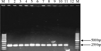
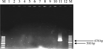
The G1 and G6 PCR primers selectively amplified the G1 and G6 genotypes of E. granulosus with specific bands of 259 (fig. 2) and 676 bp (fig. 3), respectively. The G1 primer pair did not produce any product in PCR attempts using G6 genotype DNA and vice versa.
Gel electrophoresis of the PCR products generated from the G1 genotype of Echinococcus granulosus (lanes, 1–11) on a 2% agarose gel. Lane 12, negative control; lanes M, 100 bp markers.

Gel electrophoresis of the PCR products generated from Echinococcus granulosus isolates that amplify with G1 primers, used here in PCRs with G6 primers for G6 genotype (lanes, 1–10) on a 2% agarose gel. Lane 11, E. granulosus G6 sample yields a band of about 676 bp when amplified using the G6 primers; lane 12, negative control; lanes M, 100 bp markers.

All 18 fertile cysts (7 from sheep, 1 from cattle and 10 from goats) produced identical bands with the new G1 primers. We also tried the G6 primers on all these samples but no products were observed. NCBI-BLAST searches of the public databases revealed that all ten sequences from the fertile cysts (table 1) were those of the G1 genotype.
The G6 PCR primers selectively amplified only the G6 reference sample of E. granulosus, yielding a band of 676 bp (fig. 3). Sequences from this sample (EU541210) were identical to those of the G6 genotypes in GenBank (fig. 3).
Discussion
Although DNA-based methods are useful for taxonomy at the level of genus, species and subspecies, use of such methods often requires careful attention to design of primers and preparation of pure DNA in adequate quantities (McManus & Thompson, Reference McManus and Thompson2003; Rahimi et al., Reference Rahimi, Kia, Mirhendi, Talebi, Fasihi Harandi, Jalali-zand and Rokni2007).
Mitochondrial and nuclear DNA analyses have previously been used successfully in a few molecular studies on E. granulosus in Iran. In the initial study by Zhang et al. (Reference Zhang, Eslami, Hosseini and McManus1998), the DNA sequence variation (assessed using PCR-RFLP) within regions of the mitochondrial cox1 and nad1 genes of 16 isolates of E. granulosus indicated the transmission of two strains, G1 and G6. In the study by Fasihi Harandi et al. (Reference Fasihi Harandi, Hobbs, Adams, Mobedi, Morgan-Ryan and Thompson2002), using both morphological features and molecular characterization of the ITS1 region of the rDNA, the same result was obtained. Another study also targeted the ITS1 region of the rDNA and found two common genotypes among the human, sheep and camel isolates, as above, by PCR-RFLP (Ahmadi & Dalimi, Reference Ahmadi and Dalimi2006).
In the present study, we improved DNA extraction procedures and we also designed genotype-specific primer pairs in the 12S rRNA gene, based on available sequences of E. granulosus. These indeed proved to be highly specific. Gene sequences submitted to GenBank confirmed our claim.
With these specific PCR primers, those Echinococcus genotypes which are presently assumed to have the greatest public health impact can be easily and rapidly distinguished by using as target sequence a part of the mitochondrial 12S rRNA gene. The specific PCR described here can reliably identify the E. granulosus genotypes G1 and G6. We confirmed that the G1 genotype is the most common genotype in Iranian domestic animals.
Acknowledgements
Sincere and grateful thanks are extended to Professor David Blair for his most useful and constructive comments on this work. This study was supported financially by the Research Center of Gastroenterology and Liver Disease, Shaheed Beheshti University, M.C., Tehran, Iran.