Introduction
Otosclerosis is clinically diagnosed when patients present with conductive hearing loss in the presence of a normal tympanic membrane and no previous significant middle-ear disease. Although a family history of otosclerosis and a characteristic Carhart notch on masked bone-conduction audiometry strengthens clinical suspicion, a definitive diagnosis of otosclerosis can only be established by surgery. The sensitivity of advanced imaging techniques, such as multiplanar reconstruction, has improved by over 10 per cent, leading to significantly improved diagnostic accuracy.Reference Naumann, Porcellini and Fisch 1
High-resolution computed tomography (CT) is considered the ‘gold standard’ modality for detecting otosclerosis and may aid diagnosis; this technique has a reported sensitivity of less than 95 per cent.Reference Naumann, Porcellini and Fisch 1 , Reference Virk, Singh and Lingam 2 Thin imaging slices (typically less than 1 mm) are required to detect small otosclerotic foci, although some very small foci with low density variations may still be undetectable.Reference Virk, Singh and Lingam 2 , Reference Purohit, Hermans and Op de beeck 3
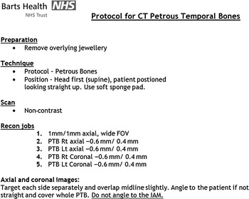
Typical imaging features included radiolucent areas in the fissula ante fenestram or hypodense areas around the otic capsule in retrofenestral disease (Figure 1).Reference Virk, Singh and Lingam 2 Imaging is also useful for excluding other middle-ear conditions that may mimic otosclerosis (e.g. congenital cholesteatoma, tympanosclerosis, ossicular discontinuity, congenital ossicular fixation and labyrinthitis ossificans). These findings are also helpful for surgical planning.Reference Swartz, Harnsberger and Mukherji 4 It is therefore essential that high-resolution CT reporting is performed accurately. The local National Health Service (NHS) Trust therefore developed a petrous temporal bone CT protocol to help ensure standardised image reporting (Figure 2).
Left axial high-resolution computed tomography images showing evidence of (a) otosclerosis at the fissula ante fenestram and (b) fenestral and retrofenestral otosclerosis.

Image showing the Barts Health NHS Trust temporal bone imaging protocol. FOV = field of view; IAM = internal auditory meatus; lt = left; PTB = petrous temporal bone; recon = reconstruction; rt = right.

Currently, more than 399 otolaryngologists in the UK subspecialise in otology (British Society of Otology spokesperson, personal communication). Undoubtedly, a reasonable number of them perform stapes surgery in district general hospitals, where specialist neuroradiologists are not available to provide clinicians with high-quality high-resolution CT reports. However, access to these reports could still be achieved via image-linking across hospital sites (particularly within the same Trust). To date, no study has measured potential differences in reporting outcomes between general radiologists and neuroradiologists.
This study aimed to compare high-resolution CT reporting for otosclerosis as performed by general radiologists and a neuroradiologist within a local NHS Trust.
Materials and methods
Study design and setting
A retrospective review of 40 high-resolution CT images of suspected otosclerosis patients obtained between 2008 and 2015 was performed within a local NHS Trust. Of the original 40 images, 4 related to suspected otosclerosis on the contralateral side (and reimaging was not performed to investigate this); therefore, 36 high-resolution CT images were available for review. All images had initially been reported on by a group of general radiologists; these were independently reviewed by a single neuroradiologist blinded to the clinical indication for imaging and the initial report findings. This study did not require ethical approval.
Main outcome measures
The primary outcome was the correct identification of otosclerosis by high-resolution CT. The secondary outcome measure was adherence to the petrous temporal bone CT protocol of the local Trust.
Statistical analysis
All statistical analyses were performed using Microsoft Office Excel 2011 (Microsoft, Redmond, Washington, USA). The association between radiology reports was calculated using a two-tailed Fisher's exact test and the relative risk was calculated. A p value of less than 0.05 was considered statistically significant.
Results
Of the 40 patients with suspected otosclerosis, the mean patient age was 40 years (range 25–69 years). Otosclerosis was surgically confirmed in 39 patients, who underwent stapedotomy plus prosthesis insertion: the other patient was found not to have otosclerosis, so did not require surgical treatment.
Of the 36 high-resolution CT images included in the study, 32 reports by general radiologists were available and all scans were reviewed by a neuroradiologist. Four findings of equivocal disease were excluded from the statistical analysis: three of these were made by general radiologists and one was by the neuroradiologist.
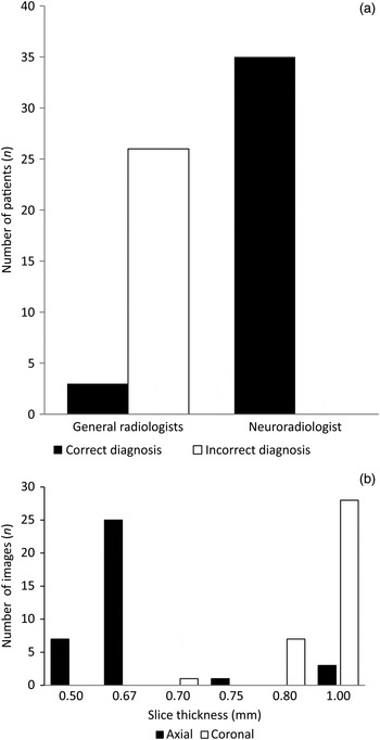
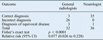
The general radiologists correctly diagnosed otosclerosis in only 3 out of 29 images (10 per cent) and the neuroradiologist correctly diagnosed otosclerosis in 35 images (100 per cent of 35; Figure 3a). Both the general radiologists and neuroradiologist correctly reported no evidence of otosclerosis in the patient without otosclerosis. A two-tailed Fisher's exact test showed a significant difference in the proportion of correct diagnoses made by the general radiologists and the neuroradiologist (p < 0.0001). The relative risk between outcomes by general radiologists and those by the neuroradiologist was 0.077 (95 per cent confidence interval, 0.026 to 0.228; Table I). None of the CT images complied with the temporal bone CT protocol (including recommendations for slice thickness). Figure 3b shows the slice thicknesses used in both axial and coronal images.
(a) Graph comparing the diagnostic outcomes for general radiologists and the neuroradiologist. (b) Graph showing the thickness of axial and coronal high-resolution computed tomography images.

Comparison of reported outcomes between general radiologists and the neuroradiologist

Discussion
Key findings
The present study shows that the correct identification rate for otosclerosis in high-resolution CT images was significantly different for general radiologists and the neuroradiologist. The relative risk calculation indicates that it is 90 per cent more likely that a correct diagnosis would be achieved if the scans were reviewed and reported exclusively by a neuroradiologist. These results are even more impressive because, unlike the general radiologists, the neuroradiologist was also blinded by the clinical indication and disease laterality.
Comparison with other studies
The present study confirmed that high-resolution CT is a sensitive, specific radiological investigation for detecting otosclerosis. For the neuroradiologist, disease detection had 100 per cent sensitivity, which is higher than in previous reports (less than 95 per cent).Reference Naumann, Porcellini and Fisch 1 , Reference Virk, Singh and Lingam 2 Despite only one patient in the present series having a negative diagnosis (i.e. no evidence of otosclerosis), the specificity was also 100 per cent, consistent with the previous reports of studies of a similar size (more than 95 per cent).Reference Lee, Wang, Lirng, Liao, Yu and Shiao 5 , Reference Lagleyre, Sorrentino, Calmels, Shin, Escude and Deguine 6 In contrast, general radiologists correctly diagnosed only 10 per cent of patients. However, false-negative high-resolution CT findings have been reported to occur secondary to otosclerotic foci of less than 1 mm, superficial foci, early sclerotic disease (where bony contours may show no irregularities) and density variations of less than 200 Hounsfield units (which do not project on imaging).Reference Virk, Singh and Lingam 2 , Reference Purohit, Hermans and Op de beeck 3 , Reference Lee, Wang, Lirng, Liao, Yu and Shiao 5 , Reference Tringali, Pouget, Bertholon, Dubreuil and Martin 7 Some of these factors may explain why detection rate was much lower for general radiologists than for the neuroradiologist.
High-resolution CT using slices less than or equal to 1 mm thick is the modality of choice for detecting otosclerosis.Reference Purohit, Hermans and Op de beeck 3 Although none of the image slices in the present study were thicker than 1 mm, none of the scans complied with Barts Health NHS Trust's petrous temporal bone CT protocol. This was probably because of poor dissemination of the protocol from the main tertiary centre (where it was developed by the Radiology Department) to relevant district general hospitals within the same local NHS Trust. The protocol recommends an axial thickness of 0.6 mm and a coronal thickness of 0.4 mm. Studies using similar parameters have shown increased detection rates of 85 per cent for fenestral disease and 44 per cent for cochlear involvement.Reference Naumann, Porcellini and Fisch 1 Over time, the diagnostic capabilities of high-resolution CT have improved significantly, and the availability of a 64- and 128-slice CT scanner within the Radiology Department means that these slice thicknesses are now readily achievable. This could improve diagnosis by general radiologists who may be less familiar with temporal bone pathology.
Clinical applicability
Adopting a good imaging protocol may help general radiology clinicians to achieve better reporting outcomes. Unfortunately, disease location was not examined in the present study, so it was not possible to stratify findings for fenestral and retrofenestral disease. It would have been interesting to compare the diagnosis rates for these sites.
Some clinicians may consider pre-operative high-resolution CT is unnecessary; however, owing to its diagnostic accuracy, this technique can help not only to confirm diagnosis before surgery but also to differentiate otosclerosis from other middle-ear disorders, thereby assisting in surgical planning and patient counselling.Reference Naumann, Porcellini and Fisch 1 In the paediatric population, where there is a greater likelihood of other congenital middle-ear disorders, confirmation of otosclerosis from imaging can be useful for counselling parents about the likelihood of surgical success. For otosclerosis, there is a 91.7–93.0 per cent success rate for achieving an air–bone gap closure of less than 10 dB.Reference Vincent, Wegner, Vonck, Bittermann, Kamalski and Grolman 8 , Reference Lippy, Burkey, Schuring and Rizer 9 This information may influence the decision to undergo surgery rather than utilise a hearing aid. Furthermore, otosclerosis proximal to the round window and cochlea can cause sensorineural hearing loss (which stapedotomy plus prosthesis insertion cannot cure) rather than conductive hearing loss (which they can). Therefore, CT reports including information on these structures may help to predict surgical outcome.Reference Purohit, Hermans and Op de beeck 3
-
• Accurate disease detection is important for diagnosis, surgical outcome, prognosis and obtaining informed consent
-
• High-resolution computed tomography is more than 95 per cent sensitive in detecting otosclerosis
-
• A neuroradiologist retrospectively reviewed high-resolution computed tomography temporal bone scans
-
• The correct otosclerosis diagnosis rate for the neuroradiologist was significantly higher than in the original reports by general radiologists
-
• Temporal bone imaging protocols are needed to standardise imaging and achieve higher disease detection rates
Limitations of the study
Although the present study showed a significant difference in the correct diagnosis rate for otosclerosis between the neuroradiologist and general radiologists, a larger study is needed to confirm these results. The main limitation of the present study was the lack of a control group (i.e. a series of normal scans). Furthermore, reports made by a group of general radiologists were compared with those made by a single neuroradiologist. This may have introduced bias because general radiologists are likely to have received different training and experiences throughout their careers, which could have contributed to inconsistent reporting. Had all reports been made by a single general radiologist, the results may have been more consistent and reliable.
Conclusion
The use of high-resolution CT for detecting otosclerotic disease is essential for diagnosis, disease staging, obtaining informed consent, surgical planning and prognosis. Use of a standardised petrous temporal bone CT protocol is recommended to provide consistently high-quality images to maximise disease detection. This study shows that use of a dedicated neuroradiologist for reporting high-resolution CT of temporal bones can improve diagnostic accuracy in patients with suspected otosclerosis.
Acknowledgements
The authors thank all staff of the Radiology Department, the Royal London, Barts Health National Health Service Trust, for their support in this study.