Introduction
The chromatin of mature mammalian sperm has a unique structure in which DNA is highly condensed in a complex with protamines that are sperm-specific proteins (Ward & Coffey, Reference Ward and Coffey1991; Ward & Zalensky, Reference Ward and Zalensky1996). In this state the DNA is transcriptionally inactive. Following penetration of the sperm into the mature oocyte, the sperm chromatin decondenses accompanied by the replacement of protamines by oocyte-supplied histones (McLay & Clarke, Reference McLay and Clarke2003). During this remodelling, profound epigenetic modification occurs in both parental nuclei (Adenot et al., Reference Adenot, Mercier, Renard and Thompson1997; Spinaci et al., Reference Spinaci, Seren and Mattioli2004).
Histone acetylation is one of the major epigenetic modifications (Bird & Wolffe, Reference Bird and Wolffe1999; Turner, Reference Turner2002). In murine (Adenot et al., Reference Adenot, Mercier, Renard and Thompson1997) and bovine (Wee et al., Reference Wee, Koo, Song, Kim, Kang, Moon, Kang, Lee and Han2006) oocytes immediately following fertilization, histone acetylation in the male nuclei precedes that of the female ones, resulting in a greater degree of histone acetylation in the male nuclei than in the female ones. Overall levels of histone acetylation are regulated by a balance between the activities of histone acetyl transferases (HATs) and deacetylases (HDACs) (Turner, Reference Turner2000). Bovine mature oocytes express these enzymes (McGraw et al., Reference McGraw, Robert, Massicotte and Sirard2003) and they are thought to function in remodelling of gamete genomes after fertilization to establish a totipotent state for normal development. Recently, Kishigami et al. (Reference Kishigami, Mizutani, Ohta, Hikichi, Thuan, Wakayama, Bui and Wakayama2006b) and Rybouchkin et al. (Reference Rybouchkin, Kato and Tsunoda2006) reported that treatment of mouse somatic cell nuclear transfer (SCNT) embryos with trichostatin A (TSA), a specific inhibitor of HDACs, improved cloning efficiency to term. This improvement is considered as a result of assisted reprogramming by TSA in terms of histone modification. In fact, TSA inhibits global deacetylation of histones that occurs in somatic cell nuclei after SCNT (Rybouchkin et al., Reference Rybouchkin, Kato and Tsunoda2006).
Herein, question arises about influences of modulating the machinery of histone acetylation during fertilization on subsequent embryonic development. In the present study, the effects of TSA during in vitro fertilization (IVF) of bovine oocytes on subsequent embryonic development were investigated.
Materials and Methods
All chemicals, except where specified otherwise, were purchased from Sigma-Aldrich.
In vitro maturation
Ovaries collected from Japanese black and F1 (Japanese Black × Holstein) cattle at a local slaughterhouse were transported to the laboratory in saline (0.85 % (w/v) NaCl). Oocytes were recovered by aspirating the follicles of 2 to 8 mm diameter with a 21-gauge needle. The cumulus-enclosed oocytes (CEOs) with compact dense cumulus cell layers were selected. Groups of 10 CEOs were matured in 50 μl drops of 25 mM HEPES-buffered TCM 199 (Earle's salts, Invitrogen) supplemented with 5% (v/v) fetal calf serum (Bio West), 0.5 mM sodium pyruvate, 0.02 AU/ml FSH (Kawasaki-Mitaka K.K.), 1 μg/ml estradiol-17β, 25 μg/ml gentamycin covered with mineral oil for 21 h at 39°C in 5% CO2 in air with high humidity.
In vitro fertilization with various concentrations of TSA
The basic procedure for IVF was performed according to the previous report (Saeki et al., Reference Saeki, Hoshi, Leibfried-Rutledge and First1990) with some modifications. Briefly, frozen–thawed Japanese black bull semen was layered on discontinuous Percoll (GE Healthcare) solution (45 and 90%) and centrifuged at 700 g for 30 min. Sedimented spermatozoa were resuspended with Brackett & Oliphant (BO) medium (Brackett & Oliphant, Reference Brackett and Oliphant1975) modified by excluding glucose and centrifuged at 700 g for 10 min. The pelleted spermatozoa were resuspended with the BO medium at concentration of 6 × 10⁶ cells/ml. Immediately before insemination, groups of 10 CEOs after IVM were transferred to 50 μl drops of the BO medium supplemented with 20 μg/ml heparin, and either with 0, 10, 100 or 1000 nM of TSA under mineral oil. TSA was at first prepared as a 1000-fold of each concentration described above in dimethyl sulphoxide (DMSO) and the stock solutions were added to the BO medium at 0.1% (v/v). Fifty Microlitres of sperm suspension was added to each drop containing CEOs, which furnished the final concentrations of spermatozoa and heparin at 3 × 10⁶ cells and 10 μg/ml, respectively. The final concentrations of TSA were 0 (control), 5, 50 and 500 nM and that of DMSO was 0.05% (v/v). CEOs and spermatozoa were co-incubated for 18 h at 39 °C in 5% CO2 in air.
In vitro culture
After IVF, presumptive zygotes were freed from cumulus cells by pipetting and cultured in a modified synthetic oviduct fluid medium with amino acids (Takahashi & First, Reference Takahashi and First1992) and further modified by excluding KH2PO4 (Kasamatsu et al., Reference Kasamatsu, Saeki, Tamari, Iwamoto, Tatemizo, Matsumoto, Hosoi and Iritani2007) until 168 hours postinsemination (hpi) at 39 °C under 5% CO2, 5% O2 and 90% N2 with high humidity. Cleavage and blastocyst development were assessed at 48 and 168 hpi, respectively.
Detection of acetylated histone by immunofluorescence
CEOs were in vitro fertilized in the absence (control) or presence of 500 nM TSA as described above. At 10 hpi, CEOs were recovered from IVF and freed from cumulus cells. Zona pellucida was removed by treatment with prewarmed pronase solution (0.5% (w/v) in PBS). Immunodetection of acetylated histone in zygotes was performed according to the method of Wee et al. (Reference Wee, Koo, Song, Kim, Kang, Moon, Kang, Lee and Han2006) with some minor modifications. Zona-free zygotes were fixed in 4% (w/v) paraformaldehyde (PFA) in PBS for 1 h at 4 °C, washed in PBS containing 0.1% Tween 20 (TPBS) for 1 h, and subsequently permeabilized with 0.5% (v/v) Triton X-100 in PBS for 2 h at 4 °C. Then, the samples were treated with blocking solution (TPBS supplemented with 1% (w/v) of bovine serum albumin) at 4 °C overnight. The antibody specific for histone H4-acetylated lysine 5 (anti-AcH4K5; 06–759, Upstate Biotechnology) was diluted 50× with blocking solution and co-incubated with samples for 3 h at 4 °C. After washing with blocking solution for 3 h, samples were incubated for 1 h in the presence of 1000× diluted Alexa Fluor 594 goat anti-rabbit IgG (A11012, Invitrogen) and then incubated with 10 μg/ml Hoechst 33342 diluted in PBS supplemented with 100 μg/ml polyvinyl alcohol (PBS–PVA) for 30 min. After washing with PBS–PVA for 30 min, samples were mounted onto a slide with a droplet of VECTASHIELD Mounting Medium (Vector Laboratories) and flattened with a coverslip. Slides were examined under a fluorescence microscope (Carl Zeiss Axiophot2) equipped with No. 2 filter (excitation G365, emission LP420) for Hoechst and No. 31 filter (excitation BP565/30, emission BP620/60) for Alexa594. Some zygotes were processed through the same immunostaining except that the primary antibody was omitted (negative control). The intensities of the nuclear immunostaining for AcH4K5 were measured using digital photography and the Image J software (National Institutes of Health). The value for the negative control was considered as background and subtracted from each measurement.
Differential staining of ICM and TE cells
Inner cell mass (ICM) and trophectoderm (TE) of blastocysts at 168 hpi were differentially stained by the method of Thouas et al. (Reference Thouas, Korfiatis, French, Jones and Trounson2001) modified by using PFA as a fixative. Briefly, zona-intact blastocysts were first incubated in 25 mM HEPES-buffered TCM-199 (Hank's salts, Invitrogen) with 1% (v/v) Triton X-100 and 100 μg/ml propidium iodide (PI) for 30 s and then immediately transferred into fixative solution of 4% (w/v) PFA in PBS with 10 μg/ml Hoechst 33342 and incubated for 30 min. After washing with PBS–PVA for 10 min, samples were mounted onto a slide and examined under a fluorescence microscope as described above using No. 2 filter for Hoechst and No. 15 filter (excitation BP546/12, emission LP590) for PI. The number of total cells was counted from the Hoechst image (blue) and the number of TE cells, which had been stained by PI as well as Hoechst, was counted from the PI image (red). The number of ICM cells was calculated by subtracting the number of TE cells from the total number of cells.
Statistical analysis
Data for the developmental rates and cell numbers were statistically analysed by multiple comparisons with the Holm method (Holm, Reference Holm1979). Data for the AcH4K5 immunostaining were analysed by t-test.
Results
Histone acetylation in zygotes during IVF with or without TSA
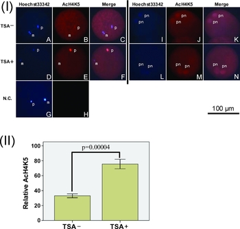
In the first experiment, bovine oocytes were fertilized with 0 or 500 nM TSA and the status of histone acetylation in parental genomes at 10 hpi was assessed by indirect immunostaining using anti-AcH4K5 antibody. Acetylation of histone H4 at K5 reflects its hyperacetylated state (Mizzen & Allis, Reference Mizzen and Allis1998; Turner, Reference Turner1998). When we examined the nuclear status of the zygotes at 10 hpi, 39% (9/23) of oocytes in the control group and 41% (14/34) in the TSA group showed the stage of enlarged sperm head (ESH). Thirty per cent (7 of 23) in the control and 32% (11 of 34) in the TSA group showed two pronuclei (2PN), which were probably female and male pronuclei. Four per cent (1 of 23) and 6% (2 of 34) were polyspermic and the rest (26 and 21%) were unfertilized oocytes in the control and TSA group, respectively. These developmental stages were similar to those reported earlier (Saeki et al., Reference Saeki, Kato, Hosoi, Miyake, Utsumi and Iritani1991). The levels of immunoreactive AcH4K5 in paternal genome at the ESH stage were significantly (p < 0.0001) higher in the TSA group than in the control (Fig. 1 (I) a–f and (II)). Negative control embryos without primary antibodies were not stained (Fig. 1 (I) g, h). At the pronuclear stage, acetylation signals were detected, but the intensity of the signals was not different between control and TSA-treated zygotes and within two pronuclei in the same ooplasm (Fig. 1 (I) i–n).
(I) Representative images of acetylation of histone H4 lysine 5 (AcH4K5) of IVF zygotes with or without TSA (500 nM). Polar bodies had been lost during staining procedure. TSA–: zygotes fertilized without TSA, TSA+: zygotes fertilized with TSA, N.C.: negative control in which the primary antibody was omitted from the staining procedure. (A–H): stage of enlarged sperm head. (I–N): pronuclear stage. m: maternal nuclei, p: paternal nuclei, pn: pronuclei. (II) The relative levels of AcH4K5 immunostaining at enlarged sperm head in the control (TSA–) and the TSA group (TSA+). Data are presented as means ± standard error obtained from nine (control) and 14 (TSA+) zygotes.

Effects of TSA during IVF on embryo development
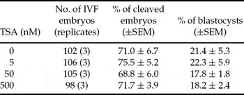
In the next experiment, IVF zygotes treated with 0, 5, 50 or 500 nM TSA were cultured to the blastocyst stage. TSA treatment during IVF did not affect the cleavage and blastocyst development after IVF (p > 0.05, Table 1).
Effects of TSA during in vitro fertilization (IVF) on postfertilization cleavage and blastocyst development.

No significant difference among the treatments. SEM, standard error of the mean.
Effects of TSA during IVF on cell number and ICM/TE allocation of embryos
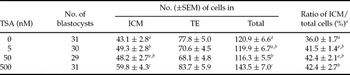
We then examined the cell number of ICM and TE of the blastocysts treated with or without TSA. TSA treatment enhanced the cell number of ICM at 5 and 500 nM (p < 0.05, Table 2). However, TSA did not increase the number of TE cells (p > 0.05). Consequently, TSA treatment altered cell allocation within blastocysts, i.e. ratios of ICM/total cells increased at 500 nM (p < 0.05).
Effects of TSA during IVF on allocation of cells in bovine blastocysts to ICM and TE.

a–cValues in the same column without common superscripts differ significantly (p < 0.05). SEM, standard error of the mean.
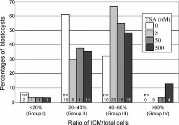
Figure 2 shows the distribution of blastocysts with or without TSA treatment according to the ratio of ICM/total cells classified into four groups (group I, <20%; group II, 20–40%; group III, 40–60%; group IV, > 60%) according to the classification by Koo et al. (Reference Koo, Kang, Choi, Park, Kim, Oh, Son, Park, Lee and Han2002). The majority of embryos in the control group (0 nM TSA) were categorized in group II, whereas in TSA groups the majority was distributed in group III.
Distribution of blastocysts derived from IVF with various concentrations of TSA according to ratio of ICM/total cells

Discussion
The DNA of mature mammalian spermatozoa are highly condensed and are associated with sperm-specific nuclear proteins called protamines. After fertilization, sperm chromatin undergoes decondensation accompanied by the replacement of protamines with oocyte-supplied histones (Perreault, Reference Perreault1992). During this period, histone H4 in the paternal nucleus is hyperacetylated compared with the female nucleus (McLay & Clarke, Reference McLay and Clarke2003; Wee et al., Reference Wee, Koo, Song, Kim, Kang, Moon, Kang, Lee and Han2006), due to a selective mechanism of rapid histone assembly from ooplasm to paternal chromatin (Spinaci et al., Reference Spinaci, Seren and Mattioli2004; Maalouf et al., Reference Maalouf, Alberio and Campbell2008). In agreement with this consensus, our results showed that AcH4K5 signals assembled at paternal nuclei while maternal chromatin remained inert on AcH4K5 assembly in the control zygotes at the stage of ESH (Fig. 1). Figure 1 clearly indicates that TSA treatment during IVF enhanced the level of AcH4K5 only in paternal nuclei, but the treatment had no observable effect on the AcH4K5 signal in maternal chromatin. These results indicate that TSA, an inhibitor of HDACs, enhanced histone acetylation status during IVF.
TSA treatment during IVF did not affect the rates of cleavage or blastocyst development (Table 1) but altered the cell number and cell allocation to ICM and TE (Table 2). TSA enhanced the number of ICM cells and resulted in enhanced ratios of ICM/total cells. Similarly, Koo et al. (Reference Koo, Kang, Choi, Park, Kim, Oh, Son, Park, Lee and Han2002) found an elevated ratio (50.1%) of the ICM/total cells in SCNT-derived bovine blastocysts compared with IVF-derived (42.6%) or artificial insemination-derived blastocysts (34.9%). They classified the bovine blastocysts into four groups (group I, <20%; group II, 20–40%; group III, 40–60%; group IV, > 60%) according to the ratio of ICM/total cells. Most SCNT blastocysts were in group III (40–60%) and IV (>60%), whereas most IVF and in vivo-derived blastocysts were in group II (20–40%). They concluded that group II embryos appear to be normal. Too high (Kang et al., Reference Kang, Park, Koo, Choi, Kim, Lee and Han2002; Koo et al., Reference Koo, Kang, Choi, Park, Kim, Oh, Son, Park, Lee and Han2002) or too low (Brison & Schultz, Reference Brison and Schultz1997; Tarin et al., Reference Tarin, Perez-Albala, Gomez-Piquer, Hermenegildo and Cano2002) ratio of ICM/total cells in blastocysts have been considered as abnormality of embryos. Therefore, higher ratio of ICM/total cells in blastocyst derived from IVF with TSA treatment (5–500 nM) in the present study may reflect the abnormalities of these embryos.
Histone acetylation is one of the major mechanisms of epigenetic reprogramming of gamete genomes to establish a totipotent and at the same time imprinted state for normal development (McLay & Clarke, Reference McLay and Clarke2003; Wee et al., Reference Wee, Koo, Song, Kim, Kang, Moon, Kang, Lee and Han2006). The histone acetylation status at specific amino acid residues determines synergistic or antagonistic interaction affinities for chromatin-associated proteins and thereby determines whether the chromatin is transcriptionally active (Turner, Reference Turner1998). There are also likely conjugated relationships between two epigenetic modalities, i.e. DNA methylation result in histone deacetylation and histone acetylation direct DNA demethylation (Cervoni & Szyf, Reference Cervoni and Szyf2001). Furthermore, and interestingly, histone acetylation may be transmitted to daughter cells as an epigenetic ‘histone code’ (Ekwall et al., Reference Ekwall, Olsson, Turner, Cranston and Allshire1997; Jenuwein & Allis, Reference Jenuwein and Allis2001). Recently, Kishigami et al. (Reference Kishigami, Mizutani, Ohta, Hikichi, Thuan, Wakayama, Bui and Wakayama2006b) and Rybouchkin et al. (Reference Rybouchkin, Kato and Tsunoda2006) reported that TSA treatment of mouse SCNT embryos improved cloning efficiency. TSA treatment can enhance histone acetylation in somatic cell nuclei after SCNT (Rybouchkin et al., Reference Rybouchkin, Kato and Tsunoda2006) and ameliorate the deficient DNA demethylation of paternal genome after round spermatid injection (ROSI) (Kishigami et al., Reference Kishigami, Thuan, Hikichi, Ohta, Wakayama, Mizutani and Wakayama2006a). Thus, the favorable effects of TSA in nuclear transfer technique may be attributed to an assisted reprogramming involving histone acetylation and DNA demethylation of transferred nuclei after SCNT and ROSI. In the case of IVF, however, our present results indicated that TSA treatment has negative effects which result in abnormal cell allocation at the blastocyst stage after IVF. Gioia et al. (Reference Gioia, Barboni, Turriani, Capacchietti, Pistilli, Berardinelli and Mattioli2005) examined effects of TSA (330 nM) during IVF of porcine oocytes and demonstrated that TSA treatment eliminated an asymmetrical mode of histone H4 acetylation between male and female pronuclei observed in porcine oocytes. However, this report did not address the subsequent development and cell proliferation of resulting embryos.
Torres-Padilla et al. (Reference Torres-Padilla, Parfitt, Kouzarides and Zernicka-Goetz2007) demonstrated that manipulating epigenetic modification of histones can influence cell fate of blastocysts. They found that murine 4-cell blastomeres with higher methylation level of specific arginine residues of H3 (H3R26) are destined to contribute predominantly to the ICM. The increase in ICM cell number in TSA-treated embryos in the present study may also be attributed to the alternative histone modification (AcH4K5). Apart from expected experimental difficulty due to the very small number of genomes in early embryos, further studies concerning the status of specific gene-associated H4K5 will be needed to elucidate mechanisms underlying the relationship between the modulation of AcH4K5 and embryonic cell differentiation. In addition, evaluating the outcome of TSA-treated embryos such as failure rate after embryo transfer should also be pursued. The possibility that TSA treatment induces better pregnancy or calving rate as in the case of SCNT (Kishigami et al., Reference Kishigami, Mizutani, Ohta, Hikichi, Thuan, Wakayama, Bui and Wakayama2006b) cannot be excluded.
In conclusion, TSA treatment during IVF of bovine oocytes alters the cell number and their allocation to ICM and TE, even though the blastocyst development with TSA was similar to that without TSA. Overriding epigenetic modification of the genome during fertilization may have a carryover effect on cell proliferation and differentiation in preimplantation embryos through an epigenetic histone code.
Acknowledgements
This study was supported by a grant from Wakayama Prefecture Collaboration of Regional Entities for the Advancement of Technological Excellence, JST.