Introduction
Head and neck squamous cell carcinomas (SCCs) represent a category of aggressive, chemoresistant malignancies which are increasing in frequency worldwide.Reference Greenlee, Murray, Bolden and Wingo1 These tumours arise from epithelia of different anatomical origin in the upper aero-digestive tract, such as the oral cavity (including tongue), pharynx and larynx.Reference Vokes, Weichselbaum, Lippman and Hong2 Neoplastic transformation of such epithelia is mediated by deregulation of crucial molecular pathways. During carcinogenesis, normal epithelia accumulate a variety of genetic alterations due to viral infections or exposure to carcinogens such as tobacco and alcohol.Reference Do, Johnson, Lee, Wu, Dong and Hong3, Reference Viswanathan and Wilson4 Hyperplasia, metaplasia, dysplasia, carcinoma in situ and, finally, invasive carcinoma are the representative stages in the progression of SCC, including those due to specific chromosomal instabilities.Reference Veltman, Bot, Huynen, Ramaekers, Manni and Hopman5
Apoptosis is a process of programmed cell death which regulates tissue homeostasis even during embryonic life. Imbalances in apoptotic cell death genes, combined with telomerase over-expression and alterations of the cell life cycle, lead to progressive loss of tissue homeostasis, reflecting the inability of cells to respond to normal apoptotic cell death signals.Reference Maellaro, Pacenti, Del Bello, Valentini, Mangiavacchi and De Felice6, Reference Goldkorn and Blackburn7 Down-regulation of apoptotic proteins (such as caspases), combined with up-regulation of anti-apoptotic factors (such as bcl2, bcl x and survivin), is responsible for excessive proliferation, inhibition of apoptosis and chemoresistance in cancers of various origins.Reference Rhee, Khuri and Shin8, Reference Zhang, Zhang, Zhou, Qiu, Chen and Chen9
Caspases represent a family of sulphydryl proteases, including 14 main members. The majority of these act as positive regulators in the apoptotic mechanism, due to their cleavage.Reference Fadeel and Orrenius10, Reference Patel, Gores and Kaufmann11 A cascade of interactions involving caspases 3, 7, 8 and 9 leads to apoptotic cell death.Reference Medema, Scaffidi and Kischel12–Reference Okada and Mak14 Caspase-8 (gene locus 2q33–34), also known as FADD-like IL-1β-converting enzyme (FLICE), comprises an N-terminal domain with sequence homology to the Fas-associated death domain protein (FADD) death effector's domain which allows association of caspase-8 with the tumour necrosis factor – Fas (TNF receptor superfamily member 6) family of receptors.Reference Vakkala, Pääkkö and Soini15 This association with cell surface death receptors indicates that caspase-8 is a proximal regulator of apoptosis. Caspase-8 is activated by association with the FADD–FAS death-induction signalling complex and releases two active subunits into the cytosol, p18 and p10.Reference Muzio, Stockwell, Stennicke, Salvesen and Dixit16 Caspase-8 activates other caspases, especially caspase-3 (gene locus 14q21), amplifying the apoptotic signal.Reference Chou, Jones and Heinrikson17
In this study, we co-evaluated protein expression of caspase-3 and -8 in tongue SCC, in order to identify potential correlations with clinicopathological parameters. To our knowledge, this is the first study of these markers within tongue SCC, based on immunohistochemical evaluation via computerised image analysis.
Materials and methods
Study group
We obtained 87 archival, paraffin-embedded tissue samples of histologically confirmed primary tongue SCC. These were derived from patients who had undergone local or extended (i.e. combined with lymphadenectomy) tumour excision, following an initial diagnostic biopsy between 1998 and 2006 at the oral and maxillofacial surgery department of St Savas Anticancer Hospital, Athens, Greece. Fifty-eight samples were derived from male patients (mean age 58.5 years) and 29 from female patients (mean age 62.5 years). The patients' survival period had ranged from 16 to 177 months (mean follow-up period, 97 months). Ten areas of apparently benign epithelia, adjacent to malignant tissue, were used as a normal control group. None of the cases demonstrated a familial history of cancer or other inheritable cancer syndromes, so all were characterised as sporadic.
The local ethical committee consented to the use of these tissues for research purposes within the head and neck surgery department.
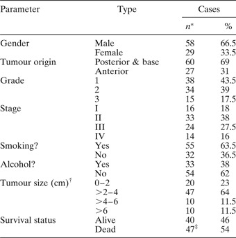
The tissue samples were fixed in 10 per cent neutral-buffered formalin. Haematoxylin and eosin stained slides of the samples were reviewed for confirmation of histopathological diagnosis. All carcinomas were graded and staged according to the histological classification criteria of the World Health Organization and the tumour–node–metastasis system for head and neck cancers. Clinicopathological data for the examined cases are shown in Table I.
Clinicopathological data for examined tongue SCC cases

* Total = 87. †Maximum diameter. ‡34 died of cancer. SCC = squamous cell carcinoma
Antibodies and immunohistochemistry
We selected and applied monoclonal antibodies, including anti-caspase-3 (3CSP03; Neomarkers, Neomarkers/LabVision, Fremont, CA, USA) at a dilution of 1:50 and anti-caspase-8 (11B6; Novocastra, Novocastra Lab, Newcastle, UK, USA) at a dilution of 1:20. These antibodies recognised N-terminal regions of the corresponding proteins.
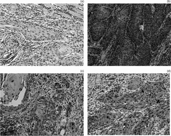
The immunohistochemistry protocol for those antigens was carried out on 3-μm thick paraffin sections of the corresponding sample blocks. Two tissue sections, initially deparaffinised in xylene and rehydrated via graded ethanol, were immunostained for each of the applied markers, using the EN Vision+ (DAKO, Glostrup, Denmark) assay and an automated staining system (I 6000; Biogenex, San Ramon, CA, USA), according to the corresponding manufacturer's instructions. This specific assay was based on a soluble, dextran-polymer system preventing endogenous biotin reaction and increasing the quality of stained slides. Briefly, after peroxidase blocking, the sections were incubated with primary antibody for 30 to 40 minutes (depending on the corresponding antibody) at room temperature and then incubated with horseradish peroxidase labelled polymer for 30 minutes. The antigen–antibody reaction was visualised using 3-3,diaminobenzidine tetrahydrochloride as a chromogen substrate. Finally, these tissue sections were slightly counterstained with haematoxylin for 30 seconds, dehydrated and mounted. For negative control slides, the primary antibodies were omitted. Predominant diffuse cytoplasmic staining and nuclear staining patterns were considered acceptable for caspase-3 and caspase-8, respectively (Figure 1).
Photomicrographs showing different patterns of caspase-3 and -8 protein expression. (a) Loss of caspase-3 expression. (b) Caspase-8 over-expression (i.e. high level). (c) Moderate expression of caspase-3. (d) Low expression of caspase-3. (3-3 diaminobenzidine tetrahydrocloride-DAB stain, original magnification 200×)

Computerised image analysis
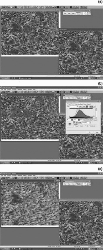
In order to evaluate the immunohistochemistry results quickly and accurately, computerised image analysis was performed using a semi-automated system. The following hardware features were used: Intel Pentium IV, Matrox II Card Frame Grabber, digital camera microwave systems (800×600) and an Olympus BX-50 microscope, Olympus, Menville, NY, USA. In addition, the following software features were used: Windows XP/Image Pro Plus, version 3.0-Media Cybernetics Media Cybernetics Inc, Bethesda, MD, USA, 1997. Measurements of caspase-3 and -8 staining intensity were performed in five optical fields per case, at a magnification of ×400 (Figure 2).
Evaluation of caspase-3 protein expression in a case of tongue squamous cell carcinoma. Snapping an image (a), digitizing and scanning it (b) and finally measuring the protein expression levels (c) represent the stages of the procedure (original magnification 200×)

Using normal epithelial samples as a control group and comparing them to the analysed tumours, we characterised staining intensity levels as high (0–120), moderate (121–160) or low (161–255). The interpretation of staining intensity values (range 0-255 gray scale levels) is demonstrated in Table II.
Results of IHC analysis and correlations with tongue SCC parameters

p values shown represent Spearmen test and Cox regression analysis for survival status.
Kappa (caspase-3 vs caspase-8) = 0.713 (0.704–0.721, 95% confidence intervals). *Total = 87. IHC = immunohistochemistry; SCC = squamous cell carcinoma; T = tumour; O = over-expression (i.e. moderate and high staining intensity; see text for details); L/N = low or negative expression
Statistical analysis
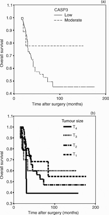
Associations between variables, including protein expression levels and clinicopathological parameters, were analysed using the Spearman test, estimated along with 95 per cent confidence interval (using Statistical Package for the Social Sciences (SPSS Inc, Chicago, Illinois, USA) version 11.0 software packages). Two-tailed p values < 0.05 were considered to be statistically significant. Cohen's inter-rater kappa was also estimated along with its 95 per cent confidence interval, in order to evaluate concordances between the examined proteins. By its definition, a kappa value of one denotes complete agreement, values of more than 0.75 are characterised as excellent agreement, values between 0.40 and 0.75 show fair to good agreement, values between zero and 0.40 show poor agreement, and a value of zero indicates that the observed agreement is equal to chance. Furthermore, Cox regression analysis (Breslow method for ties) was performed and Kaplan–Meier curves were also calculated (Figure 3). Total immunohistochemistry results and p values are shown in Table II.
Kaplan–Meier curves for overall patient survival versus (a) caspase-3 (CASP3) protein expression and (b) tumour (T) size. Cox regression analysis demonstrated that tumour size was a prognostic factor for survival in cases of tongue squamous cell carcinoma (p = 0.024).

Results
According to the image analysis process and the definition of sample groups as high, moderate or low staining intensity, high levels of caspase-8 protein expression were observed in 20/87 (23 per cent) samples, whereas no caspase-3 over-expession was observed in any of the examined samples. Moderate levels of caspase-8 and -3 protein expression were observed in 37/87 (42.5 per cent) and eight of 87 (9.5 per cent) samples, respectively. Reduced or absent caspase-8 and -3 expression was identified in 30/87 (34.5 per cent) and 79/87 (90.5 per cent) samples, respectively. Mean values for caspase-8 and -3 protein expression were significantly different (145 vs 184, respectively). Evaluating the overall expression of the examined markers, we observed that 26/87 (35.6 per cent) samples were characterised by simultaneous reduced expression, five of 87 (5.7 per cent) by simultaneous over-expression, and 46/87 (52.8 per cent) by different patterns of expression. Kappa analysis of caspase-3 and -8 expression showed a medium value (kappa = 0.713; 95 per cent CI 0.704–0.721). We observed no statistically significance correlation between caspase-8 and -3 protein expression and patients' clinicopathological parameters, nor between the overall values of the two markers (p = 0.882). Cox regression analysis showed no prognostic significance for the association between overall protein expression of either marker and patient survival probability (p = 0.174 for caspase-3; p = 0.608 for caspase-8).
Interestingly, the size of the examined tumours was strongly correlated with patients' survival status (p = 0.024), whereas the tumour stage demonstrated only borderline statistical significance (p = 0.091).
Discussion
Deregulation of apoptotic molecules (including cytoplasmic caspases, the mitochondrial-dependent factors bcl2 and bax, and survivin), is responsible for the development of chemoresistance in tongue SCCs, but not all of these agents affect the clinical prognosis.Reference de Vicente, Olay, Lequerica-Fernandez, Sanchez-Mayoral, Junquera and Fresno18 In the current study, we found that caspase-3, especially, and also caspase-8 were down-regulated in tongue SCC. We also identified simultaneous deregulation of those proteins in a significant fraction of the examined tumours. By analysing caspase-3 and -8 protein expression levels using computerised image analysis, we found different expression patterns of these factors in tongue SCC.
Some recently published studies have focused on the role of caspase-3 down-regulation in tongue SCC. Using new, in vitro agents that act as positive regulators of the apoptotic pathway, these studies found that treatment with staurosporine induced the apoptotic mechanism by activating the cascade of caspases, especially protein expression of caspase-3.Reference Zhang, Yu, Ou-Yang, Xia, Wang and Li19 Staurosporine inhibited the activity of protein kinase C alpha by reducing its membranous expression, and also acted as a negative regulator of survivin, an anti-apoptotic protein. This is an important observation because mutations of PIK3CA represent oncogenic aberrations at advanced stages of oral SCC.Reference Kozaki, Imoto, Pimkhaokham, Hasegawa, Tsuda and Omura20 Similarly, another study showed that treatment with retinoids led to increased apoptotic rates by activating the caspase-8 and -3 signalling pathway, combined with up-regulation of bax expression and down-regulation of bcl2 expression.Reference Niu, Feng, Peng, Ma, Liu and Yuan21 Another study, assessing systemic chemotherapy strategies in the treatment of cancers of different origin, concluded that 5-fluorouracil induced apoptosis was mediated by activation of a caspase-dependent pathway, especially involving caspase-1, -3 and -8.Reference Ohtani, Hatori, Ito, Takizawa, Kamijo and Nagumo22 The role of arsenic trioxide, which acts as an anti-proliferative agent in oral SCCs, has been also investigated. Tongue SCC cell cultures were used as an in vitro substrate for arsenic trioxide treatment, and this agent was observed to induce apoptosis by activating the caspase-3 proteolytic pathway; in addition, tubulins and mitochondria membrane were found to be the main targets of arsenic trioxide action.Reference Guo and Zhang23
Our analysis did not establish prognostic significance for the correlation between patients' survival status and caspase-3 and -8 protein expression levels (in contrast to the strong correlation observed between tumour size and patient survival rate). However, decreased caspase-3 expression appeared to be a crucial event in the majority of the tongue SCC samples examined. This observation is supported by another important study that investigated the potential correlation between caspase-3 activity and head and neck cancer treatment. These authors used a combination of antisense oligonucleotides and conventional chemotherapeutic agents (such as cisplatin and etoposide) in tongue SCC cell cultures, and they observed that protein expression of anti-apoptotic factors such as bcl2 and survivin was decreased, whereas caspase-3 activity was increased.Reference Sharma, Sen, Lo Muzio, Mariggio and Singh24 In multiple trials and studies of patients with locally and regionally advanced tumours, the combination of cisplatin and fluorouracil and also paclitaxel as an adjuvant treatment for head and neck SCC has produced encouraging results regarding survival benefits and clinical response.Reference Pignon, Bourhis and Domenge25, Reference Vokes, Stenson and Rosen26 Based on these results, caspase-3 deregulation is thought to be significantly involved in the response to these agents, because it is responsible for decreased apoptotic rates and for this reason is correlated with tumour viability and resistance to cell death signals.
Recently, there have also been some interesting publications regarding the potential anticancer effects of polyphenols, found in green and black tea.Reference Babich, Zuckerbraun and Weinerman27, Reference Mohan, Gunasekaran, Varalakshmi, Hara and Nagini28 These reports have suggested that there is a caspase-independent apoptotic pathway in human oral cavity cells, and also that tea polyphenols mediate the apoptotic mechanism by generation of reactive oxygen species, decreasing the bcl2/bax ratio, and also by consequent activation of caspase-3.
• This study evaluated the role of caspase-3 and -8 expression in tongue squamous cell carcinoma (SCC), using a combination of immunohistochemistry and image analysis
• The findings support the theory that simultaneous deregulation of caspase-8 and -3 is a frequent event in tongue SCC
• The authors conclude that although caspase-3 and -8 expression has no prognostic significance, activation of caspase-3, which is predominantly down-regulated in these tumours, may be a crucial process for induction of apoptosis and response to therapeutic strategies
In addition, two studies of complementary deoxyribonucleic acidsReference Yasumoto, Kirita, Takahashi, Ohnishi, Imai and Yuki29, Reference Yasumoto, Imai, Takahashi, Ohnishi, Yuki and Kirita30 have analysed the potential involvement of heat treatment (hyperthermia) and X-ray irradiation, respectively, in the regulation of apoptotic death in SCCs. Results indicate that p53 status appears to be a useful candidate for predictive indicator of the effectiveness of hyperthermic therapy, combined with caspase-3 and -9 activation and induction of apoptosis. Furthermore, X-ray irradiation was found to induce p53-dependent apoptosis via caspase-3, -8, -9 and -10 activation.
Conclusions
Simultaneous deregulation of caspase-8 and -3 protein expression is a frequent event in tongue SCCs, but it does not correlate strongly with the prognosis of the patient. In contrast, tumour size does appear to be a prognostic factor in these patients. Activation of caspase-3, which is significantly down-regulated in these tumours, appears to be the crucial process for induction of apoptosis and response to chemotherapeutic strategies.
Acknowledgments
The authors are very greatful to M Arvamis, MD, for the statistical analysis.