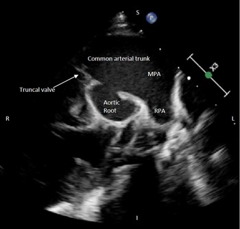
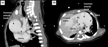
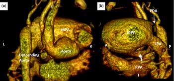
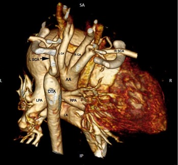
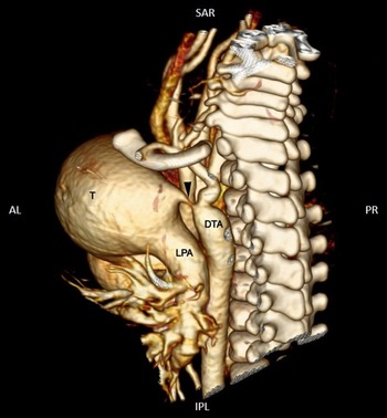
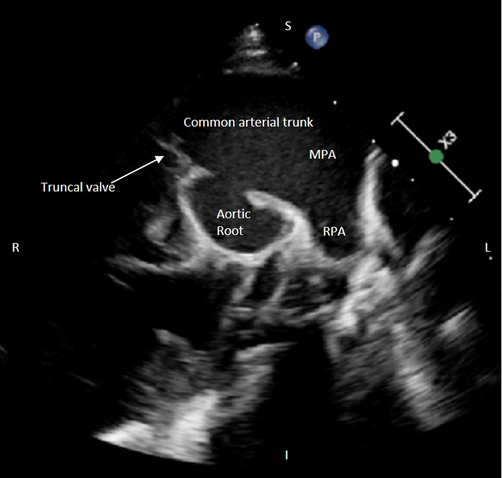
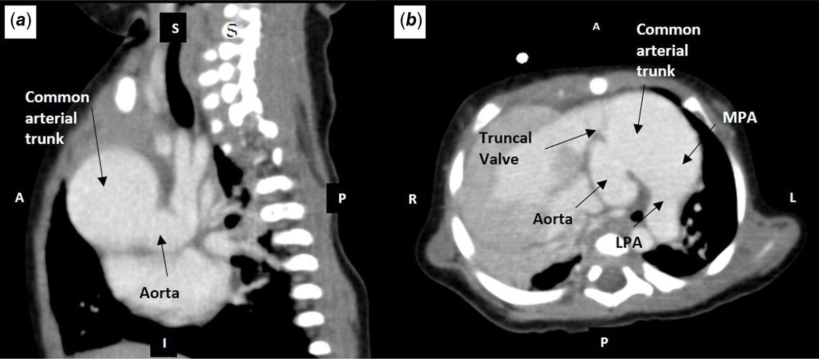
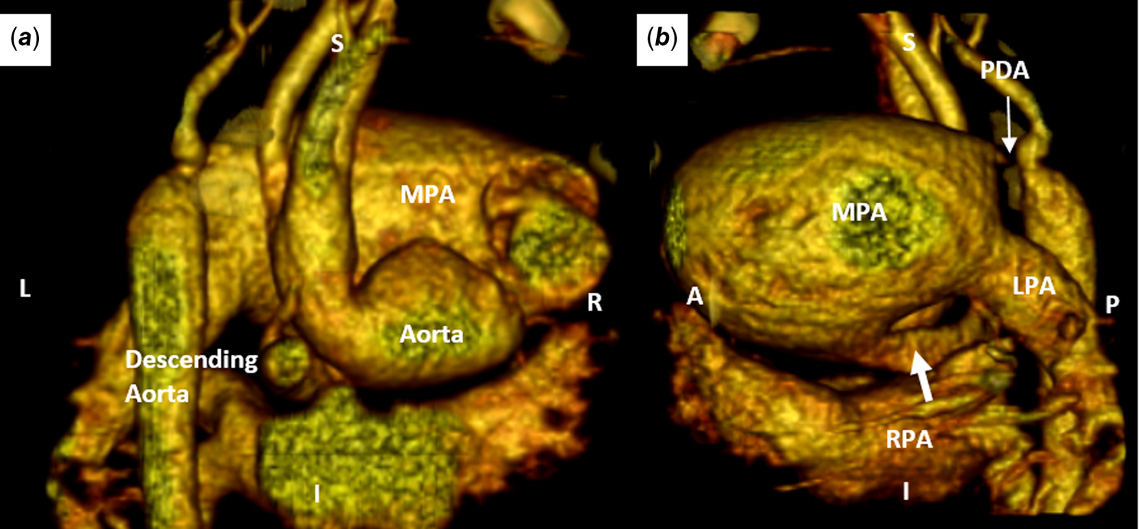
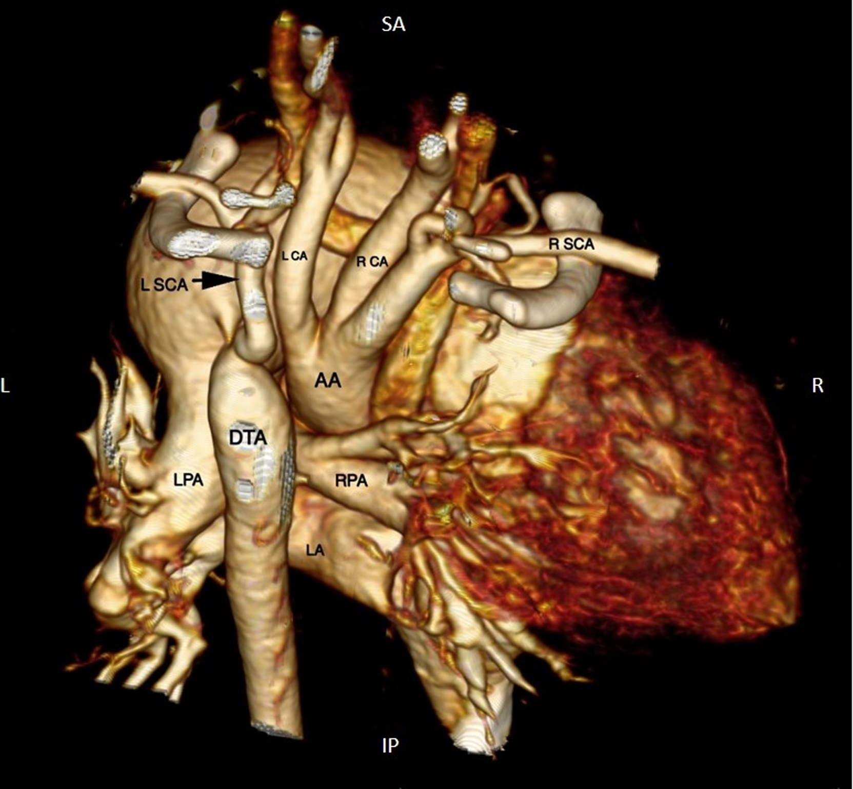
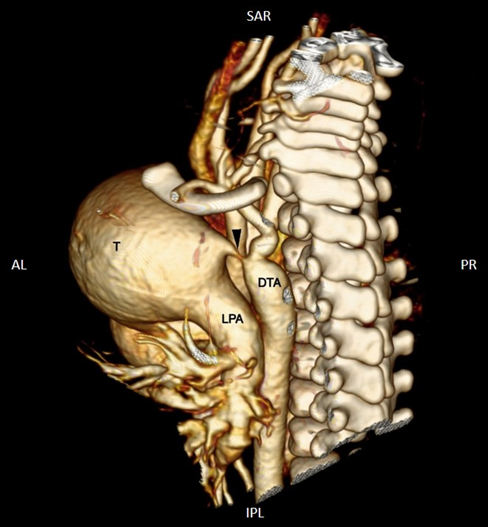
Truncus arteriosus or common arterial trunk is one of the cyanotic CHDs. Usually, the common arterial trunk arises from the base of the heart which continues as the aorta and the main pulmonary artery or its branches arise from the main trunk. Reference Gupta, Aggarwal and Shaw1 We report an unusual variation where the common arterial trunk continued as the main pulmonary artery and aorta arising from the trunk in a 3-month-old infant. He had central cyanosis with bounding right upper limb pulsation and feeble pulsations in the left arm and bilateral lower limbs. His echocardiography revealed situs solitus and dextrocardia with a common arterial trunk continuing as the main pulmonary artery with a large subtruncal ventricular septal defect (Supplementary Movie 1–4, Fig 1). CT showed a common arterial trunk continuing as the main pulmonary artery and bifurcating into the right pulmonary artery and left pulmonary artery (Figs 2 and 3, Panel A and B). The ascending aorta was seen arising from the posteroinferior surface of the common arterial trunk and giving origin to the coronary arteries (Fig 3, Panel A). The aortic arch was left-sided with ascending aorta coursing posterior and cranially giving origin to arch vessels (Fig 4). There was type B interruption of the aortic arch (Fig 5). The Left subclavian artery was seen arising from the proximal descending aorta. Small PDA was arising from the common arterial trunk supplying the descending aorta. A subset of a common arterial trunk with pulmonary arterial dominance is known to be associated with more complexity, specifically related to aortic arch anomalies and branch pulmonary artery origin. Surgically, cases with pulmonary dominance are more challenging considering associated anomalies and complexity.
Echo showing the aorta arising as a side branch from the common arterial trunk and the common arterial trunk continuing as MPA. MPA: Main pulmonary artery, RPA: right pulmonary artery, LPA: Left pulmonary artery.
CT pulmonary angiogram image of common arterial trunk: panels A and B) showing ascending aorta arising posterio-inferiorly from the common arterial trunk, MPA: main pulmonary artery, LPA: left pulmonary artery.
CT pulmonary angiogram with 3D volume-rendered CT image of common arterial trunk: panels A and B) showing ascending aorta arising posterio-inferiorly from the common arterial trunk.
CT pulmonary angiogram with 3D volume-rendered CT image of common arterial trunk: showing type B interruption of the aortic arch, AA: Ascending aorta, R SCA: Right subclavian artery, R CA: Right common carotid artery, L CA: Left common carotid artery, L SCA: Left subclavian artery, DTA: Descending thoracic aorta, LA: Left atrium.
CT pulmonary angiogram with 3D volume-rendered CT image of common arterial trunk: showing small PDA shown by a black arrow, T: common arterial trunk continuing as MPA.
Supplementary material
To view supplementary material for this article, please visit https://doi.org/10.1017/S1047951122004231
Acknowledgements
None.
Financial support
This research received no specific grant from any funding agency, commercial, or not-for-profit sectors.
Conflicts of interest
None.