Introduction
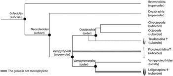
The Cephalopoda Cuvier, 1797 is a large class of molluscs, representing more than 700 species, divided into two extant subclasses: the Nautiloidea Agassiz, 1847 and the Coleoidea Bather, 1888 (Sweeney and Roper, 1998), and one extinct subclass, the Ammonoidea Zittel, 884. Coleoid cephalopods differ from nautiloids mostly through the internalization (or complete loss) of the shell (Teichert, Reference Teichert1988). Within the cohort Neocoleoidea Haas, Reference Haas1997 the shell is often called the gladius and consists of a sturdy but flexible chitinous structure within the dorsal mantle (Fuchs and Weis, Reference Fuchs and Weis2009). The cohort Neocoleoidea is subdivided into two superorders according to the number of arms: the Vampyropoda Boletzky, Reference Boletzky1992 (eight-armed coleoids), presently represented by octopods and vampire squids, and the Decabrachia Boettger, Reference Boettger1952 (ten-armed coleoids), represented by squids, cuttlefishes, and their relatives (Fig. 1). The evolutionary history of modern decabrachians and vampyropods is currently highly debated (Fuchs et al., Reference Fuchs, Keupp and Engeser2003, Reference Fuchs, Keupp and Wiese2012; Haas, Reference Haas2003; Fuchs, Reference Fuchs2006a; Fuchs and Larson, Reference Fuchs and Larson2011a, Reference Fuchs and Larson2011b; Arkhipkin et al., Reference Arkhipkin, Bizikov and Fuchs2012), as is the status of the clade Neocoleoidea (Fuchs et al., Reference Fuchs, Boletzky and Tischlinger2010b). The first unequivocal evidence of vampyropod coleoids comes from the Upper Cretaceous Lebanese Lagerstätten that yielded taxa such as Palaeoctopus newboldi Woodward, Reference Woodward1896 and Keuppia levante Fuchs et al., Reference Fuchs, Bracchi and Weis2009. However, based on molecular clock analyses, Kröger et al. (Reference Kröger, Vinther and Fuchs2011) suggested a Permian divergence (~276 Ma) between the superorders Vampyropoda and Decabrachia. This estimated divergence date is thus considerably older than paleontological estimates based on scarce, but well-preserved fossil coleoids. The fossil record of these essentially soft-bodied organisms thus provides little information on the origin and phylogenetic relationships of the extant clades (Strugnell et al., Reference Strugnell, Norman, Jackson, Drummond and Cooper2005, Reference Strugnell, Jackson, Drummond and Cooper2006).
Phylogenetic hypothesis for coleoids considering that Mesozoic gladius-bearing coleoids (in grey and bold) are related to the eight-armed coleoids (superorder Vampyropoda). In a Decabrachian hypothesis, they would be placed in the superorder Decabrachia. Phylogenetic hypothesis modified after the Tree of Life website (http://www.tolweb.org) and after Fuchs (Reference Fuchs2009) and Fuchs and Larson (Reference Fuchs and Larson2011a, Reference Fuchs and Larson2011b). Sketches after Fuchs (Reference Fuchs2009) and Fuchs and Larson (Reference Fuchs and Larson2011a, Reference Fuchs and Larson2011b).

To better constrain the origin and divergence of modern decabrachians and vampyropods, a few paleontologists are currently working on Mesozoic gladius-bearing coleoids. On the one hand, some authors suggest vampyropod affinities for Mesozoic gladius-bearing coleoids that apparently never exhibit more than eight arms (Fuchs and Larson, Reference Fuchs and Larson2011a, Reference Fuchs and Larson2011b). On the other hand, Young et al. (Reference Young, Vecchione and Donovan1998), Vecchione et al. (Reference Vecchione, Young, Donovan and Rodhouse1999), Bizikov (Reference Bizikov2008), and Donovan and Strugnell (Reference Donovan and Strugnell2010) hypothesized decabrachian affinities, explaining the absence of ten-armed Mesozoic gladius-bearing coleoids by poor preservation or by the possibility for these organisms to retract their tentacles into pockets like some modern coleoids.
Among Mesozoic gladius-bearing coleoids, three different morphotypes of gladius can be recognized: prototeuthidid, loligosepiid and teudopseid (Fuchs, Reference Fuchs2009). Each of these morphotypes is associated to a suborder, respectively Prototeuthidina Naef, Reference Naef1921; Loligosepiina Jeletzky, Reference Jeletzky1965; and Teudopseina Starobogatov, Reference Starobogatov1983 (Fig. 1). Although the phylogenetic relationships of these three suborders with modern vampyropods and decabrachians are still uncertain, the overall gladius morphology is remarkably uniform among them. This is in contrast to the extremely high morphological diversity of modern gladii (Toll, Reference Toll1998).
Most fossils of Mesozoic gladius-bearing coleoids are reported from a few localities and only rare geological horizons were peculiarly favourable to their preservation (Donovan, Reference Donovan1977). Their study is indeed limited to Lagerstätten such as the Lower Jurassic Posidonian Shales of Holzmaden (Germany), the Middle Jurassic of La Voulte-sur-Rhône (France), the Upper Jurassic of Christian Malford (UK), Solnhofen and Nusplingen Plattenkalks (Germany), and the Upper Cretaceous Lebanese Lagerstätten (e.g., Fuchs and Weis, Reference Fuchs and Weis2008; Charbonnier, Reference Charbonnier2009; Wilby et al., Reference Wilby, Hudson, Clements and Hollingworth2004; Fuchs et al. Reference Fuchs, Engeser and Keupp2007a; Fuchs and Larson, Reference Fuchs and Larson2011a, Reference Fuchs and Larson2011b; respectively). The Lebanese Lagerstätten (essentially the sites of Haqel, Hajoula, En Nammoura, and Sahel Aalma) are the unique source of fossil coleoids with soft parts for the entire Cretaceous period (Fuchs and Weis, Reference Fuchs and Weis2009) and 14 species are currently known. These sites yielded exceptionally well-preserved gladius-bearing coleoids belonging to the suborders Teudopseina and Prototeuthidina (Fuchs and Larson, Reference Fuchs and Larson2011a, Reference Fuchs and Larson2011b) and the occasional preservation of soft parts proves useful for phylogenetic and paleobiological inferences. Unfortunately, the site of Sahel Aalma is not accessible anymore because the fossiliferous layers are now covered with buildings. Therefore, only a few coleoid specimens have been described from this site in contrast with the high number of specimens sampled and described from the Haqel and Hajoula localities (Fuchs et al., Reference Fuchs, Bracchi and Weis2009; Fuchs and Larson, Reference Fuchs and Larson2011a, Reference Fuchs and Larson2011b).
The original study by Roger (Reference Roger1946) on Sahel Aalma included some of the rare coleoid specimens and it revealed the presence of only three species at this site: Dorateuthis syriaca Woodward, Reference Woodward1883, “Geoteuthis sahel-almae (Fraas) (Naef)” that Fuchs and Larson (Reference Fuchs and Larson2011a) presumed to be identical to Boreopeltis smithi Fuchs and Larson, Reference Fuchs and Larson2011a and Palaeoctopus newboldi. However, part of the Dubertret collection from Sahel Aalma, currently stored at the Muséum national d’Histoire naturelle, Paris (MNHN), has remained unstudied and has never been illustrated. These specimens certainly represent the ultimate undescribed material from Sahel Aalma. Additional MNHN specimens from Haqel, Hajoula, and En Nammoura were also included in this study.
The present work focuses on these remarkable paleontological samples and it addresses the following questions: (1) What is the overall gladius-bearing coleoid taxonomic diversity recorded for the Upper Cretaceous Lebanese Lagerstätten? (2) Can these specimens help to reconsider the paleobiological signal carried by some anatomical characters, especially those based on soft tissues? (3) Because current debates mainly focus on the phylogenetic affinities of gladius-bearing coleoids, are the Cretaceous taxa of this group related to modern vampyropods or to modern decabrachians? What characters could be used to disentangle this phylogenetic conundrum?
Geological setting
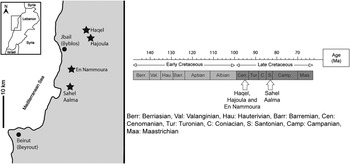
The Upper Cretaceous Lebanese Lagerstätten are mostly represented by four famous localities: Haqel, Hajoula, En Nammoura, and Sahel Aalma (Fig. 2). These “fish-shales” (Lukeneder and Harzhauser, Reference Lukeneder and Harzhauser2004) yielded a large number of well-preserved cephalopods (Fuchs and Larson, Reference Fuchs and Larson2011a, Reference Fuchs and Larson2011b), crustaceans (Audo and Charbonnier, Reference Audo and Charbonnier2013), and fishes (Gayet, Reference Gayet1980). Haqel, Hajoula, and En Nammoura are late Cenomanian in age (Forey et al., Reference Forey, Yi, Patterson and Davies2003), whereas foraminiferans indicate a late Santonian age for Sahel Aalma (~85 Ma; Ejel and Dubertret, Reference Ejel and Dubertret1966; Fig. 2). Meister (Reference Meister1993) reported the ammonites Muniericeras blanfordianum Stoliczka, 1865 and Texanites texanus Roemer, Reference Roemer1852, which corroborates a Santonian age for this site. Ten unreported ammonites from the MNHN Dubertret collection were also identified as T. texanus and M. blanfordianum, thus further supporting a Santonian age for Sahel Aalma.
Geographic location of Haqel, Hajoula, En Nammoura and Sahel Aalma (modified from Fuchs and Weis, Reference Fuchs and Weis2009) and their stratigraphic position.

Detailed sedimentological descriptions of the Lebanese Lagerstätten were provided by Audo and Charbonnier (Reference Audo and Charbonnier2013). During the Cretaceous, Lebanon and the entire Arabian Peninsula were located on the African platform in the northern part of Gondwana (Philip et al., Reference Philip, Babinot, Tronchetti, Fourcade, Ricou, Guiaud, Bellion, Herbin, Combes, Conee and Dercourt1993). The lithographic limestones from the Hajoula, Haqel, and En Nammoura Lagerstätten were probably deposited in small shallow basins corresponding to intra-shelf depressions. In this scenario, mild oscillations in relative sea-level were responsible of a spectacular sandwich of shallow-water carbonate facies alternating with finely bedded or even laminated mudstones (Ferry et al., Reference Ferry, Merran, Grosheny and Mroueh2007), thus producing favorable conditions for the preservation of a rich fauna (Fuchs and Weis, Reference Fuchs and Weis2009).
In Lebanon, deposition of micritic limestones and chalks began after the Turonian and continued until the Eocene (Ferry et al., Reference Ferry, Merran, Grosheny and Mroueh2007). The Santonian Lagerstätte of Sahel Aalma is composed of fine-grained, chalky limestones that were probably deposited in much deeper conditions than those recognized in Hajoula or Haqel. All coleoid specimens from the Lebanese Lagerstätten are preserved as compressions on slabs. Based on the assumption that vampyropod gladii derive from a belemnoid organic proostracum, an original organic composition is supposed for Mesozoic gladii (Doguzhaeva and Mutvei, Reference Doguzhaeva and Mutvei2003). Fuchs (Reference Fuchs2006a) thus suggested that the Lebanese gladii were most likely preserved in francolite as a diagenetic byproduct.
Contrary to Roger’s interpretation (1946; planche I, fig. 2), we could not find any preserved soft parts on the nautiloids described by this author. The brown fragments interpreted by Roger (Reference Roger1946) as nautiloid embryos are actually fish remains. This is also the case for other specimens of nautiloids and ammonites from the MNHN Dubertret collection.
Systematic paleontology
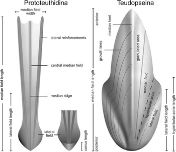
A total of 16 gladius-bearing coleoid specimens from the MNHN Dubertret, Arambourg, and En Nammoura collections have been studied, including two specimens previously illustrated by Roger (Reference Roger1946, MNHN.F.R06751, p. 14–16, pl. 4, fig. 5; MNHN.F.R06746, p. 14–16, pl. 9, figs. 1–2). We used UV-light to highlight soft tissues, using a UV wavelength of 365 nm. The gladius anatomical terminology follows Fuchs and Larson (Reference Fuchs and Larson2011a, Reference Fuchs and Larson2011b; Fig. 3). Measured characters were median field length (mfl), median field width (mfw), total body length, maximum body width, and arm length. These measurements are standard in most published studies on coleoids (e.g. Fuchs and Larson, Reference Fuchs and Larson2011a, Reference Fuchs and Larson2011b). The “arm length” corresponds to the measurement of the longest arm visible on the studied specimen. We only measured the most complete specimens (i.e., without fracturing or other disruption).
Terminology and measurements used for prototeuthidid and teudopseid gladii in dorsal view (Fuchs and Larson, Reference Fuchs and Larson2011a, Reference Fuchs and Larson2011b).

Material is housed in the Muséum National d'Histoire Naturelle, Paris (MNHN.F prefix).
Subclass Coleoidea Bather, Reference Bather1888
Superorder Vampyropoda Boletzky, Reference Boletzky1992
Suborder Prototeuthidina Naef, Reference Naef1921
Diagnosis
Slender gladius with triangular median field and ventrally closed (funnel-like) conus. Hyperbolar zones reduced or absent. Lateral field length less than the half gladius length. Median and lateral reinforcements present on the median field. Vestiges of septa and guard absent (Emended diagnosis after Fuchs et al. 2007b, p. 241; contra Jeletzky, Reference Jeletzky1966, p. 43–44).
Family Plesioteuthididae Naef, Reference Naef1921
Diagnosis
More or less slender prototeuthoids with a conus vane that is restricted to the posterior end and is bent down to form a pointed cone, grading anteriorly into the relatively broad, gradually tapering lateral plates, in which the median plate bears a simple or double median rib and narrow lateral bands clearly demarcated from the broad central part. (Naef, 1922, p. 111). The fins are very short, subterminal, resting on the conus (Fuchs and Larson, Reference Fuchs and Larson2011a).
Included genera
Plesioteuthis Wagner, Reference Wagner1859; Dorateuthis Woodward, Reference Woodward1883; Paraplesioteuthis, Naef, Reference Naef1921; Romaniteuthis Fischer and Riou, Reference Fischer and Riou1982; Rhomboteuthis Fischer and Riou, Reference Fischer and Riou1982; Boreopeltis Engeser and Reitner, Reference Engeser and Reitner1985; Senefelderiteuthis Engeser and Keupp, Reference Engeser and Keupp1999; Nesisoteuthis Doguzhaeva, Reference Doguzhaeva2005.
Occurrence
(?)Late Triassic (Rhaetian), Early Jurassic (Toarcian)–Late Cretaceous (Maastrichtian) of Europe, Central Russia, Lebanon, North America and Australia.
Genus Dorateuthis Woodward, Reference Woodward1883
Type species
Dorateuthis syriaca Woodward, Reference Woodward1883 from the Cenomanian–Santonian of Lebanon.
Included species
With certainty only Dorateuthis syriaca Woodward, Reference Woodward1883 from the Cenomanian–Santonian of Lebanon. Poorly preserved “Neololigosepia” stahleckeri Reitner and Engeser, Reference Reitner and Engeser1982 and “Maioteuthis” morroensis Reitner and Engeser, Reference Reitner and Engeser1982 both from the Barremian of the Cape Verde Islands; “Plesioteuthis” sp. Engeser and Reitner, Reference Engeser and Reitner1985 and “Maioteuthis” damesi Engeser and Reitner, Reference Engeser and Reitner1985 both from the Aptian of Heligoland (northern Germany); “Plesioteuthis” arcuata von der Marck, Reference Marck1873 from the Campanian of northwestern Germany; “Plesioteuthis” maestrichtensis Binkhorst van den Binkhorst, Reference Binkhorst van den Binkhorst1861 from the Maastrichtian of the Netherlands, all of which display a narrow gladius with continuous lateral keels, were preliminarily assigned to this genus.
Diagnosis
Gladius long and slender (ratio gladius width/length ~0.10) without a median keel. Median field triangular with a bipartite median ridge and pronounced lateral keels present continuously from anterior to posterior extremities. Lateral fields and conus strongly reduced (after Fuchs and Larson, 2011a).
Occurrence
With certainty from the Cenomanian through Santonian of Lebanon (and potentially from the Barremian up to the Maastrichtian of the Cape Verde Islands, Northern Europe and Russia).

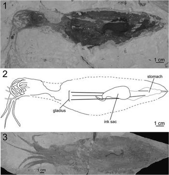
Dorateuthis syriaca (Woodward, Reference Woodward1883) specimens (1) and (2), MNHN.F.A50398; 3, MNHN.F.A50402; Santonian, Sahel Aalma, exhibiting a long and slender gladius consisting of an anteriorly diverging median field.

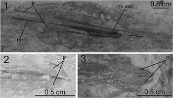
Gladius morphology of Dorateuthis syriaca (Woodward, Reference Woodward1883): (1), gladius morphology of MNHN.F.A50394; (2), lateral fields of MNHN.F.A50405; (3), lateral fields of MNHN.F.A50398. Abbreviations: lr=lateral reinforcements, mr=median ridge, lf=lateral fields.

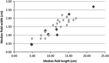
Scatter plot of the median field width versus median field length for 8 Dorateuthis syriaca (Woodward, Reference Woodward1883) studied specimens (MNHN.F.A50394, MNHN.F.A50395, MNHN.F.A50396, MNHN.F.A50398, MNHN.F.R06746, MNHN.F.A50399, MNHN.F.A50401, and MNHN.F.A50402; squares) and unpublished specimens (D. Fuchs, personal communication, 2013; circles).

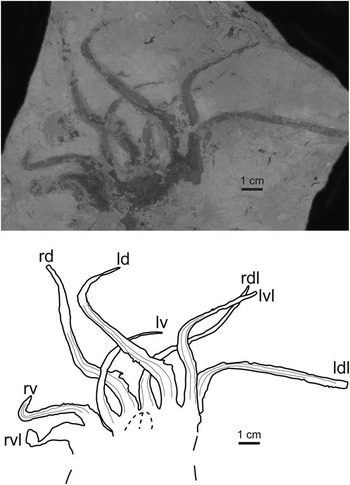
Arms of a Dorateuthis syriaca (Woodward, Reference Woodward1883) specimen, MNHN.F.A50400, Santonian, Sahel Aalma, ventral view. Abbreviations: ld=left dorsal, rd=right dorsal, lv=left ventral, rv=right ventral, ldl=left dorsolateral, rdl=right dorsolateral, lvl=left ventrolateral, rvl=right ventrolateral.

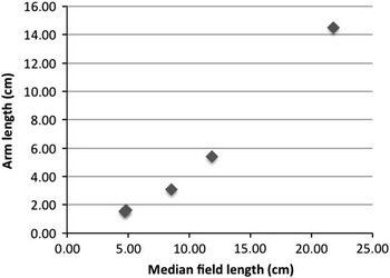
Arm length/mfl ratio for Dorateuthis syriaca (Woodward, Reference Woodward1883) specimens, MNHN.F.A50394, MNHN.F.A50395, MNHN.F.A50398, MNHN.F.R06746, and MNHN.F.A50402, Santonian, Sahel Aalma.

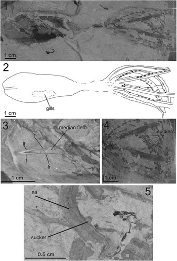
Sucker morphology of Dorateuthis syriaca (Woodward, Reference Woodward1883): (1), overall view of MNHN.F.A50397, Santonian, Sahel Aalma (dorsal view); (2), drawing of MNHN.F.A50397; (3), close-up of MNHN.F.A50397 (photographed with UV light) to show the gladius morphology; (4), close-up of the arms of MNHN.F.A50397; (5), close-up of a sucker of MNHN.F.A50398, Santonian, Sahel Aalma. Abbreviations: lf=lateral fields, lr=lateral reinforcements, na=nervous axis.

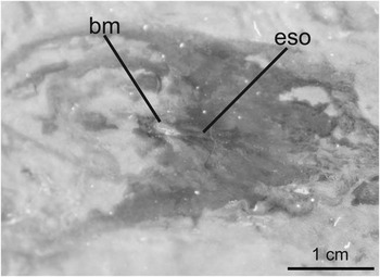
Buccal mass of a Dorateuthis syriaca (Woodward, Reference Woodward1883) specimen, MNHN.F.A50398, Santonian, Sahel Aalma (photographed under UV light). Abbreviations: bm=buccal mass, eso=esophagus.

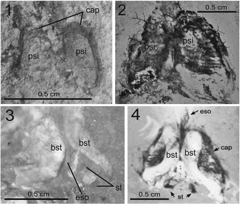
Cephalic cartilage morphology of Dorateuthis syriaca (Woodward, Reference Woodward1883): (1), MNHN.F.A50405, Santonian, Sahel Aalma; (2), MNHNL CRE047, Cenomanian, Haqel; taken from Fuchs and Larson, Reference Fuchs and Larson2011a); (3), MNHN.F.A50403, Santonian, Sahel Aalma (photographed under UV light); (4), cephalic cartilage of a Dorateuthis syriaca specimen (taken from Fuchs and Larson, Reference Fuchs and Larson2011a). Abbreviations: psi=pear-shaped imprints, cap=eye capillaries, bst=boomerang-shaped structures, eso=esophagus, st=statocysts.

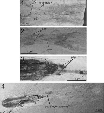
(1), posterior salivary glands of a Dorateuthis syriaca (Woodward, Reference Woodward1883) specimen, MNHN.F.A50396, Santonian, Sahel Aalma; (2), posterior salivary glands of a D. syriaca specimen, MNHN.F.A50394 (Santonian, Sahel Aalma), laterally to the esophagus; (3), posterior salivary glands of a D. syriaca specimen (NHMW1998z0105/0000, Santonian, Sahel Aalma; modified from Lukeneder and Harzhauser, Reference Lukeneder and Harzhauser2004); (4), posterior salivary glands of Glyphiteuthis abisaadiorum (taken from Fuchs and Weis, Reference Fuchs and Weis2009), CRE042, Cenomanian, Hajoula (modified from Fuchs and Weis, Reference Fuchs and Weis2009). Abbreviations: psg=posterior salivary glands, cc=cephalic cartilage, eso=esophagus.

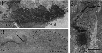
(1), gills of a Dorateuthis syriaca (Woodward, Reference Woodward1883) specimen, MNHN.F.A50397, Santonian, Sahel Aalma; (2), ink sac and stomach of MNHN.F.A50402, Santonian, Sahel Aalma; (3), ink sac of MNHN.F.A50401, Santonian, Sahel Aalma. Abbreviations: gl=gills lappets, is=ink sac, st=stomach.

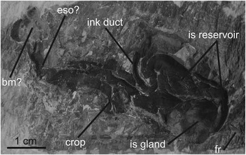
Digestive system of a Dorateuthis syriaca (Woodward, Reference Woodward1883) specimen, MNHN.F.R06746, Santonian, Sahel Aalma. Abbreviations: bm=buccal mass, eso=esophagus, is reservoir=ink sac reservoir, is gland=ink sac gland, fr=fish remains (stomach).

Type locality
Sahel Aalma, Lebanon.
Occurrence
early Late Cenomanian–Late Santonian of Lebanon; type horizon Late Santonian.
Description
Thirteen specimens (MNHN.F.A50394–MNHN.F.A50405, MNHN.F.R06751, and MNHN.F.R06746; all from the Santonian of Sahel Aalma) from the Dubertret collection can be assigned to D. syriaca based on their similar gladius morphology. They all exhibit a long and slender gladius with an anteriorly diverging median field (Fig. 4). Conspicuous lateral reinforcements extending from posterior to anterior extremities are visible and diverge at an acute angle of ~6°. The thickness of the lateral reinforcements hardly increases anteriorly. This slight thickening is visible on the dorsal and ventral gladius surfaces. On most of specimens, a delicate median ridge is present and it is thinner than the lateral reinforcements. Interestingly, the specimen MNHN.F.A50394 shows a relatively strong median ridge with almost the same thickness as the lateral reinforcements (Fig. 5.1).
Three gladii show weak imprints of the lateral fields: MNHN.F.A50405 (Fig. 5.2) is a gladius of a small individual (mfl ~28 mm). Its lateral fields are relatively long (6 mm, i.e., ~20 % of mfl) and large (3.4 mm, i.e., ~12% of mfl). On MNHN.F.A50397, perceptible lateral fields are 4.5 mm long and 1.8 mm wide (the specimen is illustrated and discussed hereafter). The third gladius showing lateral fields (MNHN.F.A50398, Fig. 5.3) is of much larger dimensions (mfl ~151 mm). Compared to its large size, its lateral fields are relatively short and narrow (8.1 mm long and 3.5 mm wide, i.e., ~5% and 2% of mfl, respectively). None of the 13 studied specimens yielded long and wide lateral fields as reported on the holotype by Woodward (Reference Woodward1883). The size of the lateral fields appears partly more stable and independent relative to mfl size. Thus, the lateral fields probably do not have an isometric growth relative to gladius size (i.e., the size of the lateral fields does not necessarily increase in the same proportion as the gladius). No specimen shows evidence of lateral fields with a conus.
Mfl and mfw measurements of eight specimens (MNHN.F.A50394, MNHN.F.A50395, MNHN.F.A50396, MNHN.F.A50398, MNHN.F.R06746, MNHN.F.A50399, MNHN.F.A50401, and MNHN.F.A50402) are illustrated in Figure 6. This figure also includes unpublished measurements of Dorateuthis syriaca specimens from Haqel, Hajoula, and Sahel Aalma (Fuchs, personal communication, 2013) and these individuals all appear to be conspecific. The mean mfw/mfl ratio (~0.13) of the MNHN specimens is also consistent with the 0.10 ratio mentioned in the emended diagnosis of the genus Dorateuthis by Fuchs and Larson (Reference Fuchs and Larson2011a). This ratio has also been estimated for the holotype of D. syriaca published by Woodward (Reference Woodward1883) and it is similar (~0.11). Overall, these observations support an assignment of MNHN.F.A50394–MNHN.F.A50405, MNHN.F.R06751, and MNHN.F.R06746 specimens to D. syriaca.
Five of the studied specimens (MNHN.F.A50397, MNHN.F.A50398, MNHN.F.R06746, MNHN.F.A50400, and MNHN.F.A50403) unequivocally show eight arms only. Those of specimen MNHN.F.A50400 are particularly well preserved (Fig. 7). An additional strongly elongated ventrolateral arm pair (tentacles) as drawn by Woodward (Reference Woodward1883) for the holotype, or assumed by Naef (Reference Naef1922, p. 72), is not visible on any studied specimen and is therefore not supported. Difference in arm thickness is frequently observed but it most probably results from the curvature, orientation, and differential crushing of the arms. Only two specimens (MNHN.F.A50394 and MNHN.F.A50400) exhibit differences in arm thickness that apparently do not result from taphonomic processes. MNHN.F.A50400 shows exceptionally well-preserved arms whose length can be categorized into three groups (Fig. 7). MNHN.F.A50394 apparently also displays three types of arm length. This observation is in accordance with Lukeneder and Harzhauser (Reference Lukeneder and Harzhauser2004) and Fuchs and Larson (Reference Fuchs and Larson2011a) who described three coexisting types of arms with different lengths in D. syriaca. Fuchs and Larson (Reference Fuchs and Larson2011a) suggested that the shortest and most delicate arms are ventral and that the longest and thickest are in dorsal position. The ventrolateral and dorsolateral arm pairs seem to be equal in length and thickness. These types of arm pairs were tentatively identified on MNHN.F.A50400 and are reported in Figure 7. A dorsal arm pair that is longer than the other arms is also a well-known feature of the Jurassic Plesioteuthis prisca (Fuchs, Reference Fuchs2006b). On MNHN.F.A50398 (Fig. 4), four delicate “filaments” are barely perceptible but represent the distal part of highly curved arms. In most of the studied specimens, arms are narrower and more delicate on their distal part than on their proximal part.
The arm length/mfl ratio is variable among five studied Dorateuthis syriaca specimens from Sahel Aalma (MNHN.F.A50394, MNHN.F.A50395, MNHN.F.A50398, MNHN.F.R06746, and MNHN.F.A50402; Fig. 8). Arms on small specimens (i.e., mfl ~5–10 cm) are comparatively shorter (arm length/mfl ratio ~0.3–0.4) than those of medium-sized specimens (mfl ~12 cm, arm length/mfl ratio ~0.45) and of the largest specimen (mfl ~22 cm, arm length/mfl ratio ~0.65). These values are in agreement with measurements by Fuchs and Larson (Reference Fuchs and Larson2011a) who documented ratios of 0.4 for “juvenile specimens,” 0.45 for “a possibly adolescent specimen” and at least 0.5 for “large specimens.” We therefore agree with the hypothesis of Fuchs (Reference Fuchs2006a), who suggested that the differences in relative arm length and thickness result from allometric growth. This hypothesis is also consistent with observations made on modern cephalopods. Indeed, changes in the relative size of various body components during growth occur in most cephalopods (Boyle and Rodhouse, Reference Boyle and Rodhouse2005). For instance, changes in the proportions of arms or mantle are conspicuous among squids during the paralarval and juvenile stages. Allometric differences among somatic, reproductive, and digestive components are variable and are linked to ontogeny or nutrient availability (Boyle and Rodhouse, Reference Boyle and Rodhouse2005).
As for Dorateuthis syriaca individuals studied by Fuchs and Larson (Reference Fuchs and Larson2011a), none of the studied specimens yielded evidence of cirri or decabrachian characters such as hooks or horny rings. This contrasts with Woodward (Reference Woodward1883) who mentioned the presence of hooks on the holotype. Naef (Reference Naef1922, p. 72) and Lukeneder and Harzhauser (Reference Lukeneder and Harzhauser2004, p. 4) reported faint traces of sucker-like structures on D. syriaca specimens from Sahel Aalma. Fuchs and Larson (Reference Fuchs and Larson2011a) did not report suckers for D. syriaca specimens from Lebanon. Interestingly, one D. syriaca specimen (MNHN.F.A50397, Fig. 9.1–9.3) yielded unambiguous, exceptionally well-preserved suckers (Fig. 9.1, 9.2, 9.4). When flattened with a lateral orientation, they show a large base, indicating that they were sessile and not pedunculate. Moreover, the suckers are organized in two rows forming transverse pairs (Fig. 9.4). Another studied specimen, but less well preserved (MNHN.F.A50398; Fig. 4), also exhibits an apparent sucker-like structure (Fig. 9.5). This is the first time that unambiguous evidence of suckers is documented for specimens of D. syriaca.
The preservation of the buccal mass as a white spot located anteriorly on the head is a common feature in D. syriaca (Fuchs and Larson, Reference Fuchs and Larson2011a). However, only MNHN.F.A50398 yields reliable evidence of such a character (Fig. 10). On this specimen, the beaks are also discernible in the center of the buccal mass. Posteriorly to the buccal mass, a distinct cord is visible and probably represents the esophagus (Fig. 10). On MNHN.F.A50403, the esophagus appears as a cord of yellowish staining. Lukeneder and Harzhauser (Reference Lukeneder and Harzhauser2004) and Fuchs and Larson (Reference Fuchs and Larson2011a) already described these organs in prototeuthidid coleoids from Lebanon.
One studied specimen (MNHN.F.A50405) exhibits a pair of pear-shaped imprints (Fig. 11.1) located on the longitudinal axis of the body and between the anterior margin of the gladius and the supposed position of the arm base. Their outer margins are weakly curved and the inner margins appear straight and close to each other, but not connected. Fuchs and Larson (Reference Fuchs and Larson2011a) previously described such structures (Fig. 11.2) on specimens of Dorateuthis syriaca. According to these authors, these structures, when present, always appear in pairs and thus suggest a median connection. A pair of boomerang-shaped structures is also perceptible on MNHN.F.A50403. Those “boomerangs” are better seen under UV light (Fig. 11.3). Fuchs and Larson (Reference Fuchs and Larson2011a) described similar structures within the pear-shaped imprints (Fig. 11.4). We never found both characters together, probably due to unsufficient preservation. However, we agree with the hypothesis of Fuchs and Larson (Reference Fuchs and Larson2011a) who suggested that the “boomerangs” and the “pears” represent the dorsal and ventral parts of the cephalic cartilage, respectively. Indeed, these structures are very similar to the ones that can be observed in modern coleoids (Nixon, Reference Nixon1998).
As illustrated by Fuchs and Larson (Reference Fuchs and Larson2011a) on specimens of D. syriaca from Lebanon, a structure located close to the posterior side of the cephalic cartilage of MNHN.F.A50403 is barely perceptible. It is characterized by a white rectangle in between two black rectangles (Fig. 11.3). Fuchs and Larson (Reference Fuchs and Larson2011a; Fig. 11.4) interpreted the black rectangles as statocysts, i.e., balance sensory receptor. In modern coleoids, the cephalic cartilage completely surrounds two statocysts, which are located at the same place than the observed structures (Nixon, Reference Nixon1998). However, the preservation is not good enough to allow a definitive anatomical assignment.
As Fuchs and Larson (Reference Fuchs and Larson2011a) did, we suggest that the cephalic cartilage of D. syriaca resembles the one of Vampyroteuthis Chun, Reference Chun1903 in having lateral projections (Nixon, Reference Nixon1998, fig. 3c). Although the cephalic cartilage of MNHN.F.A50396 (Fig. 12.1) appears quite similar to that of the extant Gonatus fabricii Lichtenstein, Reference Lichtenstein1818 (Nixon, Reference Nixon1998, fig. 3b), its structure has been disrupted by taphonomic processes. Furthermore, the phylogenetic value of this character is still poorly known, even in living taxa (Nixon, Reference Nixon1998).
MNHN.F.A50396 and MNHN.F.A50394 exhibit two pill-shaped brown structures identified as the posterior salivary glands (Fig. 12.1 and 12.2, respectively). These two ovoid structures are symmetrical on MNHN.F.A50396 indicating that their original location along the sagittal plan is preserved on this specimen. Two faint traces of channels can also be distinguished and apparently interconnect the ovoid structures and the buccal mass (Fig. 12.1). By comparison with present-day coleoids, these pill-shaped structures are similar in form and location to the paired posterior salivary glands (Mangold and Bidder, Reference Mangold and Bidder1989a; Mangold and Young, Reference Mangold and Young1998). This is also in accordance with the interpretation of Lukeneder and Harzhauser (Reference Lukeneder and Harzhauser2004) of similar but less well-preserved structures from a Dorateuthis syriaca specimen from Sahel Aalma (Fig. 12.3). Fuchs and Larson (Reference Fuchs and Larson2011a) did not observe such structures on other D. syriaca specimens. However, these pill-shaped structures strongly ressemble those interpreted as eye capsules by Fuchs and Weis (Reference Fuchs and Weis2009, illustrated here in Fig. 12.4) and by Fuchs and Larson (Reference Fuchs and Larson2011b) for teudopseid coleoids from the Cenomanian of Haqel and Hajoula. Based on this resemblance, the identification as “eye capsules” of the pill-shaped brown structures observable in Fuchs and Weis (Reference Fuchs and Weis2009) and Fuchs and Larson (Reference Fuchs and Larson2011b) appears unlikely to us. Indeed, the pill-shaped brown structures observed in MNHN.F.A50394 and MNHN.F.A50396 rather represent posterior salivary glands, simply because D. syriaca is not supposed to possess eye capsules (as stated by Fuchs and Larson, Reference Fuchs and Larson2011a, p. 241 and Fuchs and Larson, Reference Fuchs and Larson2011b, p. 827). Consequently, the similar structures observable in Fuchs and Weis (Reference Fuchs and Weis2009) and Fuchs and Larson (Reference Fuchs and Larson2011b) should also be posterior salivary glands rather than eye capsules.
Furthermore, some of the pill-shaped structures (on MNHN.F.A50394 and MNHN.F.A50405, and on teudospeid specimens described in Fuchs and Weis, Reference Fuchs and Weis2009 and Fuchs and Larson, Reference Fuchs and Larson2011b) appear to be too distant from each other to represent eye capsules. Indeed, if the cephalic cartilage simply consists of eye capsules in teudopseid coleoids as stated by Fuchs and Larson (Reference Fuchs and Larson2011a), these eye capsules should presumably be closer to each other. Moreover, MNHN.F.A50396 exhibits a cephalic-like structure (Fig. 12.1) relatively distant from the pill-shaped structures. Both structures are anatomically dissociated. Consequently, these observations call for the identification as posterior salivary glands for these pill-shaped structures. As a whole, our anatomical re-interpretation means that a true cephalic cartilage has not been found yet in teudopseid coleoids and that it would be documented in D. syriaca only. Paired posterior salivary glands are known in modern decabrachians (Sepiida Zittel, Reference Zittel1895) but also in modern vampyropods (Octopoda Leach, Reference Leach1818). Glands observed in D. syriaca are quite similar to those of the modern Sepia officinalis Linnaeus, Reference Linnaeus1758 as described in Mangold and Bidder (Reference Mangold and Bidder1989a) and Mangold and Young (Reference Mangold and Young1998). However, this organ probably carries little phylogenetic information. Incidentally, when the pill-shaped structures are located close to the buccal mass (as illustrated in Fuchs and Larson, Reference Fuchs and Larson2011b), they may correspond to the anterior salivary glands rather than to eye capsules (Mangold and Young, Reference Mangold and Young1998).
Feather-like structures occur in MNHN.F.A50397 along the lateral mantle margin and may represent gill remains (Fig. 13.1). They markedly differ from the surrounding matrix by their darkened colour. A series of at least 16 long lappets branching off from a longitudinal axis is visible. This latter is similar to the branchial membrane in modern coleoids (Mangold and Bidder, Reference Mangold and Bidder1989b). Our observation reinforces the hypothesis of Fuchs and Larson (Reference Fuchs and Larson2011a, p. 239) who stated that “these structures might represent remains of the cartilaginous gill skeleton […] but it clearly reflects the primary efferent gill vessel and its outer branches (=secondary efferent vessels, branchial laminae)”. The gills of D. syriaca resemble those of Plesioteuthis prisca (Bandel and Leich, Reference Bandel and Leich1986; Haas, Reference Haas2002) in that they have a large number of branchial laminae. The incirrate octopods Keuppia levante and Keuppia hyperbolaris Fuchs et al., Reference Fuchs, Bracchi and Weis2009 from Hajoula apparently show only six or seven branchial laminae (Fuchs et al., Reference Fuchs, Bracchi and Weis2009). Branchial laminae have also been reported from the belemnoid “Phragmoteuthis” conocauda Quenstedt, Reference Quenstedt1849, but their total number remains unclear (Reitner, Reference Reitner2009). Gill morphology of Dorateuthis syriaca, as well as of Plesioteuthis prisca, ressembles that of modern teuthids in having numerous gill blades (Reitner, Reference Reitner2009). However, this can be seen as a homoplasy. Indeed, according to Reitner (Reference Reitner2009), gill type is most probably due to lifestyle and has no phylogenetic significance.
Almost all specimens described here exhibit a relatively large 3-D black structure located at mid-length of the mantle (Fig. 13.2). Similar structures have been interpreted as the ink sac (Fuchs, Reference Fuchs2006a; Fuchs and Larson, Reference Fuchs and Larson2011a, Reference Fuchs and Larson2011b) and we agree with this interpretation. According to Fuchs (Reference Fuchs2006a), the preservation of an ink sac is common in the Cretaceous coleoids from the Lebanese Lagerstätten, but its presence is phylogenetically uninformative. It also appears that in fossil coleoids, the stomach is frequently characterized by fish remains (Fig.13.3). Nearly all coleoids have an ink sac, except some deepwater species and the night-active shallow-water octopus Ameloctopus Norman, Reference Norman1992, presumably because ink ejection has been counter-selected in dark environments (Boyle and Rodhouse, Reference Boyle and Rodhouse2005). The observed glands (Fig. 14) appear to be quite similar to the one seen in Loligo Lamarck, Reference Lamarck1798 and even more in Sepia Linnaeus, Reference Linnaeus1758 where the reservoir spreads on the back and reaches the bottom of the sac (Mangold et al., Reference Mangold, Bidder and Portmann1989b). On the contrary, in Octopus Cuvier, Reference Cuvier1797, the glands fully occupy the bottom of the sac (Mangold et al., Reference Mangold, Bidder and Portmann1989b). Remarkably, the presence of a crop is suggested in MNHN.F.R06746 by a distinct bulge within the black structure (Fig. 14). Contrary to the potential crop described by Lukeneder and Harzhauser (Reference Lukeneder and Harzhauser2004) and Fuchs and Larson (Reference Fuchs and Larson2011a), we report here an anteriorly-located crop that is distant from the stomach, as observed in modern Octopus (Mangold and Bidder, Reference Mangold and Bidder1989a and Mangold and Young, Reference Mangold and Young1998). Specimen MNHN.F.R06746 also exhibits a peculiar ventral trilobated structure at the anterior part of the black structure (Fig.14). Roger (Reference Roger1946) interpreted this feature as a rectal bulb and this assumption cannot be discarded. However, an alternative interpretation could be the remains of a buccal mass. Indeed, this trilobate morphology is quite similar to the buccal mass in extant coleoids (Mangold and Bidder, Reference Mangold and Bidder1989a; Mangold and Young, Reference Mangold and Young1998).
Up to now, a well-preserved continuous digestive tract consisting of a buccal mass, an esophagus, a crop and a stomach, has only been described for specimens of D. syriaca (Lukeneder and Harzhauser, Reference Lukeneder and Harzhauser2004; Fuchs and Larson, Reference Fuchs and Larson2011a), and this digestive tract thus cannot be compared with other known fossil cephalopods.
Remarks
Dorateuthis syriaca is the most abundant coleoid in the Haqel and Hajoula localities (Fuchs and Larson, Reference Fuchs and Larson2011a). Roger (Reference Roger1946, p.14) stated that D. syriaca was also relatively frequent in Sahel Aalma, making this taxon the most abundant Cretaceous coleoid in Lebanon. In his original description, Woodward (Reference Woodward1883, p.3) stated that the gladius of D. syriaca is marked by “three equidistant ridges, one median and two lateral, which converge together at the very acute distal extremity,” and also by “two extremely delicate lateral expansions.” Naef (Reference Naef1922, p. 72) presumed a strong median rib and doubted the presence of lateral fields. However, Roger (Reference Roger1946, p. 14) reported long and wide lateral fields and therefore regarded the gladius as close to the Jurassic Leptoteuthis (suborder Teudopseina). Recently, Fuchs (Reference Fuchs2006a, p. 7) redescribed D. syriaca from the Cenomanian of Haqel and Hadjoula and concluded that if lateral fields are present, they must be extremely short and narrow. Interestingly, the gladius of D. syriaca is similar in shape to the gladius of the Early Jurassic Paraplesioteuthis sagittata Münster, Reference Münster1843; the Middle Jurassic Romaniteuthis gevreyi Roman, Reference Roman1928; and the Late Jurassic Senefelderiteuthis tricarinata, Münster, Reference Münster1846. They all show a slender gladius that is flanked by lateral reinforcements. According to Fuchs and Larson (Reference Fuchs and Larson2011a), D. syriaca mainly differs from these Jurassic taxa by the absence of well-defined lateral fields. However, three studied specimens (MNHN.F.A50397, MNHN.F.A50398, and MNHN.F.A50405; Figs. 5.2 and 5.3) exhibit well-defined lateral fields, suggesting that these characters were partly missing on the specimens described by Fuchs and Larson (Reference Fuchs and Larson2011a). These authors (p. 237, fig. 2.7) described a small D. syriaca specimen from Sahel Aalma with lateral fields equivalent to ~10% of the mfl and 6% of the mfw. Therefore, it appears that the “absence of true lateral fields” as stated by Fuchs and Larson (Reference Fuchs and Larson2011a) cannot be used to distinguish D. syriaca from Paraplesioteuthis sagittata, Romaniteuthis gevreyi, and Senefelderiteuthis tricarinata. Instead, we suggest that D. syriaca differs from P. sagittata and R. gevreyi in having smaller lateral fields.
Fuchs et al. (Reference Fuchs, Klinghammer and Keupp2007b) considered the genus Senefelderiteuthis as a junior synonym of the genus Dorateuthis, but recently Fuchs and Larson (Reference Fuchs and Larson2011a) re-established Senefelderiteuthis and assigned “Dorateuthis” tricarinata to this genus, based on the presence of well-defined lateral fields. However, MNHN specimens of D. syriaca exhibiting well-defined lateral fields show that the differences between D. syriaca and Senefelderiteuthis tricarinata are tenuous for this feature. According to Fuchs and Larson (Reference Fuchs and Larson2011a), D. syriaca also differs from S. tricarinata by the presence of a median ridge. However, it could be argued that the lack of a delicate median ridge in S. tricarinata is possibly a preservation artefact. In agreement with Fuchs et al. (Reference Fuchs, Klinghammer and Keupp2007b), we find difficult to distinguish S. tricarinata and D. syriaca based on gladius features. However, S. tricarinata apparently shows longer arms than D. syriaca (Fuchs and Larson, Reference Fuchs and Larson2011a).
The gladius of D. syriaca is also similar to the gladius of the Jurassic coleoids Plesioteuthis subovata Münster, Reference Münster1846 and Plesioteuthis prisca Rüppell, Reference Rüppel1829. According to Fuchs and Larson (Reference Fuchs and Larson2011a), these two taxa can be distinguished from D. syriaca by the presence of a distinct median ridge and the absence of continuous lateral reinforcements. For Fuchs et al. (Reference Fuchs, Klinghammer and Keupp2007b), these two qualitative characters clearly separate Dorateuthis from Plesioteuthis and other plesioteuthidid genera. However, the sketch of the gladius of P. prisca in Fuchs and Larson (Reference Fuchs and Larson2011a, fig. 7.4, p. 243) shows the presence of continuous lateral keels. This illustration has thus to be corrected.
Furthermore, we observed in MNHN.F.A50394 a strong median ridge presenting almost the same thickness as the lateral reinforcements (Fig. 5.1). This contrasts with Fuchs and Larson (Reference Fuchs and Larson2011a), who considered D. syriaca as being characterized by a weak median ridge. Such a strong median ridge is in better agreement with the description of D. syriaca by Naef (Reference Naef1922, p. 72). Based on this new observation, we suggest that a “pronounced median keel” (Fuchs and Larson, Reference Fuchs and Larson2011a) is not a truly reliable character to distinguish P. prisca and P. subovata from D. syriaca. In addition, biometric values are similar for both P. prisca and D. syriaca (Fig. 6; Fuchs et al., Reference Fuchs, Klinghammer and Keupp2007b, p. 244, fig. 2). Based on soft-part morphology, P. prisca can be distinguished from D. syriaca by shorter arms (Fuchs and Larson, Reference Fuchs and Larson2011a).
We conclude that diagnostic characters that unambiguously identify D. syriaca are rare. According to Fuchs and Larson (Reference Fuchs and Larson2011a), D. syriaca was characterized by the presence of a delicate median ridge and the absence of true lateral fields. However, two of the specimens studied here show the opposite features and suggest that the gladius characters used to diagnose D. syriaca are highly variable among specimens according to their preservation. Even though P. subovata may be distinguished from D. syriaca by larger lateral fields (Fuchs et al., Reference Fuchs, Klinghammer and Keupp2007b, fig. 3C), the gladius differences between D. syriaca and P. prisca are much more tenuous.

Diagnosis
Gladius with clearly reduced, opened, and spoon-shaped conus. Lateral fields and hyperbolar zones 55% and less of the median field length. Hyperbolar zones located between lateral and median field as well-developed broad furrows. Anterior median field more or less pointed (after Fuchs and Weis, 2009).
Families included
Trachyteuthididae, Naef, Reference Naef1921; Palaeololiginidae, Naef, Reference Naef1921; Teudopsidae, Regteren Altena, Reference Regteren Altena1949; Muensterellidae, Roger, Reference Roger1952.
Family Trachyteuthididae Naef, Reference Naef1921
Diagnosis
Teudopseid vampyropods with granules (tubercles) on the dorsal surface of the gladius.
Included genera
Glyphiteuthis Reuss, Reference Reuss1854; Actinosepia Whiteaves, Reference Whiteaves1897; Glyphidopsis Fuchs and Larson, Reference Fuchs and Larson2011b.
Occurrence
Middle Jurassic (Callovian)–Late Cretaceous (Maastrichtian) of Europe, Central Russia, Lebanon, Cuba, Chile, Antarctica, Australia, North America, and Mexico.

Other species
Glyphiteuthis boutillieri (Lennier, Reference Lennier1866); Glyphiteuthis libanotica (Fraas, Reference Fraas1878); Glyphiteuthis abisaadiorum Fuchs and Weis, Reference Fuchs and Weis2009; Glyphiteuthis rhinophora Fuchs et al., Reference Fuchs, Stinnesbeck, Ifrim, Giersch, Gutierrez and Frey2010a; Glyphiteuthis freijii Fuchs and Larson, Reference Fuchs and Larson2011b.
Diagnosis
Trachyteuthidid gladius with hyperbolar zone length/mfl ratio of 0.33–0.39. Anterior median field sharply pointed and with a mid-dorsal keel-like elevation. Angle of diverging inner asymptotes 20°–40°. Head not fused with the dorsal mantle. Cephalic cartilage ring-shaped. Two pairs of lobate fins.
Occurrence.—Late Cenomanian of Lebanon and France, early/middle Turonian of Czech Republic and Mexico, Late Santonian of Lebanon.

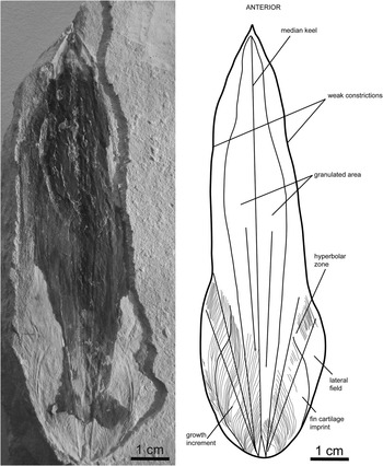
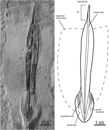
Gladius morphology of Glyphiteuthis libanotica (Fraas, Reference Fraas1878), MNHN.F.A50406, Cenomanian, Haqel, ventral view. All growth increments have not been drawn.

Occurrence
Early late Cenomanian of Haqel and Hajoula (Lebanon), (?)Santonian of Sahel Aalma (Lebanon).
Description
The studied gladius (MNHN.F.A50406; from the Cenomanian of Haqel) is unique and relatively damaged (Fig. 15), but a morphological description and a taxonomic identification are possible. This specimen is 103 mm in mfl and is seen in ventral view. The anterior median field is sharp. Halfway along the gladius, the mfw is 21 mm (~20% of the mfl). The width continuously decreases anteriorly, but at ~70% of the mfl from its posterior extremity, the median field is weakly constricted. Based on distinct growth increments, the gladius can be subdivided into a median field, a pair of comparatively wide and long hyperbolar zones, and a pair of lateral fields. Hyperbolar zone length represents ~37% of the mfl. Outer asymptotes are distinct, but inner asymptotes are barely perceptible. The outer asymptotes diverge at an angle of 25°. Inner asymptotes appear to diverge at an angle of ~15°. Laterally, outspread lateral fields form a rounded posterior gladius end. A granulation is visible as a negative relief on the median field. Halfway along the gladius, the granulated area is relatively wide and diverges at an angle of 10°. The granulated area also does not seem to reach the anterior, posterior, and lateral margins of the gladius. The granulation is very thin on the posterior part of the gladius and it becomes more conspicuous toward the anterior part. Imprints of the growth increments are well preserved on the posterior part of the gladius. The posterior end shows an M-like shape. Within the same posterior area, two symmetrical half-ovoid structures are visible and may represent imprints of fin cartilages (i.e., fin attachments). There is no evidence of soft parts attached to the gladius.
Remarks
Potential affinities with the suborders Loligosepiina and Prototeuthidina can be excluded as these two suborders are characterized by a gladius with a triangular anteriorly diverging median field and the absence of a prominent median keel (Fuchs and Larson, Reference Fuchs and Larson2011b). Compared to the Jurassic Trachyteuthis and Actinosepia, the specimen described here is more slender and anteriorly sharper. The Cretaceous genus Glyphidopsis strongly differs from the studied specimen in having a hyperbolar zone length representing more than 50% of the mfl. The studied specimen very likely pertains to the genus Glyphiteuthis because of the granulation of the dorsal surface and of the hyperbolar zone length/mfl ratio (~0.37) that is similar to that of Glyphiteuthis libanotica (0.39) documented by Fuchs and Weis (Reference Fuchs and Weis2009). Placement of Glyphiteuthis within the family Trachyteuthididae is commonly accepted owing to the presence of granules on the dorsal gladius surface.
The specimen described here is very likely conspecific with Glyphiteuthis libanotica. This species differs from Glyphiteuthis ornata by its dorsal granulation that is significantly wider. Additionally, G. ornata exhibits a row of very coarse granules on the top of the median keel, whereas G. libanotica show a homogeneous granulation (Fuchs and Larson, Reference Fuchs and Larson2011b). The homogeneous and large granulated surface of the studied specimen justifies its attribution to G. libanotica. G. libanotica and Glyphiteuthis freijii can be distinguished by a marked constriction close to the end of the anterior median field in G. freijii. Glyphiteuthis abisaadiorum is generally distinguished from G. libanotica by its narrower gladius morphology with a mfw/mfl ratio ~0.16 at half way along the gladius (Fuchs and Weis, Reference Fuchs and Weis2009). This ratio is ~0.20 for the studied specimen and is identical to the ratio obtained by Fuchs and Larson (Reference Fuchs and Larson2011b) for G. libanotica. In addition, G. abisaadiorum does not exhibit any constriction of the anterior median field.
Glyphiteuthis libanotica was reported from the early late Cenomanian of Haqel. Roger (Reference Roger1946, p. 17, fig. 10) also studied two fragmentary gladii from Sahel Aalma that probably belong to G. libanotica, but their current repository is unknown and thus preventing confirmation of the potential temporal range extension of this taxon up to the Santonian. Detailed information on the presumed existence of this species in the Santonian of Sahel Aalma is given in Fuchs and Larson (Reference Fuchs and Larson2011b).
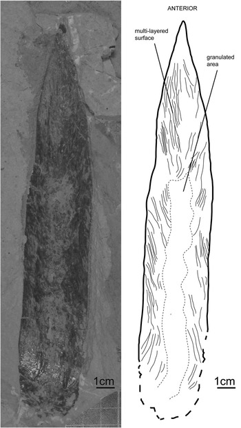
Gladius morphology of Glyphiteuthis sp., MNHN.F.A50407, Cenomanian, En Nammoura, ventral view.

Description
The specimen (MNHN.F.A50407; from the Cenomanian of En Nammoura), seen in ventral view, exhibits a mfl of 221 mm, but it is only partly preserved. The anterior tip of the median field is angular. The posterior conus area is apparently missing. The lateral fields and hyperbolar zones are not preserved. The maximum mfw (~33 mm) is located close to the presumed anterior end of the hyperbolar zones and it decreases continuously anteriorly. Halfway along the gladius, the mfw is ~14% of the incomplete mfl and thus was originally smaller. The gladius is distinctly multilayered and exhibits circular depressions close to the median axis. These depressions likely correspond to the negative relief of the granules on the dorsal surface. The granulated area does not reach the lateral gladius margin and terminates at about two-thirds of the mfl. There is no evidence of constriction of the anterior median field and median keel. The lateral margins appear to be curved.
Remarks
Similarly to Glyphiteuthis libanotica, an affinity with the suborders Loligosepiina and Prototeuthidina can be excluded. Compared to the genera Trachyteuthis and Actinosepia, the gladius of the studied specimen is more slender and anteriorly sharper. Glyphidopsis strongly differs from the specimen described here in having a relatively larger gladius. Due to the granulation of its dorsal surface, the studied specimen very likely pertains to the genus Glyphiteuthis.
Although mfw/mfl ratios have to be interpreted with caution, the studied specimen differs from G. libanotica and G. ornata by its overall narrower gladius morphology. Indeed, the original mfw/mfl ratio halfway along the gladius was <0.14, whereas the same ratio is ~0.20 for G. libanotica and G. ornata (Fuchs and Weis, Reference Fuchs and Weis2009). The constriction of the anterior median field observed in G. libanotica, G. ornata, and G. freijii is absent in the specimen described here. The studied gladius resembles G. abisaadiorum in having a slender median field (mfw/mfl ratio halfway along the gladius ~0.16 for G. abisaadiorum; Fuchs and Weis, Reference Fuchs and Weis2009). It also exhibits curved lateral margins and numerous laminated layers as observed in G. rhinophora (Fuchs et al., Reference Fuchs, Stinnesbeck, Ifrim, Giersch, Gutierrez and Frey2010a). However, G. rhinophora mainly differs from the studied specimen by its nose-shaped anterior part of the gladius and a prominent median keel (Fuchs et al., Reference Fuchs, Stinnesbeck, Ifrim, Giersch, Gutierrez and Frey2010a). G. abisaadiorum differs from the studied specimen in having straighter lateral margins (Fuchs and Larson, Reference Fuchs and Larson2011b) and an inconspicuous but distinct mid-dorsal keel (Fuchs and Weis, Reference Fuchs and Weis2009). The specimen described here displays an additional interesting feature: the granulated area seems to occupy only the posterior two-thirds of the visible mfl (Fig. 16). On the contrary, the granulated area in other Glyphiteuthis species always extends much more anteriorly.
Specimen MNHN.F.A50407 is incompletely preserved but its combination of characters is apparently unique within the genus Glyphiteuthis. Glyphiteuthis sp. share more similarities with G. rhinophora and G. abisaadiorum than with any other Glyphiteuthis species. In any case, it differs from G. libanotica. This specimen being unique, we prefer not to assign it to a particular species for the time being.
Family Palaeololiginidae Naef, Reference Naef1921
Diagnosis
Teudopseid gladius rachis-like anteriorly, leaf-like posteriorly. Median field narrow with a pronounced median keel. Hyperbolar zones weakly curved. Ratio hyperbolar zone length/mfl 0.23–0.42 (after Fuchs and Larson, 2011b).
Other genera
Rachiteuthis Fuchs, Reference Fuchs2006a; Marekites Kostak, Reference Kostak2002; (?) Styloteuthis Fritsch, Reference Fritsch1910.
Occurrence
Late Jurassic (Tithonian) of SW Germany–Late Cretaceous (Cenomanian) of Lebanon.
Genus Rachiteuthis Fuchs, Reference Fuchs2006a
Type species
Rachiteuthis donovani Fuchs, Reference Fuchs2006a.
Diagnosis after Fuchs (Reference Fuchs2006a)
Palaeololiginid with a spear or shaft-like gladius; lateral fields short and spread out with a weak hyperbolar zone; conus reduced; free rachis elongated; distinct lateral ridges and a median unornamented keel extends from the posterior to the anterior extremity; body torpedo-shaped; insertions of triangular fins relatively broad; slender arms.
Occurrence
Early late Cenomanian.

Gladius morphology of Rachiteuthis acutali n. sp., holotype, MNHN.F.A50408, Cenomanian, Hajoula, dorsal view. All growth increments have not been drawn.

Diagnosis
Palaeololiginid with a spear-like gladius. Hyperbolar zones between the median field and the lateral fields poorly developed. Prominent mid-dorsal keel and lateral reinforcements. Slender and narrow anterior tip, appearing to be the prolongation of the mid-dorsal keel. Without the tip, hyperbolar zone length/mfl ratio ~0.3.
Description
The holotype (MNHN.F.A50408; from the Cenomanian of Hajoula), seen in dorsal view, shows a well-preserved slender gladius consisting of a rachis-like median field and well-developed lateral fields. A median unornamented keel extends from posterior to anterior extremities terminating with a characteristically sharp and narrow tip at the extreme anterior part of the gladius. The holotype mfl is 95 mm, including the tip. The maximum mfw is ~10 mm at two-thirds of the mfl from the posterior end. The hyperbolar zones between the median field and the lateral fields are poorly developed. The anterior sharp median field bears a prominent mid-dorsal keel as well as lateral reinforcements. The hyperbolar zone length is ~25 mm and represents ~26% of the mfl, including the tip. Without the tip, the hyperbolar zone length represents ~30% of the mfl. Inner asymptotes are distinct, but outer asymptotes are barely perceptible. Inner asymptotes diverge at an angle of 7°. The elongated tip is 14 mm in length, i.e., ~15% of the mfl, and appears to be the prolongation of the mid-dorsal keel. Growth increments are well preserved on the posterior part of the gladius. Soft parts are poorly preserved on the specimen, but the outline of the mantle can be distinguished under UV light, as well as poorly preserved arm-like structures.
Etymology
After the typical tip of this species (L., acutalis, ended by a tip).
Holotype
MNHN.F.A50408, Hajoula (Lebanon), late Cenomanian.
Occurrence
Hajoula, Lebanon, late Cenomanian.
Remarks
Affinity of the holotype (Fig. 17) with the suborders Loligosepiina and Prototeuthidina can be excluded since the representatives of these two clades have a gladius with a triangular, diverging median field and without a prominent median keel. The gladius morphology of the holotype is in accordance with Naef’s (Reference Naef1921) diagnosis of the family Palaeololiginidae (suborder Teudopseina). The specimen described here is characterized by an elongated rachis in contrast to Palaeololigo oblonga Naef, Reference Naef1921, but it also differs by the presence of a tip on the anterior part of the gladius. A comparison with Marekites vinarensis Fritsch, Reference Fritsch1910 and Styloteuthis convexa Fritsch, Reference Fritsch1910 (both from the Turonian of Bohemia) is more problematic as the anterior extension of what resembles a rachis-like median field remains unknown for these two taxa (Fuchs and Larson, Reference Fuchs and Larson2011b). Comparison of the soft-part anatomy with known palaeololiginids is not possible owing to the poor preservation of specimen MNHN.F.A50408. The holotype is very similar to R. donovani, but it slightly differs from it by its hyperbolar zone length/mfl ratio (0.3 vs 0.23–0.26, respectively). Additionally, illustrated specimens of R. donovani (Fuchs, Reference Fuchs2006a; Fuchs and Larson, Reference Fuchs and Larson2011a) lack a slender and narrow anterior tip. The erection of a new species is thus justified based on its diagnostic tip.
Interestingly, the typical tip of R. acutali n. sp. resembles the pointed anterior extremity described in the Jurassic Teudopsis bunelii Eudes-Deslongchamps, Reference Eudes-Deslongchamps1835, but also in Jurassic muensterellid and palaeloliginid coleoids (Fuchs and Weis, Reference Fuchs and Weis2010; Fuchs and Larson, Reference Fuchs and Larson2011b). The latter are assumed to be ancestral to the genus Rachiteuthis (Fuchs and Larson, Reference Fuchs and Larson2011b). However, Rachiteuthis donovani does not display a pointed anterior extremity or a tip (Fuchs, Reference Fuchs2006a; Fuchs and Larson, Reference Fuchs and Larson2011b) and a small tip is observed in the teudopseid Glyphiteuthis rhinophora (Fuchs et al., Reference Fuchs, Stinnesbeck, Ifrim, Giersch, Gutierrez and Frey2010a). It can be therefore hypothesized that a pointed anterior extremity or a tip is a character that may have appeared independently in different clades. The presence of a tip in R. acutali n. sp. could thus be seen as a convergence with other species of the palaeololiginid/muensterellid lineage and G. rhinophora.
Phylogenetic implications
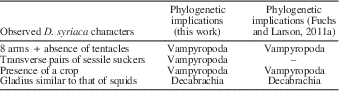
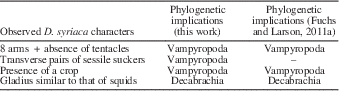
The study of Dorateuthis syriaca specimens has allowed us to infer the phylogenetic position of this taxon relative to modern coleoids (see also Fuchs et al., Reference Fuchs, Klinghammer and Keupp2007b; Fuchs and Larson, Reference Fuchs and Larson2011a, Reference Fuchs and Larson2011b). We thus detected four characters phylogenetically significant at the superorder level including one that has never been described before this work. A synthesis of these features is given in Table 1. Based on these characters, we suggest that D. syriaca (suborder Prototeuthidina) is closely related to modern vampyropods. The decabrachian-like characters are considered here as convergent with modern squids.
Synthesis of the characters with phylogenetic significance at the superorder level observed in the studied Dorateuthis syriaca specimens
Number of arms
The key feature of the Decabrachia is the presence of 10 arms with the fourth pair modified in tentacles. Supporters of the “Decabrachia Theory” usually explain the absence of preserved tentacles on gladius-bearing coleoids as reflecting the ability of modern coleoids to retract them into pockets (Young et al., Reference Young, Vecchione and Donovan1998; Vecchione et al., Reference Vecchione, Young, Donovan and Rodhouse1999; Bizikov, Reference Bizikov2008; and Donovan and Strugnell, Reference Donovan and Strugnell2010). However, no evidence of such pockets exists among the studied specimens. Additionally, to our knowledge, such pockets have not been described yet in fossil coleoids (as discussed in Fuchs and Larson, Reference Fuchs and Larson2011b, p. 829). It can also be argued that the absence of tentacles may simply result from a preservation artifact. Kear et al. (Reference Kear, Briggs and Donovan1995) studied the influence of buoyancy on preservation probability: in coleoids that are positively buoyant after death (cuttlefishes and some oceanic squids), the tentacles easily slip out and hang loose in the water. These authors therefore considered that the prototeuthidid Plesioteuthis had tentacles, which are never preserved because of the post-mortem positive buoyancy of the individuals. However, given the absence of unambiguous tentacles in numerous published specimens of exceptionally well-preserved coleoids representing juvenile to adult stages, the invariable post-mortem tentacles loss prior to fossilization suggested by Kear et al. (Reference Kear, Briggs and Donovan1995) or the loss during ontogeny (only known in the modern decabrachian Octopoteuthis Rüppell, Reference Rüppel1844; Mangold et al. Reference Mangold, Bidder and Portmann1989a) appears unlikely. We therefore suggest that the absence of tentacles on the studied specimens indicates that D. syriaca is closely related to modern vampyropods (Table 1). The taxa Mastigophora Owen, Reference Owen1856 and Plesioteuthis have been described with tentacles (Vecchione et al., Reference Vecchione, Young, Donovan and Rodhouse1999; Naef, Reference Naef1922, p. 118). However, agreeing with Haas’s interpretation (2002) of only eight arms in Mastigophora, we consider that Naef (Reference Naef1922) misinterpreted the slightly elongated dorsal arm pair as tentacles and that the only faint traces present on Mastigophora specimens (Vecchione et al., Reference Vecchione, Young, Donovan and Rodhouse1999) cannot be easily interpreted as tentacles.
Suckers
Among extant coleoids, all decabrachian species have pedunculate suckers that alternate to form oblique pairs (Young et al., Reference Young, Vecchione and Donovan1998). Vampyropods present suckers that form oblique or transverse pairs, depending on the species. Additionally, the suckers are sessile with a wide base in incirrate octopods (Mangold et al., Reference Mangold, Bidder and Portmann1989a). Thus, in light of the transverse pairs of suckers observed in MNHN.F.A50397 (Fig. 9.4), Dorateuthis syriaca appears closely related to modern vampyropods (Table 1). Furthermore, the fact that visible suckers are sessile and cirri are absent suggests an affinity with incirrate octopods.
Modern decabrachians possess suckers with a bilateral symmetry whereas extant vampyropods have suckers with a radial symmetry (Young and Vecchione, Reference Young and Vecchione1996). Unfortunately, the sucker symmetry is not discernible on the studied specimens. Suckers of modern decabrachians also possess chitinous rings or teeth (or hooks). Coleoid hooks are well-known in the Lebanese Lagerstätten, but always in association with phragmocone-bearing belemnoids and never with gladius-bearing coleoids (Fuchs and Larson, Reference Fuchs and Larson2011b, p. 829).
Within the suborder Octopoda Leach, Reference Leach1818, suckers are arranged all along the arm as a single (Eledone Leach, Reference Leach1817) or double (Octopus) row. Fuchs et al. (Reference Fuchs, Klinghammer and Keupp2007b) presumed a single row of suckers in the Jurassic Plesioteuthis prisca. However, Voight (Reference Voight1993) showed that the arrangment of suckers on octopodid arms is not a character having phylogenetic significance.
Crop
Mangold and Young (Reference Mangold and Young1998) emphasized the phylogenetic value of the digestive system for the main coleoid groups. Indeed, they considered that the presence of a crop is characteristic of octopods. If the observation of a crop is validated, it would also support that Dorateuthis syriaca is closely related to modern vampyropods (Table 1).
Gladius
Donovan and Toll (Reference Donovan and Toll1988) and Doyle et al. (Reference Doyle, Donovan and Nixon1994) considered Dorateuthis syriaca and Plesioteuthis prisca as members of the superorder Decabrachia. Indeed, they suggested a close relationship with modern squids, based on a proposed homology of the gladii of extant ommastrephids and that of the genera Dorateuthis and Plesioteuthis. Similarly, Reitner and Engeser (Reference Reitner and Engeser1982) suggested a phylogenetic relationship between the Jurassic plesioteuthidid Maioteuthis and modern squids. According to Young et al. (Reference Young, Vecchione and Donovan1998), the gladius of the genus Plesioteuthis (and therefore that of Dorateuthis syriaca) is so similar to that of the ommastrephids that it indicates either a phylogenetic link or a convergence between extant squids and the genera Plesioteuthis and Dorateuthis. We agree that the gladius of D. syriaca, as well as other genera of the suborder Prototeuthidina, is very similar to that of modern squids (Mangold et al., Reference Mangold, Bidder and Portmann1989a; Boyle and Rodhouse, Reference Boyle and Rodhouse2005). However, in light of the other characters we discussed previously, and in accordance with numerous authors (Bandel and Leich, Reference Bandel and Leich1986; Engeser and Keupp, Reference Engeser and Keupp1999; Haas, Reference Haas2002; Lukeneder and Harzhauser, Reference Lukeneder and Harzhauser2004; Fuchs, Reference Fuchs2006a, Reference Fuchs2006b; Fuchs et al., Reference Fuchs, Klinghammer and Keupp2007b; Fuchs and Larson, Reference Fuchs and Larson2011a), it is more likely that the similarities of the gladius of prototeuthidid coleoids with that of modern squids indicate evolutionary convergences.
To summarize, we failed to find a single unequivocal decabrachian character in the specimens of Dorateuthis syriaca studied here. We consider that gladius-bearing coleoids belonging to the suborder Prototeuthidina are only superficially similar to living teuthids and that the soft-part morphology of Dorateuthis (eight arms, transverse and sessile suckers, presence of a crop; Table 1) suggests close phylogenetic relationships with the eight-armed vampyropods. We thus support the hypothesis of a vampyropod affinity as initially proposed by Bandel and Leich (Reference Bandel and Leich1986) and later endorsed by Engeser and Keupp (Reference Engeser and Keupp1999), Haas (Reference Haas2002), Fuchs (Reference Fuchs2006a), and Fuchs and Larson (Reference Fuchs and Larson2011a).
Conclusions
The study of 16 gladius-bearing coleoid specimens from the Upper Cretaceous Lebanese Lagerstätten leads to a better understanding of their anatomy and of their taxonomic diversity. Four taxa of gladius-bearing coleoids were identified: the previously known Dorateuthis syriaca and Glyphiteuthis libanotica, but also Rachiteuthis acutali n. sp. and an original form of Glyphiteuthis sp. This increases the number of known Cretaceous gladius-bearing coleoid species from Lebanon from 9 to 11.
Despite the exceptional preservation characterizing these Lebanese fossils, the anatomical and phylogenetic interpretations of soft-part characters remain difficult. However, the quality of preservation of specimens from Sahel Aalma helped us to determine a group of important soft-part characters that have a phylogenetic significance at the superorder level for D. syriaca. These are the absence of tentacles, the presence of a crop, and the presence of sessile and transverse pairs of suckers. Based on these characters, we conclude that D. syriaca (suborder Prototeuthidina) is closely related to modern vampyropods. No evidence supports a close phylogenetic relationship of the studied gladius-bearing coleoids from the Upper Cretaceous Lebanese Lagerstätten with modern decabrachians. Consequently, the evolutionary history of present-day squids and cuttlefishes still remains unclear.
Acknowledgments
We thank D. Fuchs (Berlin) who kindly shared unpublished data on D. syriaca and contributed to numerous discussions. We are grateful to S. Hageman and M. Yacobucci for their editorial comments, as well as the two anonymous reviewers for their constructive suggestions that improved the quality of the manuscript.