Thyroid hormone (TH) coordinates short-term and long-term energy requirements by regulating metabolic processes essential for normal growth, development and maintenance of cellular functions (Ref. Reference Oetting and Yen1). Thyroid gland produces and releases the prohormone thyroxin (T4) into circulation, a process that is regulated by thyrotropin releasing hormone and thyroid stimulating hormone. T4 is further taken up into cells and transformed into the active form triiodothyronine (T3) by iodothyronine deiodinases D1 and D2, which are differentially expressed in mammalian tissues (Ref. Reference Mullur, Liu and Brent2). As a key metabolic regulator, T3 is considered a hormetic agent, which is able to induce physiological (i.e. calorigenic effect), beneficial (i.e. organ protection) responses in the low-concentration range, whereas potentially harmful responses are triggered at high-concentration ranges (i.e. thyrotoxicosis) (Refs Reference Calabrese3, Reference Videla4, Reference Bernardes5). Recently, Mourouzis et al. have drawn attention that T3 or T4 play a critical role for the repair after injury in several organs in the experimental setting (Ref. Reference Mourouzis, Politi and Pantos6). Studies by our group have demonstrated that the in vivo administration of a single dose of T3 (0.1 mg/kg; 20 μg/animal) to rats affords protection against liver injury induced by1 h ischemia/20 h reperfusion protocol (Ref. Reference Fernández7), representing an example of pharmacological preconditioning (PC) that might have clinical application. In general terms, organ PC refers to the development of an increased tolerance to injuring stimuli, which is induced by previous maneuvers triggering beneficial molecular and functional changes (Ref. Reference Selzner, Boehnert and Selzner8). In addition to the PC action of THs against organ injury, restoring normal TH levels in specific cases has resulted in several beneficial effects, which include (i) the T3-dependent preservation of liver regenerative capacity and antiinflammatory responses after partial hepatectomy in the rat when combined with methylprednisolone (Ref. Reference D'Espessailles9); (ii) the improvement induced by T4 in mental, motor and neurological outcomes in infant <28 weeks of gestation, which exhibit lower plasma TH levels than in term-born infants (Ref. Reference van Wassenaer and Kok10); (iii) the diminution in the cognitive side effects of lithium and electroconvulsive therapy by T3 administration to patients with bipolar disorders (Ref. Reference Tremont and Stern11); and (iv) the higher number of organs being functionally acceptable and in the early and intermediate graft survival found in brain-dead donors (Ref. Reference Cooper12). However, because of discrepancies observed in the benefits of routine TH administration in brain-dead potential organ donors, additional large-scale, prospective, multicentre, doubled-blind and placebo-controlled studies are needed to optimise TH therapy (Refs Reference Dikdan, Mora-Esteves and Koneru13, Reference Mi14). In this review article, the cytoprotective signalling mechanisms associated with T3 action are addressed, which may constitute hormetic responses underlying organ PC against injury or the repair and recovery after injury.
Signalling pathways in T3-induced calorigenesis and liver reactive oxygen species generation
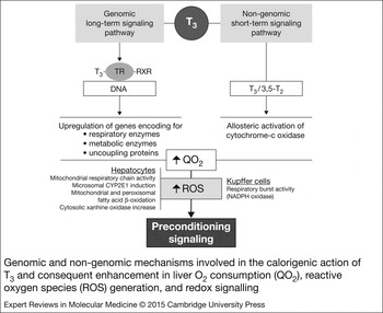
Acceleration of energy metabolism by T3 is achieved through genomic and non-genomic pathways (Fig. 1). In the genomic pathway, T3 binds to nuclear thyroid hormone receptors (TRs) expressed in multiple isoforms in almost all tissues (Ref. Reference Oppenheimer, Schwartz and Surks15), namely, TRα1, TRβ1 and TRβ2 (Ref. Reference Oetting and Yen1). These TRs bind as heterodimers with retinoid X receptor to specific thyroid hormone response elements present in the promoter region of target genes DNA (Fig. 1). Upon T3 binding, co-repressors are released and co-activators and several additional complexes are recruited, constituting a bridge with RNA-polymerase-II and the basal transcription machinery, leading to enhanced promoter activity and synthesis of respiratory, metabolic enzymes and uncoupling proteins (Refs Reference Oetting and Yen1, Reference Mullur, Liu and Brent2). Consequently, higher rates of oxidative phosphorylation and ATP production are achieved, ATP levels being partially balanced by uncoupling protein induction (Ref. Reference Lanni16), and the enhancement in mitochondrial O2 consumption (QO2) establishes higher rates of reactive oxygen species (ROS) generation (Refs Reference Fernández and Videla17, Reference Venditti, De Rosa and Di Meo18) (Fig. 1). These responses have a latency of hours to days, contrary to those triggered by non-genomic pathways.
Genomic and non-genomic mechanisms involved in the calorigenic action of T3 and consequent enhancement in liver O2 consumption (QO2), reactive oxygen species (ROS) generation, and redox signalling. CYP2E1, cytochrome P450 isoform 2E1; RXR, retinoid X receptor; 3,5-T2, 3,5-diiodothyronine; TR, thyroid hormone receptors.

Non-genomic mechanisms of T3/T4 action have been described to be dependent on plasma membrane receptor on integrin αVβ3 and cytoplasmic TRβ1 and TRα-derived polypeptide (TRΔα1), which show rapid onsets of action (seconds–minutes), and are carried out by alternate signalling pathways (Refs Reference Oetting and Yen1, Reference Mullur, Liu and Brent2, Reference Davis, Leonard and Davis19). Among them, T3 and 3,5-diiodothyronine (3,5-T2) can directly stimulate oxidative phosphorylation, as evidenced by the rapid activation of cytochrome-c oxidase through binding to its Va subunit that abolishes the allosteric inhibition by adenosine-5,-triphosphate (ATP) (Ref. Reference Arnold, Goglia and Kadenbach20) (Fig. 1).
Both genomic long-term and non-genomic short-term signalling mechanisms determine the increase in mitochondrial QO2 with consequent ROS generation observed in hyperthyroid state, which may be contributed by induction of other enzymatic processes. In the liver, these include enhanced microsomal activity secondary to the induction of the highly pro-oxidant cytochrome P450 isoform 2E1 (CYP2E1) (Ref. Reference Fernández21), coupled to higher activities of reduced nicotinamide adenine dinucleotide phosphate (NADPH)-cytochrome P450 reductase (Ref. Reference Ram and Waxman22), NADPH oxidase (Ref. Reference Fernández23), and of the NADPH-generating system glucose-6-phosphate dehydrogenase (Ref. Reference Simon-Giavarotti24), thus supporting ROS generation (Fig. 1) (Ref. Reference Fernández23). Besides liver mitochondrial and microsomal ROS production, T3 enhances ROS generation because of xanthine oxidase increase (Ref. Reference Huh25) and that coupled to enhanced fatty acid β-oxidation because of liver peroxisomal proliferation (Refs Reference Fernández and Videla17, Reference Just and Hartl26) (Fig. 1). The above mentioned hepatic mechanisms generating ROS after T3 administration are presumed to occur primarily in hepatocytes, with Kupffer cells playing a contributory role because of T3-induced increase in the respiratory burst activity, a process mainly associated with ROS production because of NADPH oxidase activation (Ref. Reference Tapia27) (Fig. 1). Data discussed support the development of a pro-oxidant state in the liver by T3 administration, a finding that is in line with the concept of redox signalling, a phenomenon in which ROS affect cellular signalling and eventually gene expression (Ref. Reference Forman, Maiorino and Ursini28). In this respect, hydrogen peroxide (H2O2) is considered as a major ROS able to achieve redox signalling, which can be accomplished by reversible oxidation of cysteine residues in signalling proteins (Ref. Reference Forman, Maiorino and Ursini28), H2O2 being significantly enhanced in hyperthyroid state (Refs Reference Fernández and Videla17, Reference Venditti, De Rosa and Di Meo18) (Fig. 1).
At the cellular level, oxidative stress induces a wide spectrum of responses, depending on the cell type, the level of ROS attained, and the duration of the exposure (Ref. Reference Videla29). In this context, the moderate increase in ROS levels in a define time period elicits an imbalance capable of redox regulation with beneficial responses, as found for T3-induced liver oxidative stress. However, conditions in which large levels of ROS are achieved, severe oxidation of biomolecules and dysregulation of signal transduction and gene expression are induced resulting in cell death, as reported in organs subjected to ischemia-reperfusion (IR) (Ref. Reference Videla29).
T3-induced redox signalling and liver PC
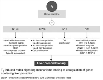
The sinusoidal lining of the liver contains non-parenchymal cells including Kupffer cells, endothelial cells and stellate cells, which are important in liver homeostasis and in the pathogenesis of liver disease. Kupffer cells are primarily involved in phagocytosis and processing of ingested materials, antigen presentation and secretion of biologically active mediators upon activation, representing a key component of innate immunity, the initial, rapid response to dangerous stimuli (Ref. Reference Bilzer, Roggel and Gerbes30). T3 administration led to Kupffer cell hyperplasia (Ref. Reference Gomes31), an effect that was paralleled by enhancement in the rate of carbon phagocytosis and the associated carbon-induced O2 consumption, as evidenced in the isolated perfused rat liver (Ref. Reference Tapia27). The latter parameter is related to the respiratory burst activity of Kupffer cells, which is largely dependent on the activity of the ROS-generator NADPH oxidase and abolished by the Kupffer cell inactivator gadolinium chloride (GdCl3) (Ref. Reference Tapia27). In addition to the respiratory burst, T3-induced GdCl3-sensitive liver O2 uptake may involve enhancement in mitochondrial respiration. This contention is supported by the existence of nuclear binding proteins for T3 in Kupffer cells (Ref. Reference Sellitti, Oliver and Latham32), which may trigger the genomic signalling mechanism (Fig. 1) contributing to higher rates of liver macrophage O2 consumption and ROS generation. Under these conditions, T3 increases the hepatic DNA binding of transcription factors nuclear factor-κB (NF-κB), signal transducer and activator of transcription 3 (STAT3) and activating protein 1 (AP-1) (Fig. 2), an effect that is suppressed by GdCl3 and antioxidants when given prior to T3 (Refs Reference Tapia33, Reference Tapia34, Reference Fernández35), suggesting that T3 triggers their redox activation by actions exerted at the Kupffer cell level, which may be involved in liver PC. It is important to note that T3-induced liver PC against IR injury is observed 48 h after T3 administration, time period in which oxidative stress triggered by T3 has already relapsed to a normal status (Ref. Reference Fernández7). However, if noxious stimuli such as IR (Ref. Reference Troncoso, Smok and Videla36) or acute lindane treatment (Ref. Reference Videla37) are applied following 24 h after T3 administration under full oxidative stress development, exacerbation of liver injury is observed because of oxidative stress potentiation. Similarly, severe hyperthyroidism achieved by high T3/T4 dosages and/or prolonged periods of treatment is associated with thyrotoxic myopathy, cardiac dysfunction, potentiation of xenobiotic hepatotoxicity and higher susceptibility of hyperthyroid tissues to prolonged exercise (Ref. Reference Venditti and Di Meo38).
T3-induced redox signalling mechanisms leading to upregulation of genes conferring liver protection. AP-1, activating protein-1; GCL, glutamate cysteine ligase; GST, glutathione-S-transferases; HO-1 Heme-oxygenase 1; iNOS, inducible nitric oxide synthase; mEh-1, microsomal epoxide hydrolase; MnSOD, manganese superoxide dismutase; MRP-2/3, multidrug resistance protein 2/3; NF-κB, nuclear factor-κB; NQO-1, NADPH-quinone oxidoreductase 1; Nrf2, nuclear factor erythroid-2-related factor-2; STAT3, signal transducer and activator of transcription-3; Thr, thioredoxin.

Activation of NF-κB, AP-1 and STAT3
In the liver, Kupffer cells are more sensitive for the redox control of transcription factor activation than hepatocytes (Ref. Reference Tran-Thi, Decker and Baeuerle39), because of the highly pro-oxidant state prevailing in Kupffer cells, which is related to NADPH oxidase activity and that most of the antioxidant mechanisms, are confined to parenchymal cells (Ref. Reference Vrba and Modriansky40). Consequently, NF-κB and AP-1 redox activation by T3 upregulate the expression of several mediators released by Kupffer cells, including tumour necrosis factor-α (TNF-α) (Ref. Reference Tapia33) and interleukin 6 (IL-6) (Ref. Reference Tapia34). The effects of these cytokines are exerted through their interaction with specific surface receptors in target cells within the liver to mediate signalling from cell membrane to the nucleus (Ref. Reference Tsukamoto41). Coupling of TNF-α with TNF-α receptor 1 (TNFR-1) results in the activation of the downstream kinases inhibitor of κB (IκB) kinase (IKK), c-Jun N-terminal kinase (JNK) and p38 (Ref. Reference Hayden and Ghosh42). In the case of T3-induced TNF-α response, liver IKK phosphorylation and NF-κB activation are achieved, effects that are abolished by α-tocopherol or GdCl3, supporting the role of ROS and Kupffer cell activation in T3-dependent upregulation of hepatic gene expression (Refs Reference Fernández43, Reference Fernández44). Therefore, enhanced expression of the NF-κB-responsive genes encoding for antioxidant enzymes, antiapoptotic proteins and acute-phase proteins type I is achieved, representing hepatoprotective responses triggered by the underlying oxidative stress status developed (Fig. 2). In this respect, oxidative stress-induced activation of NF-κB pathway in lymphocytes was associated with the rise of serum immunoglobulins in hyperthyroid patients, a pathway that is also involved in immune response (Ref. Reference Nandakumar45).
Besides signalling through NF-κB activation, T3-induced IL-6 upregulation triggers the acute-phase response (APR) of the liver (Ref. Reference Tapia34). The APR is an important pathophysiologic reaction, in which normal homeostatic mechanisms are replaced by new set points contributing to defensive or adaptive capabilities. This reaction is characterised by (i) being focused mainly on the liver; (ii) the involvement of IL-1 like cytokines (IL-1, TNF-α) and IL-6 like cytokines (IL-6, oncostatin M); (iii) the activation of transcription factors NF-κB, AP-1, STAT 3/5, or CCAAT/enhancing-binding protein β (C/EBPβ); and (iv) the enhancement in the plasma levels of a number of positive acute-phase proteins (APPs), such as clotting factors, transport proteins, antiproteases and complement factors, with diminution of negative APPs (albumin) (Refs Reference Samols, Agrawal, Krushner, Oppenheim and Feldman46, Reference Gruys47). Coupling of IL-6 with gp130 receptor subunit and the subsequent activation of the gp130-associated Janus kinase (JAK)/STAT3 system triggers the expression of APPs (Ref. Reference Erns and Jenkins48). This cascade is upregulated by T3 administration, with induction of both types of APPs (Ref. Reference Tapia34) (Fig. 2), which may be contributed by the T3-induced TNF-α/IKK/NF-κB pathway controlling type I APP genes (Refs Reference Fernández43, Reference Fernández44). Although T3 or T4 were shown to upregulate the hepatic synthesis of the APPs α1-acid glycoprotein, α2-(acute phase) globulin (Ref. Reference Griffin and Miller49), ceruloplasmin (Ref. Reference Fitch, Song and Leversom50), angiotensinogen, complement component 4A and serum amiloid A protein precursor (Ref. Reference Lin51), the mechanisms underlying these effects were not assessed. In addition, T3 also upregulated the expression of the APP ferritin post-transcriptionally by increasing iron-induced displacement of iron regulatory protein from the iron-responsive element present in ferritin mRNA (Ref. Reference Leedman52). Hence, upregulation of the hepatic APR may represent a defence mechanism against the detrimental effects of oxidative stress occurring in the liver and in extrahepatic tissues with a calorigenic response (Ref. Reference Tapia34), considering that the goal of the APR is to restore homeostasis (Refs Reference Samols, Agrawal, Krushner, Oppenheim and Feldman46, Reference Gruys47).
The in vivo effects of T3 as a primary hepatic mitogen leading to hepatocyte proliferation in intact liver are well established (Refs Reference Francavilla53, Reference Bungay54). In this process, resting hepatocytes in the G0 phase of the cell cycle are primed by TNF-α and IL-6 before they can respond to growth factors, with the concomitant activation of NF-κB, STAT3 and AP-1, to enter the G1 phase and initiate cell cycle progression (Ref. Reference Fausto, Campbell and Riehle55). T3 administration triggered the expression of liver cyclin-dependent kinase 2 (CDK-2) (Ref. Reference Fernández35) and cyclin D1, with enhanced retinoblastoma protein phosphorylation (Ref. Reference Pibiri56), thus favouring G1 to S phase transition and hepatocyte proliferation. This was evidenced by the increase in nuclear cell proliferation-associated protein Ki-67 and in proliferating cell nuclear antigen (Ref. Reference Fernández35). The mechanisms of the cell proliferating action of T3 may involve Kupffer cell-dependent generation of the priming cytokines TNF-α and IL-6, leading to JNK/AP-1 and/or JAK/STAT3 activation, respectively (Ref. Reference Fernández35) (Fig. 2). Recently, T3 was reported to stimulate hepatocyte proliferation through the activation of the protein kinase A (PKA)-dependent β-catenin pathway, a finding that may be of therapeutic importance in the setting of liver transplantation, cell therapy, or hepatic tissue engineering (Ref. Reference Fanti57). Furthermore, the mitogenic action of T3 on the liver constitutes a powerful stimulus for the proliferation of transplanted hepatocytes (Ref. Reference Oren58) and is associated with nodule regression and reduction in hepatocellular carcinomas, an anticarcinogenic effect probably related to re-differentiation of preneoplastic cells (Ref. Reference Ledda-Columbano59).
Data discussed indicate that the redox activation of hepatic transcription factors NF-κB, STAT3 and AP-1 by T3 administration represent PC mechanisms against IR liver injury (Fig. 2). These include, (i) NF-κB activation through IKK phosphorylation, with the expression of antioxidant, antiapoptotic and type I APPs; and (ii) STAT3 activation triggering the transcription of type I and II APPs and cell proliferation. Hepatocyte and Kupffer cell proliferation may be also favoured by AP-1 activation via JNK phosphorylation, which is an important factor compensating for liver cells lost during IR-induced hepatocellular necrosis. The protective effect of T3 against IR liver injury is also associated with downregulation of adhesion molecules ICAM-1 and VCAM-1 expression, as well as that of endothelin-1, as an antiinflammatory response (Ref. Reference Taki-Eldin60). These findings, however, were observed in animals subjected to a high T3 dose (0.5 mg/kg) and IR in a 50% hepatectomy model (Ref. Reference Taki-Eldin60), a drastic condition that may dysregulate liver signalling pathways to abolish pro-inflammatory responses.
Activation of nuclear factor erythroid 2-related factor 2
Nuclear factor erythroid 2-related factor 2 (Nrf2) is a transcription factor that is sensitive to a rather low oxidative stress status (Ref. Reference Gloire, Legrand-Poels and Piette61) as that induced by acceleration of oxidative metabolism by T3 (Fig. 1). Increased ROS production promotes the dissociation of Nrf2 from its negative regulator Kelch-like ECH-associating protein 1 (Keap-1) in the cytosol, leading to Nrf2 translocation into the nucleus and its interaction with antioxidant response elements to trigger target gene transcription after heterodimerisation with small Maf proteins (Ref. Reference Zhang62). Liver Nrf2 is transiently activated by T3 administration in a redox- (Ref. Reference Romanque63) and Kupffer cell-dependent processes (Ref. Reference Videla64), with concomitant antioxidant protein expression (Ref. Reference Videla and Berhardt65) (Fig. 2).
Induction of Nrf2-controlled phase-II detoxification enzymes by T3 constitutes and additional hepatoprotective mechanism owing to their roles in xenobiotic biotransformation of potentially toxic epoxides, quinones and electrophiles. These include (i) microsomal epoxide hydrolase 1 (mEh1) transforming alkene epoxides and arene oxides into dihydrodiol derivatives; (ii) NADPH-quinone oxidoreductase 1 (NQO1) catalysing the two electron reduction of quinones into hydroquinones; and (iii) glutathione-S-transferases conjugating electrophillic metabolites with reduced glutathione (Ref. Reference Cornejo, Vargas and Videla66) (Fig. 2). Dihydrodiol derivatives produced by mEh1 and hydroquinones formed by NQO1 are subjected to glucuronidation and/or sulfation, which, in addition of glutathione conjugates, constitute readily excretable forms of reactive metabolites. In this respect, T3 upregulated the expression of phase-III transporters that is also Nrf2-dependent, namely, sinusoidal multidrug resistance protein 3 (MRP-3) retro-transporting xenobiotic conjugates and bile acids, and canalicular MRP-2 excreting anionic drugs and xenobiotic conjugates (Ref. Reference Cornejo, Vargas and Videla66) (Fig. 2). Furthermore, T3 and T4 also induce P-glycoprotein expression, an efflux pump exporting hydrophobic bulky drugs, which is achieved through the genomic signalling pathway (Ref. Reference Kurose67). These findings, and the induction of phase-I enzymes by T3 (Ref. Reference Fernández, Videla, Miquel, Quintanilha and Weber68), suggest the development of a higher hepatic capacity for xenobiotic processing and elimination in hyperthyroid state, triggering the elimination of toxic metabolites arising from oxidative stress and xenobiotic biotransformation, thus affording PC. In agreement with this contention, increased rates of glucuronidation of paracetamol and oxazepan (Refs Reference O'Connor and Feely69, Reference Scott70) and T3 sulphate formation (Ref. Reference Chopra71) are observed in hyperthyroid state, with decreased plasma concentrations of digoxin (Ref. Reference Nishio72), cyclosporine A (Ref. Reference Jin73), antipyrine, methimazole, or propylthiouracyl (Ref. Reference Vessel74) being reported in hyperthyroid condition.
Upregulation of AMP-activated protein kinase signalling
Because of the multiplicity of processes involved in liver functioning, namely, most pathways of intermediary metabolism, plasma protein synthesis, xenobiotic biotransformation, excretion and secretion of several metabolites and mediators, an extremely high energy requirement is needed under physiological and pathological conditions. This is primarily met by fatty acid (FA) oxidation, with the exception of hyperglycemic states, which imposes on the liver a strict dependency of O2 availability and high susceptibility to hypoxic conditions (Ref. Reference Videla, Fernández, Kuehn and Lozada75), liver energy supply being further enhanced to effectively support PC mechanisms. These include ATP demands for (i) the expression of antioxidant, antiapoptotic, antiinflammatory and acute-phase response proteins, in addition to phase-II detoxification enzymes and phase-III transporters; (ii) the repair of oxidised biomolecules (poly-unsaturated FAs in phospholipids, DNA) or the resynthesis of those that cannot be repaired (proteins); and (iii) the promotion of hepatocyte and Kupffer cell proliferation (Fig. 2), which are needed to cope with the damaging processes set in by IR, a phenomenon drastically depleting cellular ATP. These considerations let to the proposal that upregulation of AMP-activated protein kinase (AMPK) could constitute the metabolic basis of T3 liver PC (Ref. Reference Videla76). AMPK is a downstream component of a protein kinase cascade sensing energy dynamics by reducing anabolic pathways, to prevent excessive ATP utilisation, and stimulation of catabolic processes, to enhance ATP production (Ref. Reference Viollet77).
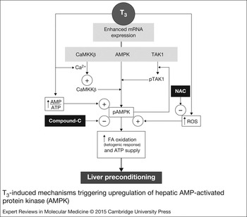
T3 administration to rats was found to upregulate liver AMPK in association with (i) enhanced AMPK mRNA expression; (ii) higher AMPK Thr 172 phosphorylation; and (iii) increased AMP/ATP ratios promoting allosteric AMPK activation (Ref. Reference Vargas78) (Fig. 3). AMPK Thr 172 phosphorylation induced by T3 is related to enhanced mRNA expression of the upstream kinases Ca2+-calmodulin-dependent protein kinase kinase-β (CaMKKβ) and transforming growth factor-β-activated kinase-1 (TAK1), with increased protein levels of CaMKKβ and higher TAK1 phosphorylation (Ref. Reference Vargas78) (Fig. 3). The CaMKKβ component of the AMPK cascade requires increased cytosolic Ca2+ concentrations for operation (Ref. Reference Viollet77), a requisite that is met by T3, as previously shown by different experimental approaches (Refs Reference Wallach79, Reference Hummerich and Sobol80, Reference Yehuda-Shniadman81).
T3-induced mechanisms triggering upregulation of hepatic AMP-activated protein kinase (AMPK). CaMKKβ, calcium-calmodulin-dependent kinase kinase-β; FA, fatty acid; NAC, N-acetylcysteine; pAMPK, phosphorylated AMPK; ROS, reactive oxygen species; TAK1, transforming growth factor-β-activated kinase 1 (pTAK1, phosphorylated TAK1).

Recent studies by our group revealed that T3-induced ROS production has a causal role in upregulating liver AMPK signalling (Ref. Reference Videla82). This is evidenced by suppression of T3-induced hepatic mRNA and protein levels of AMPK, CaMKKβ and TAK1 by pretreatment with N-acetylcysteine (NAC) prior to T3, an antioxidant abrogating the concomitant enhancement in liver and plasma 8-isoprostane levels achieved by T3 (Ref. Reference Videla82). In agreement with these findings, ROS trigger cellular AMPK activation under different experimental conditions such as (i) direct in vitro H2O2 addition to cell cultures; (ii) in vitro and in vivo conditions underlying ROS production; and (iii) ROS generation coupled to free-radical chain reactions induced by polyphenols, with responses being time and concentration dependent (for specific references see Ref. Reference Videla76). Besides, the higher AMPK phosphorylation potential induced by T3 is associated with a significant elevation in hepatic levels of phosphorylated acetyl-CoA carboxylase (pACC) and cyclic AMP response element binding protein (pCREB) (Ref. Reference Videla82). The phosphorylation of these signalling mediators triggers FA oxidation (FAO) as evidenced by the consequent ketogenic response (Fig. 3), which underlies upregulated expression of the FAO-related enzymes carnitine palmitoyltransferase-1α, acyl-CoA oxidase-1 and acyl-CoA thioesterase (Ref. Reference Videla82). Enhancement of FAO by T3 is in agreement with the higher liver ATP turnover reported (Refs Reference Vargas78, Reference Ismail-Beigi83), which may comply with high-energy requiring processes such as liver PC. In accordance with this view, T3 was shown to stimulate hepatic FAO coupled with the induction of autophagy, a stress-related process degrading cellular components to produce FAs to generate ATP or amino acids to synthesise proteins for cell survival (Ref. Reference Sinha84).
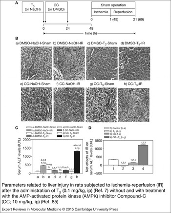
The relevance of T3-induced liver AMPK upregulation on hepatic PC against IR injury was assessed by the in vivo administration of Compound-C (CC), a selective inhibitor of AMPK (Refs Reference Handa85, Reference De Lange86), along with T3 administration, according to the experimental design shown in Figure 4A. As can be observed, sham-operated groups of rats subjected to drug vehicles (group a), T3 (group c), CC (group e) and T3 plus CC (group g) exhibited normal liver histology (Fig. 4B) and serum alanine aminotransferase (ALT) levels (Fig. 4C). IR induced liver injury as evidenced by substantial distortion of liver architecture with extensive areas of hepatocyte necrosis and polymorphonuclear leukocyte infiltration, either in the absence [Fig. 4B (group b)] or presence [Fig. 4B (group f)] of CC treatment, with concomitant 249 and 318% increases in serum ALT levels, compared with the respective control groups (Fig. 4C). On the contrary, T3 administration suppressed liver IR injury as shown by the absence of significant histological alterations [Fig. 4B (group d)] and normal serum ALT levels (Fig. 4C), effects that were abolished by CC [Figs 4B (group h) and 4C]. These findings demonstrate that T3-induced AMPK upregulation has a causal role on liver PC against IR injury, with 266% increase in serum ALT being observed, when net effects elicited by IR in CC plus T3 treated rats (h–g) are compared with those in untreated animals (b–a) (Fig. 4D). In addition to AMPK upregulation, T3-induced liver PC is also reliant upon oxidative stress development (Ref. Reference Fernández87) and Kupffer cell functioning (Ref. Reference Tapia88).
Parameters related to liver injury in rats subjected to ischemia-reperfusion (IR) after the administration of T3 (0.1 mg/kg, ip) (Ref. Reference Fernández7) without and with treatment with the AMP-activated protein kinase (AMPK) inhibitor Compound-C (CC; 10 mg/kg, ip) (Ref. Reference De Lange86). (A) Experimental design, (B) liver histology (hematoxylin-eosin; magnification × 400), (C) serum alanine aminotransferase (ALT) levels and (D) net changes in serum ALT levels induced by IR, calculated by subtracting average values obtained in sham-operated groups from individual values found in IR groups. Considering that PC by T3 requires 48 h for both development and termination of liver oxidative stress (Ref. Reference Fernández7) and AMPK upregulation (data not shown), the experimental design involved eight experimental groups, namely, (a) DMSO at time 0 and 24 h (CC vehicle)-NaOH at time 0 (T3 vehicle)-sham operation at time 48 h, (b) DMSO-NaOH-IR (I h ischemia and 20 h reperfusion in the 48–69 h time interval), (c) DMSO-T3 (0.1 mg/kg at time 0)-sham, (d) DMSO-T3-IR, (e) CC (10 mg/kg at time 0 and 24 h)-NaOH-sham; (f) CC-NaOH-IR, (g) CC-T3-sham and (h) CC-T3-IR. Values in (C) and (D) represent means ± SEM for 3–4 animals per experimental group. Significance studies were carried by two-way ANOVA and the Newman-Keuls, test, shown by the letters identifying each experimental (C) or by the number identifying the calculated net effects (D).

T3-induced PC in extrahepatic tissues
PC against IR injury by thyroid hormones is not restricted to the liver, but it is also achieved in the heart, kidney and brain of experimental animals. It is interesting to note that several studies have suggested that some biological pathways induced by TH are conserved however some organ-specific PC protective mechanisms have been put forward as described below. Long-term administration of T4 was shown to exert cardioprotection against IR, as assessed by an enhancement in postischemic recovery function (Ref. Reference Pantos89) or the reduction of myocardial infarct size (Ref. Reference Kumar, Taliyan and Sharma90). The underlying protective mechanisms involve (i) a significant reduction in the cell death-associated p38 mitogen-activated protein kinase and an enhancement in protein kinase Cδ, known to mediate cardioprotection (Ref. Reference Pantos89); (ii) inhibition of mitochondrial permeability pore opening during reperfusion (Ref. Reference Kumar, Taliyan and Sharma90); (iii) reduction of ROS levels preventing oxidative stress development, as afforded by a combined T3 plus T4 protocol (Ref. Reference de Castro91), a finding confirmed by antioxidant intervention in right ventricle dysfunction in pulmonary arterial hypertension (Ref. Reference Redout92); (iv) upregulation of the cardiac expression of the antiapoptotic protein heat shock protein 70 by T4 (Ref. Reference Pantos93), which is in agreement with T3-induced liver Bcl2 (Ref. Reference Fernández44); and (v) TH-induced Akt/mTOR upregulation and AMPK activation supporting cardiac energetics (Ref. Reference Mourouzis94). T3 also increases the expression of micro RNAs, namely, miRNA-30a and miRNAs of the 29 family, which have antiapoptotic and antifibrotic effects, respectively (Ref. Reference Nicolini95), constituting emerging mechanisms of T3 cardioprotection that deserve investigation under IR conditions. Considering that treatment with TH (Ref. Reference Hajje96) or TH analogs (Ref. Reference Talukder97) may exacerbate cardiac injury, concentrations and timings are crucial factors determining the outcome of the therapies.
T3 or T4 also exert kidney PC against IR injury, which is related to (i) upregulation of HO-1 expression (Ref. Reference Li98); (ii) oxidative stress abrogation (Refs Reference Li98, Reference Ferreyra99, Reference Kim100); (iii) downregulation of pro-inflammatory cytokine (Ref. Reference Li98) and poly(ADP-ribose) polymerase expression, an enzyme promoting acute tubular necrosis (Ref. Reference Ferreyra99), with antiapoptotic response (Ref. Reference Kim100); (iv) improvement in renal ATP levels (Ref. Reference Sutter101); and (v) enhancement in IL-10 and interleukin-1 receptor antagonist expression reducing neutrophil infiltration at the late phase of renal injury (Ref. Reference Wang102). These mechanisms were observed concomitantly with progress in renal function, as evidenced by increased inulin clearance (Ref. Reference Sutter101) and lower proteinuria (Ref. Reference Ferreyra99) and serum creatinine and urea nitrogen levels (Ref. Reference Li98).
Ischemic stroke caused by thrombotic or embolic occlusion decreasing blood flow in the middle cerebral artery triggers brain injury and adult disability with limited treatment options (Ref. Reference Genovese103). In this scenario, T4 administration to rats significantly reduced IR-induced infarct area and volume of total hemisphere infarction, with attenuation of reactive gliosis, downregulation of iNOS and COX-2, and restoration of the expression of neutrotrophins (Ref. Reference Genovese103). Interestingly, T3 also affords neuroprotection against glutamate toxicity, both in astrocytes and neurons (Refs Reference Losi, Garzon and Puia104, Reference Mendes-de-Aguiar105). In this respect, it is important to point that TH or TH derivatives were shown to exhibit additional effects that can be considered as neuroprotective. These include the nongenomical (i) activation of the Na+/H+ exchanger and Ca2+-ATPases to avoid intracellular acidosis and Ca2+ toxicity, respectively; and (ii) stimulation of astrocyte glutamate uptake to reduce its excitotoxicity (Ref. Reference Lin106). Besides, the genomic action of T3 increases the expression of the antiapoptotic protein seladin-1 and that of transthyretin, which favours blood-brain barrier T4 transfer. Taken as a whole, the above mentioned effects of TH encourage further studies for beneficial actions on the nervous system at injury risk (Ref. Reference Lin106).
Comparable with the liver (Refs Reference Vargas78, Reference Videla82), T3 was reported to upregulate the expression of AMPK in skeletal muscle (Refs Reference De Lange86, Reference Park107, Reference Irrcher108, Reference Yamauchi109) and heart (Ref. Reference Heather110), as a response to physiological energy needs during performance, which may be of importance in cardioprotection under IR conditions. In addition to IR injury, thyroid hormones play a critical role in the repair and functional recovery of several organs subjected to different types of injury (Ref. Reference Mourouzis, Politi and Pantos6). These include mechanical injury in rat carotid artery (Ref. Reference Fukuyama111) and mouse hippocampal slices (Ref. Reference Shulga112), rat sciatic nerve transection (Ref. Reference Panaite and Barakat-Walter113), rat lung hyperoxia injury (Ref. Reference Bhargava114), mouse (Ref. Reference Safer, Crawford and Holick115) and guinea pig (Ref. Reference Kassem116) skin wound, chemotherapy-induced toxicity in rat ovarian granulose cells (Ref. Reference Verga Falzacappa117), or serum starvation in neonatal rat cardiomyocytes (Ref. Reference Kuzman118).
Concluding remarks
T3 action leads to hormetic responses that in the low-concentration range have beneficial characteristics, including the calorigenic effect with consequent ROS generation and redox signalling affording cell protection, which may be contributed by redox-independent mechanisms. These responses underlie genomic and non-genomic signalling pathways (Fig. 1) that are integrated and exhibit hierarchical organisation. Indeed, redox signalling induced by T3 through non-genomic redox pathways are secondary to genomic mechanisms, as shown by the lack of effects of α-tocopherol and N-acetylcysteine pretreatment on T3-induced liver mRNA expression of mitochondrial glycerol-3-phosphate dehydrogenase and adenine nucleotide translocator-2, target proteins of T3 action at the transcriptional level (Ref. Reference Videla119), whereas that triggered by non-genomic redox mechanisms (Fig. 2) is abolished (Refs Reference Fernández87, Reference Tapia88, Reference Videla119). Integration of genomic and non-genomic mechanisms of T3 action is observed by the upregulation of the expression of uncoupling proteins (UCPs) through the classical genomic pathway (Ref. Reference Lanni16), reinforcing antioxidant responses set in by non-genomic mechanisms (Fig. 2). In fact, upregulation of UCPs may diminish mitochondrial ROS generation because of mild uncoupling that decreases membrane potential (Ref. Reference Lanni16), and/or allows the transport of peroxidised unsaturated fatty acid anions to the outer side on the inner mitochondrial membrane, thus decreasing oxidative damage to mitochondrial molecules (Ref. Reference Goglia and Skulachev120). Cell protection and survival by thyroid hormone involve enhancement in the homeostatic potential of cells, which includes antioxidant, antiapoptotic, antiinflammatory and cell proliferation responses, besides higher detoxification capabilities and energy support. These responses constitute the molecular basis for TH-induced PC of the liver that exerts protection against IR injury, a strategy also observed in extrahepatic organs of experimental animals and with other types of injury, which awaits application in the clinical setting. Animal studies have clearly indicated that the dose and timings of TH treatment are crucial as biological pathways that are activated can generate opposing – or at least conflicting – signals to cell survival, suggesting that integration of signals ultimately provides a balanced biological outcome. This contention is particularly important, considering that most pharmacological PC protocols available at present time have not been incorporated into clinical practice, with the exception of methylprednisolone that shows promising results in terms of improved clinical outcome (Refs Reference Dikdan, Mora-Esteves and Koneru13, Reference Mi14, Reference Aldrighetti121, Reference Bahde and Spiegel122). Moreover, ischemic PC or intermittent clamping are limited to short ischemia in young patients or prolonged ischemia, pathological liver and elderly patients, respectively, whereas no human data is available for ischemic postconditioning (Refs Reference Selzner, Boehnert and Selzner8, Reference Bahde and Spiegel122). Although T3 is an important determinant in energy expenditure, this hormone is not indicated as an antiobesity drug reducing liver steatosis in euthyroid obese patients, because of the development of side effects (Refs Reference Duntas and Biondi123, Reference Santini124). However, naturally occurring iodothyronines such as 3,5-T2 have a potential pharmacological use (Ref. Reference Coppola125). This proposal is supported by preliminary studies showing that euthyroid subjects given 3,5-T2 (300 μg/day for 3 weeks) exhibit increased basal metabolic rate and decreased body weight, with no deleterious side effects on the thyroid axis and at the cardiac level (Ref. Reference Antonelli126). Nonetheless, the influence of 3,5-T2 on organ PC against IR injury remains to be studied.
Acknowledgements and funding
This article was supported by Grant 1120034 from FONDECYT (Chile). The authors thank Prof. Yedy Israel (Faculty of Medicine, University of Chile) for kindly reviewing the manuscript.