Introduction
Hypoplastic left heart syndrome with aortic atresia represents a high-risk subset among hypoplastic left heart syndrome variants, demonstrating worse outcomes compared to those with aortic stenosis despite advances in surgical management. Reference Fetcu, Schaeffer and Klawonn1 This disparity has led to investigations into specific anatomical and physiological factors unique to this population. Of particular interest is the role of the hypoplastic left ventricle, which, while not contributing to systemic output, remains as a non-functional chamber that may influence right ventricular geometry and function through mechanical interactions. Reference Wisler, Khoury and Kimball2 Tricuspid valve annular dilation after the Norwood procedure is associated with valve regurgitation and right ventricular dysfunction. While the relationship between tricuspid valve size and right ventricular function has been explored, to our knowledge, the simultaneous assessment of both tricuspid valve and mitral valve dimensions in hypoplastic left heart syndrome patients with aortic atresia remains limited. The mitral valve, though connected to a non-functional left ventricle, may provide insights into left ventricular volume and its potential impact on right ventricular performance. The ratio of mitral valve to tricuspid valve diameters could serve as a novel parameter reflecting the balance between the hypoplastic left ventricle and the functional right ventricle. Understanding serial changes in atrioventricular valve dimensions and their relationship to right ventricular function has important clinical implications. This knowledge may enable early identification of patients at risk for right ventricular dysfunction, allowing for timely interventions before irreversible changes occur. Reference King, Ayer and Celermajer3 Furthermore, it provides insights into the mechanisms of ventricular-ventricular interactions in univentricular physiology, particularly how the hypoplastic left ventricle influences right ventricular performance. Reference Wang, Kelle and Hyun4 These findings may guide both the timing of interventions and surgical strategies throughout staged palliation, ultimately improving patient outcomes.
Therefore, this study aimed to evaluate the serial changes of tricuspid and mitral valve diameters following the Norwood procedure in patients with hypoplastic left heart syndrome and aortic atresia, and to compare these parameters between patients with normal right ventricular function and those with impaired right ventricular function.
Materials and methods
Patients and data collection
This single-centre retrospective study was conducted on neonates with hypoplastic left heart syndrome and aortic atresia who underwent the Norwood procedure at the Technical University of Munich, German Heart Center, between January 2001 and December 2020. Patients operated on beyond 28 days of age and those initially managed with bilateral pulmonary artery banding were excluded from the analysis. Clinical data were extracted from our institutional single ventricle database, in which outcomes of all palliative stages were documented, including the bidirectional cavopulmonary shunt and total cavopulmonary connection. Follow-up was determined as the period from the Norwood procedure to the most recent clinical assessment, with censoring applied at the time of death for non-survivors. Patients were categorised into two groups based on echocardiographic right ventricular function assessment: those with normal right ventricular function and those with impaired right ventricular function.
Surgical techniques
All Norwood procedures were performed under cardiopulmonary bypass with deep hypothermic circulatory arrest. Selective cerebral perfusion was introduced in 2008. Through a median sternotomy, complete atrial septectomy was performed via right atriotomy. The ductus arteriosus was ligated, and the main pulmonary artery was transected at the bifurcation. Following ductal tissue excision, the aortic arch was opened longitudinally to the sinotubular junction. Direct anastomosis was created between the proximal pulmonary trunk and the ascending aorta. The descending aorta was enlarged using a Y incision, and arch reconstruction was completed using a patch of surgeon-selected material. The pulmonary artery bifurcation was closed with a patch. Pulmonary blood flow was established with either a modified Blalock-Taussig-Thomas shunt or right ventricle-to-pulmonary artery conduit, based on surgeon preference. Reference Cleuziou, Kasnar-Samprec and Hörer5
Echocardiography
Serial echocardiograms were reviewed by experienced paediatric echocardiographers. Tricuspid valve and mitral valve diameters were measured according to established guidelines Reference Lopez, Colan and Frommelt6 from apical four-chamber views during early diastole at maximum leaflet excursion, from inner edge to inner edge at the hinge points. Anteroposterior diameters were obtained from parasternal long-axis views when feasible. Left ventricular volume was measured from apical four-chamber views using the area-length method when the left ventricular cavity was visualisable. Right ventricular function was evaluated according to the methodology established at our institution for single-ventricle patients. Reference Margossian, Schwartz and Prakash7 An experienced paediatric echocardiographer (C.R.) reviewed archived echocardiography data and assessed right ventricular function using an integrative approach combining quantitative measurements with qualitative assessment. End-systolic and end-diastolic areas were measured to calculate fractional area change, and ejection fraction was estimated. Additional parameters, including tricuspid annular plane systolic excursion and tissue Doppler imaging, were incorporated when available, recognising the load-dependent nature of these measurements in univentricular physiology. Reference Alphonso, Angelini and Barron8 Right ventricular function was graded as: normal (ejection fraction ≥ 50%), mildly impaired (ejection fraction 40–49%), moderately impaired (ejection fraction 30–39%), or severely impaired (ejection fraction < 30%). The final grading incorporated visual assessment by the experienced echocardiographer. Reference Piber, Ono and Palm9,Reference Fetcu, Osawa and Klawonn10 Right ventricular dysfunction was defined as any degree of functional impairment (mild, moderate, or severe) compared to normal function. Tricuspid regurgitation was assessed based on colour Doppler jet width: trivial (<2 mm), mild (2–4 mm), moderate (4–6 mm), or severe (>6 mm). Severe tricuspid regurgitation was defined as moderate or severe regurgitation for clinical outcome analyses.
Statistical analysis
Continuous variables were expressed as medians with interquartile ranges, and categorical variables as numbers and percentages. Group comparisons used the Mann–Whitney U test for continuous variables and Fisher’s exact test or chi-square test for categorical variables. Serial changes were visualised using scatter plots with locally estimated scatterplot smoothing curves. Right ventricular function was classified at the individual examination level. Each echocardiogram was independently categorised as normal or dysfunctional, allowing the same patient to contribute to both groups at different time points. Linear mixed-effects models with random patient intercepts accounted for within-patient correlation and assessed associations between right ventricular dysfunction and valve dimensions, adjusting for time post-Norwood, age at surgery, and shunt type to account for the unbalanced repeated measurements. Stage-specific analyses compared valve dimensions at each palliation stage using Wilcoxon rank-sum tests. Statistical significance was set at p < 0.05. All statistical analyses were performed using SPSS version 28.0 for Windows (IBM, Ehningen, Germany) and R statistical software (R Foundation for Statistical Computing, Vienna, Austria).
Results
Patient characteristics
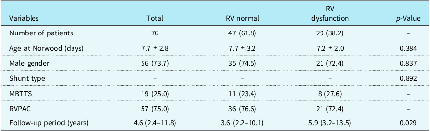
A total of 150 patients with hypoplastic left heart syndrome and aortic atresia underwent the Norwood procedure. Of these, 47 died before total cavopulmonary connection. Of the 103 survivors, 76 with sufficient serial echocardiographic data comprised the study cohort (Table 1). Age at surgery and shunt type distribution were similar between the right ventricular function groups.
Patient baseline characteristics

Table 1. Long description
The table presents patient baseline characteristics for 76 patients who underwent the Norwood procedure, divided into two groups based on right ventricular function: normal and dysfunction. The table has 7 rows and 5 columns. The columns are labeled Variables, Total, RV normal, RV dysfunction, and p-Value. The rows detail the number of patients, age at Norwood procedure in days, gender distribution, shunt type distribution, MBTTS and RVPAC percentages, and follow-up period in years. Notable trends include a similar age at surgery and shunt type distribution between the two groups. The follow-up period is longer for patients with RV dysfunction at an average of 5.9 years compared to 3.6 years for those with normal RV function.
Data are presented as mean ± standard deviation, median (interquartile ranges), or n (%).
RV = right ventricle, MBTTS = modified Blalock-Taussig Thomas shunt, RVPAC = right ventricle-to-pulmonary artery conduit.
RV dysfunction was defined as any degree of impairment on at least one echocardiographic examination during follow-up.
Echocardiographic data
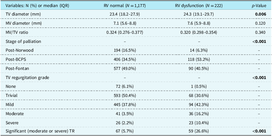
Serial echocardiographic follow-up comprised 1,399 examinations with a median follow-up of 4.6 years (IQR 2.4–11.8). Right ventricular dysfunction was observed in 222 examinations (15.9%), predominantly at the post-bidirectional cavopulmonary shunt (53.2%) and post-Fontan (40.5%) stages (Table 2).
Echocardiographic measurements by RV function

Table 2. Long description
The table presents echocardiographic measurements comparing right ventricular (RV) normal and RV dysfunction groups. It includes data on TV diameter, MV diameter, MV/TV ratio, stage of palliation, and TV regurgitation grade. The RV normal group has 1,177 examinations, while the RV dysfunction group has 222. Notable differences include a higher median TV diameter in the RV dysfunction group (24.3 millimeters) compared to the RV normal group (23.4 millimeters), with a p-value of 0.006. The stage of palliation shows significant differences, with the post-Bidirectional Cavopulmonary Shunt (BCPS) stage being more prevalent in the RV dysfunction group (53.2 percent) compared to the RV normal group (34.5 percent). TV regurgitation grades also differ, with higher grades of regurgitation more common in the RV dysfunction group.
RV = right ventricle, IQR = interquartile ranges, TV = tricuspid valve, MV = mitral valve, BCPS = bidirectional cavopulmonary shunt, TR = tricuspid regurgitation.
Bold indicates p < 0.05.
Valve dimensions and right ventricular function
Echocardiographic measurements stratified by right ventricular function are shown in Table 2. Patients with right ventricular dysfunction demonstrated significantly larger tricuspid valve diameters compared to those with normal right ventricular function (median 24.3 mm versus 23.4 mm, p = 0.006). Mitral valve diameters showed no significant difference between groups. The mitral/tricuspid valve ratio was similar between groups (0.320 versus 0.324, p = 0.340). A sensitivity analysis reclassifying mildly impaired right ventricular function with normal function confirmed the primary findings (24.3 versus 23.5 mm, p = 0.014; mixed-effects model: 0.641 mm, p = 0.038).
Serial changes in valve dimensions
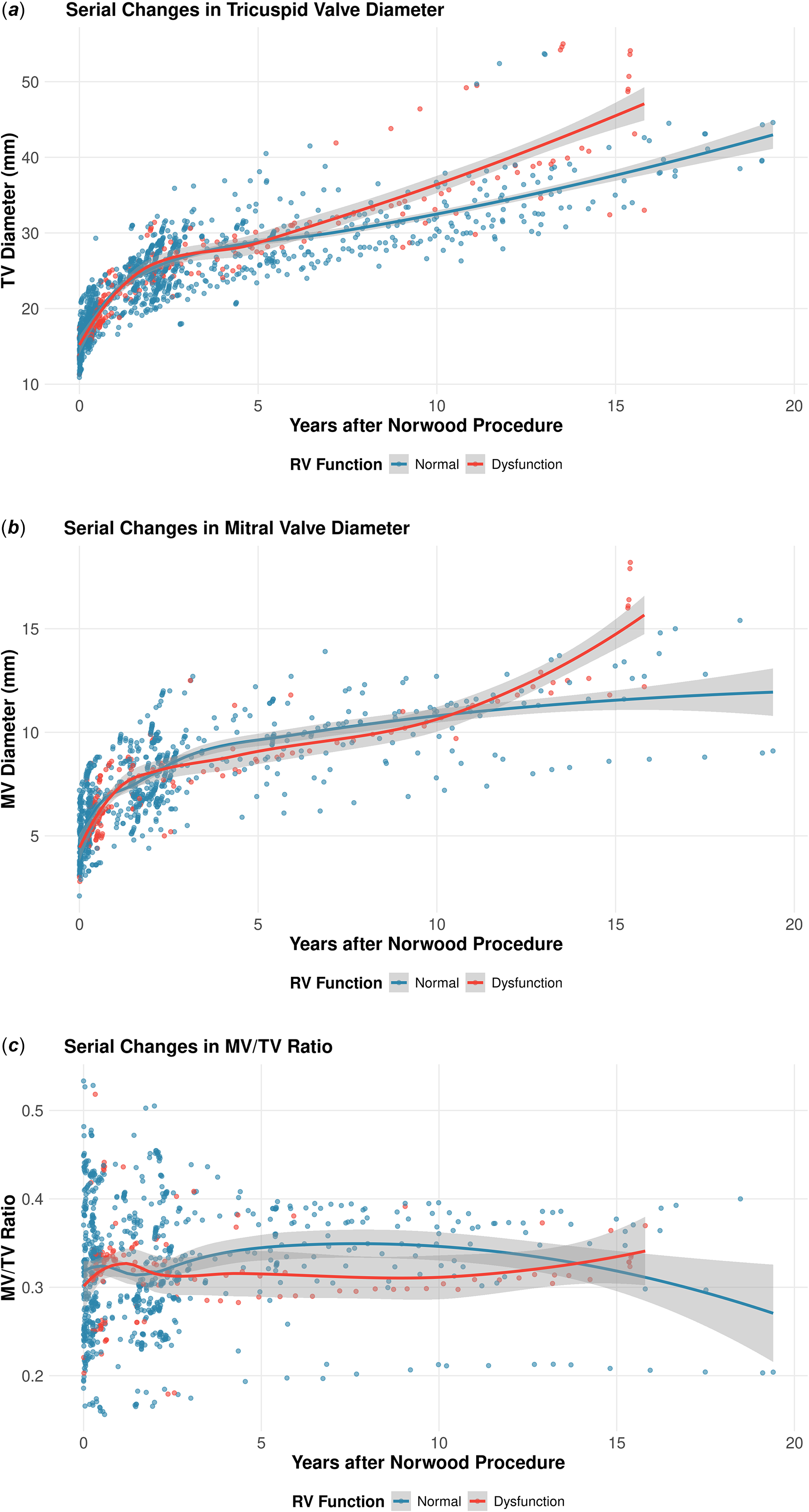
Figure 1(a) demonstrates the serial changes in tricuspid valve diameter over time. Both groups showed progressive increase in tricuspid valve diameter, with the right ventricular dysfunction group maintaining consistently larger diameters throughout follow-up. Figure 1(b) shows similar patterns for mitral valve diameter, with progressive growth over time. The mitral/tricuspid valve ratio (Figure 1(c)) remained remarkably stable at approximately 0.32 throughout the follow-up period, with no significant difference between the groups.
Serial changes in valve dimensions. Scatter plots with locally estimated scatterplot smoothing curves showing ( a ) tricuspid valve diameter, ( b ) mitral valve diameter, and ( c ) mitral/tricuspid valve ratio over time in patients with normal (blue) versus impaired (red) right ventricular function.

Figure 1. Long description
Three scatter plots with locally estimated scatterplot smoothing curves showing tricuspid valve diameter, mitral valve diameter, and mitral/tricuspid valve ratio over time in patients with normal versus impaired right ventricular function. The first scatter plot (a) displays tricuspid valve diameter in millimeters on the y-axis and years after the Norwood procedure on the x-axis, with blue dots representing normal right ventricular function and red dots representing impaired right ventricular function. The second scatter plot (b) shows mitral valve diameter in millimeters on the y-axis and years after the Norwood procedure on the x-axis, with the same color coding for right ventricular function. The third scatter plot (c) illustrates the mitral/tricuspid valve ratio on the y-axis and years after the Norwood procedure on the x-axis, again with blue and red dots indicating normal and impaired right ventricular function, respectively. The locally estimated scatterplot smoothing curves provide a visual trend of the data points over time.
Mixed-effects model analysis
Mixed-effects models confirmed that right ventricular dysfunction was independently associated with larger tricuspid valve diameter (Supplementary Table S1). After adjusting for time since Norwood procedure, age at surgery, and shunt type, patients with right ventricular dysfunction had tricuspid valve diameters that were 0.618 mm larger than those with normal function (p = 0.029). Both the tricuspid valve and the mitral valve diameters increased significantly over time in all patients (tricuspid valve: 0.0042 mm/day, p < 0.001; mitral valve: 0.0015 mm/day, p < 0.001), while the mitral/tricuspid valve ratio remained stable over time regardless of right ventricular function.
Tricuspid valve regurgitation
Tricuspid regurgitation was present in nearly all examinations, with severity distribution varying significantly between right ventricular function groups (p < 0.001). In the right ventricular dysfunction group, severe tricuspid regurgitation was observed in 26.6% of examinations compared to 5.7% in the normal function group (p < 0.001). The distribution of tricuspid regurgitation grades differed substantially: the normal right ventricular function group showed predominantly trivial (50.4%) and mild (37.8%) regurgitation, while the dysfunction group had higher proportions of moderate (16.2%) and severe (10.4%) regurgitation.
Valve dimensions by palliation stage
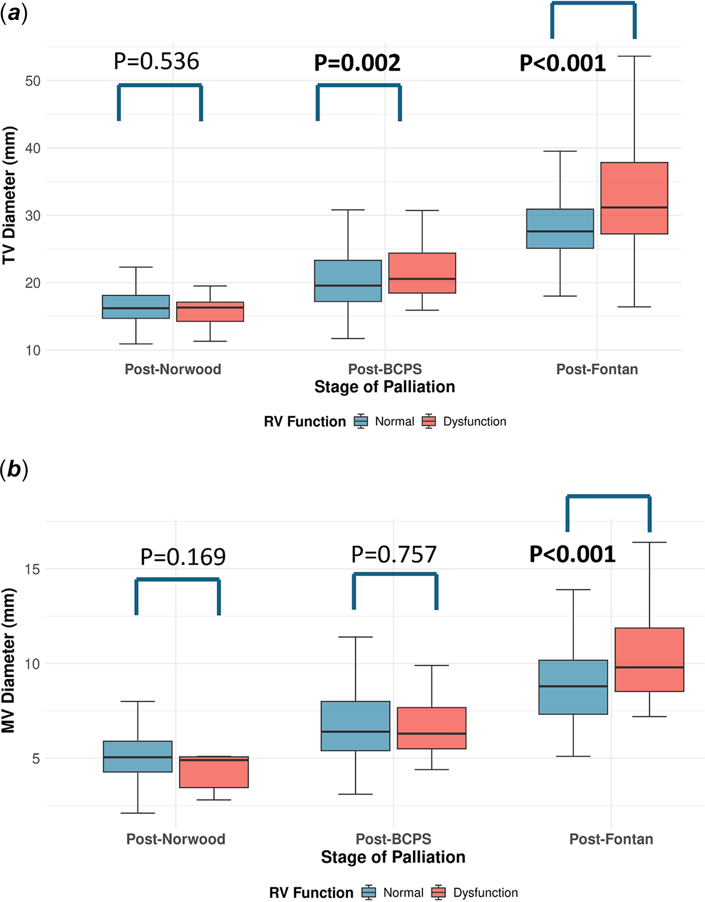
Figure 2 illustrates the distribution of valve diameters across palliation stages (Supplementary Table S2). Both the tricuspid valve and the mitral valve diameters increased progressively from the post-Norwood to the Post-Fontan stages. Tricuspid valve diameter differences between right ventricular function groups evolved: no significant difference at the Post-Norwood stage (0.1 mm difference, p = 0.536), a significant difference at the post-bidirectional cavopulmonary shunt (20.6 mm versus 19.6 mm, 1.0 mm difference, p = 0.002), and a highly significant difference at the Post-Fontan stage (31.2 mm versus 27.6 mm, 3.6 mm difference, corresponding to a 13.0% larger diameter in the right ventricular dysfunction group, p < 0.001). Similarly, mitral valve diameter differences became significant only at the post-Fontan stage (9.8 mm versus 8.8 mm, 1.0 mm difference, p < 0.001), while showing no significant differences at earlier stages.
Valve diameters by palliation stage. Box plots showing ( a ) tricuspid valve and ( b ) mitral valve diameters in patients with normal right ventricular function (blue) and right ventricular dysfunction (red) at each palliation stage. P-values indicate between-group comparisons. BCPS = bidirectional cavopulmonary shunt; RV = right ventricular.

Figure 2. Long description
A box-and-whisker plot showing tricuspid valve diameters in patients with normal and dysfunctional right ventricular function at different palliation stages. The plot has three groups: Post-Norwood, Post-BCPS, and Post-Fontan. The x-axis represents the stage of palliation, and the y-axis represents the tricuspid valve diameter in millimeters. Each group has two box plots: one for normal right ventricular function (blue) and one for dysfunctional right ventricular function (red). The Post-Norwood group shows similar diameters for both functions. The Post-BCPS group shows a slight increase in diameter for dysfunctional function. The Post-Fontan group shows a significant increase in diameter for dysfunctional function. P-values indicate statistical significance between groups. All values are approximated.
Discussion
In this study of 76 hypoplastic left heart syndrome with aortic atresia patients, the tricuspid valve diameter was larger in those with right ventricular dysfunction, independent of growth. The mitral/tricuspid valve ratio remained constant (∼0.32) throughout staged palliation regardless of right ventricular function. While the stable ratio suggests proportional annular growth, mitral valve diameter is an imperfect surrogate for left ventricular volume, and the small absolute differences preclude definitive conclusions from annular dimensions alone. Comprehensive volumetric assessment using three-dimensional echocardiography or cardiac magnetic resonance imaging would be needed to directly evaluate ventricular interactions in this population.
Recent evidence indicates that coronary abnormalities in hypoplastic left heart syndrome with aortic atresia—particularly ventriculocoronary connections present in many mitral stenosis/aortic atresia cases—contribute to right ventricular dysfunction through impaired diastolic perfusion and coronary steal. Reference Kobayashi, Brown and Gauvreau11 This relationship is bidirectional, with right ventricular diastolic dysfunction independently predicting coronary microvascular impairment, creating a vicious cycle. Reference Rickers, Wegner and Silberbach12 While tricuspid valve dilatation results primarily from right ventricular geometric remodelling, coronary insufficiency accelerates this process through supply-demand mismatch.
Clinical implications
The association between tricuspid valve enlargement and right ventricular dysfunction has important clinical implications for hypoplastic left heart syndrome management. Serial tricuspid valve diameter measurements may help identify patients at risk for right ventricular dysfunction before clinical deterioration occurs. While the absolute difference in tricuspid valve diameter between groups was modest (0.9 mm in univariate analysis), this difference persisted after adjusting for multiple confounders in mixed-effects modelling (0.618 mm, p = 0.029), which accounts for the repeated-measures structure of our data. The clinical significance lies not in the absolute millimetre difference, but in the consistent pattern observed across palliation stages: tricuspid valve enlargement was consistently associated with right ventricular dysfunction across palliation stages, suggesting a potential surveillance marker. This aligns with studies showing that early detection of right ventricular dysfunction is critical, as systolic dysfunction strongly predicts pre-Fontan mortality. Reference Salaets, Gewillig, Van De Bruaene and Mertens13 The high prevalence of severe tricuspid valve regurgitation was significantly greater in the right ventricular dysfunction group, suggesting that tricuspid valve regurgitation and annular dilation are closely linked phenomena requiring integrated management strategies. This prevalence reflects our exclusive focus on the aortic atresia subtype, which has worse outcomes. Reference Stasik, Goldberg, Bove, Devaney and Ohye14 The progressive nature creates a vicious cycle where volume overload leads to further annular dilation and worsening regurgitation. Reference Ross, Mir, Burkhart, Holzapfel and Lee15 Recent studies demonstrate that timely tricuspid valve intervention can improve right ventricular function and reduce mortality. Reference Ponzoni, Azzolina and Vedovelli16 The challenge lies in determining optimal timing, balancing surgical risks against the potential for irreversible right ventricular damage. Reference Pigula and Mettler17
Timing of interventions
Stage-specific analysis revealed tricuspid valve diameter differences emerged at bidirectional cavopulmonary shunt (1.0 mm, p = 0.002) and accelerated at Fontan (3.6 mm, p < 0.001), while mitral valve changes appeared only post-Fontan (p < 0.001). This temporal pattern—from 0.1 mm post-Norwood to 3.6 mm post-Fontan—demonstrates accelerating tricuspid valve remodelling during the interstage period, when the right ventricle experiences combined volume and pressure overload. Reference Salaets, Gewillig, Van De Bruaene and Mertens13,Reference Colen, Kutty and Thompson18 These findings suggest the bidirectional cavopulmonary shunt stage represents a critical window for surveillance and potential intervention, as tricuspid valve repair at Fontan completion shows better outcomes than delayed intervention. Reference Ono, Cleuziou and Pabst von Ohain19
Future prospective
Prospective studies with standardised longitudinal protocols are needed to establish whether tricuspid valve enlargement precedes right ventricular dysfunction and clinical deterioration. Advanced imaging modalities, including three-dimensional echocardiography and cardiac magnetic resonance imaging (MRI), combined with strain imaging and artificial intelligence-based predictive models, could enhance early detection of right ventricular dysfunction and optimise intervention timing. Development of paediatric transcatheter tricuspid valve interventions offers promise for reducing surgical morbidity. Reference Doan, Pignatelli, Parekh and Parthiban20 Long-term studies beyond Fontan completion are essential to establish the relationship between early valve changes and late outcomes, while investigation of molecular mechanisms may identify novel therapeutic targets for preventing tricuspid valve dilation and preserving right ventricular function. Reference Cersosimo, Gavazzoni and Inciardi21
Limitations
Several limitations should be acknowledged in interpreting our findings. First, this single-centre retrospective study spanning 20 years may have been influenced by evolving surgical techniques and echocardiographic protocols, with potential inter-observer variability affecting measurements; additionally, the potential influence of inotropic support on echocardiographic parameters in the early postoperative period cannot be entirely excluded. Second, right ventricular function assessment relied on ejection fraction estimation with integrative visual grading, which has known limitations in univentricular physiology; furthermore, the binary classification of right ventricular function may have attenuated the observed associations. Three-dimensional echocardiography or cardiac magnetic resonance imaging would provide a more comprehensive functional assessment. Third, the unbalanced measurement frequency and longer follow-up in the dysfunction group may have increased the likelihood of detecting right ventricular dysfunction. The lack of systematic coronary anatomy assessment also limits causal inference regarding the relationship between coronary abnormalities and right ventricular dysfunction. Fourth, as our cohort excludes patients who died before total cavopulmonary connection, survivorship bias is inherent; patients with the most severe tricuspid valve dilation and right ventricular dysfunction may have been disproportionately excluded. Finally, we did not systematically evaluate other factors potentially influencing tricuspid valve remodelling, including genetic variations, inflammatory markers, or detailed haemodynamic assessments. Additionally, the impact of tricuspid valve intervention on valve and ventricular remodelling could not be assessed due to limited post-intervention echocardiographic data.
Conclusions
In hypoplastic left heart syndrome with aortic atresia, right ventricular dysfunction is associated with progressive tricuspid valve enlargement but not with changes in the mitral/tricuspid ratio. While the stable ratio suggests proportional annular growth, a comprehensive volumetric assessment is needed to clarify ventricular-ventricular interactions. Tricuspid valve diameter may serve as an early marker of right ventricular remodelling, with differences between function groups emerging at the bidirectional cavopulmonary shunt stage—a critical window for surveillance and intervention.
Supplementary material
The supplementary material for this article can be found at https://doi.org/10.1017/S1047951126113365.
Acknowledgements
None.
Financial support
This study was supported by grants from the Förderverein des Deutschen Herzzentrums München.
Competing interests
The authors declare that they have no known competing financial interests or personal relationships that could have appeared to influence the work reported in this paper.
Ethical standard
The authors assert that all procedures contributing to this work comply with the ethical standards of the relevant national guidelines on human experimentation and with the Helsinki Declaration of 1975, as revised in 2008, and has been approved by the Institutional Review Board of the Technical University of Munich (approval number 2025-232-S-NP, May 05, 2025). The requirement for individual patient consent was waived due to the retrospective design.