Introduction
The genus Ozolaimus was established by Felix Dujardin in 1845 for a nematode Ozolaimus megatyphlon found in a young Iguana Laurenti, 1768 from the collection of the Paris Museum of Natural History (Dujardin, Reference Dujardin1845). The characteristic feature of the genus, reflected in its name, was the division of the pharyngeal corpus into two parts: a wide, ampoule-shaped anterior and a slender posterior (Greek óζóς = nod and λαιμóς = throat, larynx). Dujardin considered it possible that it was the same as Ascaris megatyphlon, a species described by Rudolphi from the intestine of Iguana tuberculata (Laurenti, 1768) (now a synonym of the green iguana Iguana iguana Linnaeus, 1758) (Rudolphi, Reference Rudolphi1819). Dujardin assumed that the long, sac-like pharynx and the following narrow and long oesophagus of Rudolphi's species corresponded with the anterior and posterior pharyngeal parts of O. megatyphlon. Schneider (Reference Schneider1866) had considered both Ascaris megatyphlon Rudolphi, Reference Rudolphi1819 and Ozolaimus megatyphlon Dujardin, Reference Dujardin1845 as junior synonyms and members of the Oxyuris Rudolphi, 1803 genus, having included a brief description of Oxyuris megatyphlon (Rudolphi, Reference Rudolphi1819) in his monograph. Later, two additional species were added to the genus, of which Oxyuris monhystera Linstow, Reference Linstow1902 possessed a narrow, cylindrical pharynx not divided into sections, and Oxyuris cirrata Linstow, Reference Linstow1906 was characterized by a two-parted pharynx similar to that of O. megatyphlon (Linstow, Reference Linstow1902, Reference Linstow1906). Such apparent differences in pharynx morphology within the genus could not be ignored for long. Gedoelst (Reference Gedoelst1916), in an additional note to his work on the parasite fauna of the Belgian Congo (now Democratic Republic of the Congo), included a new genus Macracis with the type species M. monhystera ( = O. monhystera). Unfortunately no morphological diagnosis was given by the author. Nevertheless, both generic names became entrenched in the literature, referring to closely related genera with different pharynx morphology: two-parted in Ozolaimus and simple in Macracis (Baylis & Daubney, Reference Baylis and Daubney1926; Yorke & Maplestone, Reference Yorke and Maplestone1926; Dosse, Reference Dosse1939; Skrjabin et al., Reference Skrjabin, Schikhobalova and Mosgovoy1951, Reference Skrjabin, Schikhobalova and Lagodovskaja1960). Thus, Chitwood (Reference Chitwood1931) gave a detailed description of M. monhystera with a uniform pharynx width, and redescribed Ozolaimus megatyphlon (Rudolphi, Reference Rudolphi1819) from I. tuberculata, and Reference OrtleppOrtlepp (1933a), based on pharynx morphology, excluded Ozolaimus from the subfamily Oxyurinae Hall, 1916 and proposed the subfamily Ozolaiminae, which was later raised to the rank of family (Pereira, Reference Pereira1935). At the same time, some authors have attempted to synonymize both genera, arguing that pharynx morphology cannot be considered as an important diagnostic feature because of its strong variability due to fixation (Thapar, Reference Thapar1926; Bravo-Hollis & Brenes, Reference Bravo-Hollis and Brenes1960; Inglis et al., Reference Inglis, Díaz Ungría and Coles1960).
Currently, Ozolaimus ( = Macracis) is included within the Pharyngodonidae family and comprises four valid species: O. megatyphlon, O. monhystera, O. cirratus and O. ctenosauri ( = O. prolixa) (Moravec et al., Reference Moravec, Salgado-Maldonado and Mayen-Peña1996). The new species Ozolaimus linstowi n. sp. is described and illustrated herein with molecular phylogenetic analysis also provided.
Materials and methods
Collection and examination of nematodes
Specimens of O. linstowi n. sp. were recovered from a female Iguana iguana Linnaeus, 1758 (Squamata: Iguanidae) which was brought to Moscow Zoo from an unknown location in Central Mexico, maintained in captivity in a separate cage for 2 years and then died. Following a post-mortem examination a massive infection of nematodes was located in the large intestine, which was removed and transferred to clean physiological saline. Nematodes were fixed in hot (60–70°C) 4% formaldehyde and processed to glycerol according to Seinhorst (Reference Seinhorst1959). Some specimens were frozen for molecular analysis. Measurements and drawings were made with the aid of a camera lucida. Several nematodes were prepared for SEM by dehydration through a graded ethanol series and acetone, and dried in a critical-point drier. After coating with gold/palladium they were examined in JSM-6380LA (JEOL, Tokyo, Japan) and TM3000 (Hitachi, Tokyo, Japan) electron microscopes.
Molecular analysis
To increase the number of comparable sequences of oxyurid nematodes used for the phylogenetic analysis, sequence data for two Pseudonymus Diesing, 1857 species (Thelastomatoidea: Pseudonymidae), Hammerschmidtiella diesingi Hammerschmidt, 1838 and Hammerschmidtiella cristata Spiridonov, 1984 (Thelastomatoidea: Thelastomatidae) were also obtained. Specimens of Pseudonymus islamabadi Basir, 1941 and Pseudonymus spirotheca Györy, 1856 were recovered from water scavenger beetles Hydrophilus piceus Linnaeus, 1758 (Coleoptera: Hydrophilidae) collected by S.E. Spiridonov and E.A. Guzeeva in June 2012 in the territory of the Astrakhan State Biosphere Reserve at the Volga River delta. Specimens of H. diesingi and H. cristata were recovered from Oriental cockroaches Blatta orientalis Linnaeus, 1758 (Blattodea: Blattidae) and Madagascar cockroaches Gromphadorhina portentosa (Schaum, 1853) (Blattodea: Blaberidae), respectively, collected by E.A. Guzeeva in June 2013 at Moscow Zoo.
Nematode specimens were kept at − 18°C prior to DNA extraction. The DNA was extracted according to Holterman et al. (Reference Holterman, van der Wurff, van den Elsen, van Megen, Bongers, Holovachov, Bakker and Helder2006). The worm-lysis solution was prepared immediately before DNA extraction, containing 950 μl of a mixture of 2 ml of 1 m NaCl, 2 ml of 1 m Tris–HCl (pH 8) plus 5.5 ml of deionized water plus 10 μl of mercaptoethanol and 40 μl of proteinase K (20 mg/ml). Single nematodes were transferred to 25 μl of sterile water and, after addition of 25 μl of worm-lysis solution, the tube was incubated at 65°C for 90 min. The tubes with homogenate were then incubated at 99°C for 5 min to deactivate proteinase K and 0.8–1.2 μl of homogenate was used as the polymerase chain reaction (PCR) template.
PCR reactions were performed using Encyclo Plus PCR kit (Evrogen, Moscow, Russia) according to the manufacturer's manual. Primer pairs D2A (5′-ACA AGT ACC GTG AGG GAA AGT TG-3′) and D3B (5′-TCG GAA GGA ACC AGC TAC TA-3′) were used to amplify D2–D3 expansion segments of large subunit (LSU) rDNA fragment (Nunn, Reference Nunn1992). PCR cycling parameters included primary denaturation at 94°C for 5 min, followed by 34 cycles of 94°C for 60 s, 50°C for 60 s and 72°C for 1 min, followed by post-amplification extension at 72°C for 10 min.
Two pairs of primers were used to amplify small subunit (SSU) rDNA. A pair of nematode-specific primers nem18SF (5′-CGC GAA TRG CTC ATT ACA ACA GC-3′) and nem18SR (5′-GGG CGG TAT CTG ATC GCC-3′) was used to amplify the 5′ portion of SSU rDNA (Floyd et al., Reference Floyd, Rogers, Lambshead and Smith2005). PCR cycling parameters included primary denaturation at 95°C for 5 min followed by five cycles of 94°C for 30 s, 47°C for 30 s and 72°C for 40 s, and 35 cycles of 94°C for 25 s, 54°C for 30 s and 72°C for 40 s, followed by post-amplification extension at 72°C for 5 min. Another pair of primers, 24F (5′-AGR GGT GAA ATY CGT GGA CC-3′) and Q39 (5′-TAA TGA TCC WTC YGC AGG TTC ACC TAC-3′), was used to obtain the remaining 3′ end of SSU rDNA (Blaxter et al., Reference Blaxter, De Ley, Garey, Liu, Scheldemann, Vierstraete, Vanfleteren, Mackey, Dorris, Frisse, Vida and Thomas1998). PCR cycling parameters included primary denaturation at 95°C for 5 min followed by 35 cycles of 94°C for 60 s, 53°C for 90 s and 72°C for 90 s, followed by post-amplification extension at 72°C for 6 min.
PCR products were visualized in agarose gel and bands were excised for DNA extraction with Promega columns (Wizard® SV Gel and PCR Clean-Up System; Promega, Madison, Wisconsin, USA). Samples were sequenced directly, using the same primers as used for primary PCR reactions.
Nematode sequences were deposited in GenBank (National Center for Biotechnology Information; NCBI) as: KJ632667 (O. linstowi n. sp.) for D2–D3 LSU rDNA; and KJ632671 (O. linstowi n. sp.), KJ632668 (P. islamabadi), KJ632669 (P. spirotheca), KJ632670 (H. diesingi) and KM670445 (H. cristata) for SSU rDNA.
For comparative purposes and phylogenetic construction, other sequences from GenBank were also used (see fig. 6). Sequence alignments were generated using Clustal X (Thompson et al., Reference Thompson, Gibson, Plewniak, Jeanmougin and Higgins1997) under default values for gap opening and gap extension penalties. All alignments were analysed using PAUP* 4.0b10 (Swofford, Reference Swofford1998) for maximum parsimony (MP), maximum likelihood (ML) and neighbour joining (NJ) methods. MrBayes v3.2.1. (Ronquist et al., Reference Ronquist, Teslenko, van der Mark, Ayres, Aaron, Höhna, Larget, Liu, Suchard and Huelsenbeck2012) was used for Bayesian analysis (Bayesian inference; BI). Bayesian analysis was run for 2 × 10⁶ generations using the GTR+G+I model with relative burn-in (discarding the first 25% of samples). Average standard deviation of split frequencies at the end of the analyses was lower than 0.01.
Ozolaimus linstowi n. sp
Type host and locality. Large intestine of Iguana iguana Linnaeus, 1758 from Central Mexico.
Type material. Holotype male (No. 1249), paratype male (No. 1225) and paratype female (No. 1226) deposited in the collection of the Centre of Parasitology, Institute of Ecology and Evolution, Russian Academy of Sciences. Paratype male (No. UGMD 104292) and paratype female (No. UGMD 104293) deposited in the nematological collection of the Museum voor Deirkunde, Ghent University (Ghent, Belgium).
Etymology. The species is named after Otto Friedrich Bernhard von Linstow (1842–1916), a German high-ranking medical officer and helminthologist, author of numerous publications devoted to Aschelminthes and parasitic nematodes and, in particular, author of two species of the present genus, of which one was described as a form with a straight, slender pharynx (i.e. Ozolaimus monhystera Linstow, Reference Linstow1902 ( = Oxyuris monhystera; Macracis monhystera)).
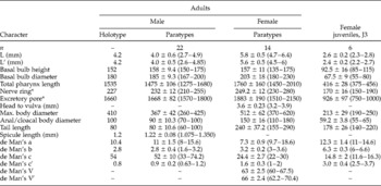
Measurements. See table 1.
Morphometrics (in μm unless otherwise stated) of adults and female juveniles of Ozolaimus linstowi n. sp.; mean±standard deviation (range), except for de Man ratios.

n, Number of specimens examined; L, total body length (head to tail tip); L′, body length from head to anal/cloacal aperture; a, total body length divided by maximum body diameter; b, total body length divided by pharyngeal length (the pharynx is defined as head end to the pharyngo-intestinal junction); c, total body length divided by tail length; c′, tail length divided by body diameter at the anal/ cloacal aperture; V, position of vulva from anterior end expressed as percentage of body length; V′, position of vulva from anterior end expressed as percentage of distance from head to anal aperture; *from anterior extremity.
Description
General
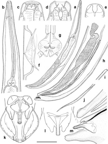
Small to medium-sized nematodes, males 3–4.9 mm and females 4.7–6.4 mm in length. Body narrow initially, widening gradually and reaching maximum diameter near mid-body, then tapering posterior towards anus. Cuticle with fine annulation 4–5 μm apart. Cephalic capsule rounded, smooth, slightly swollen laterally but apically blunt, clearly defined from the rest of the body. Four cephalic papillae present. Amphids rounded, prominent, with slit-like apertures. Oral opening dorso-ventrally elongate immersed in head end tissues. Buccal capsule 30 μm long, dorso-ventrally elongate with thickened cuticular walls at its base. Pharyngeal corpus very long, widened at anterior end behind buccal capsule then slender, almost cylindrical; isthmus very short, not clearly expressed, 30–35 μm long; basal bulb well developed, pear-shaped with three cardial lobes projecting into a gut lumen (fig. 1b, g). Intestine swollen anteriorly, slowly attenuating until the rectal valve, surrounded at its junction with rectum by well-developed muscular circular sphincter. Rectum 150–160 μm long. Nerve ring encircles the pharyngeal corpus at its anterior end. Excretory pore located just below the basal bulb level, leading to narrow excretory duct initially surrounded by valve-like structure, then widening into a funnel-like expansion (fig. 1f). Distinctively marked sexual dimorphism is present in organization of the cuticular structures surrounding oral opening and cuticular armature of buccal capsule.
Ozolaimus linstowi n. sp., male: (a) entire view; (b) anterior end, lateral view; (c, d) optical section through buccal capsule (lateral and ventral views, respectively); (e) dorsal complex of two cuticular plates situated in buccal capsule; (f) distal part of excretory system, lateral view; (g) basal bulb; (h) posterior end, lateral view; (i, j) spicule proximal and distal ends, respectively; (k) cloaca region, ventral view; (l) optical section through genital cone, ventral view; (m) optical section though genital cone, lateral view. Scale bars: a = 750 μm; b, h = 240 μm; c, d, i, j = 50 μm; e = 15 μm; f = 125 μm; g = 100 μm; k, l = 45 μm; m = 60 μm.

Adults
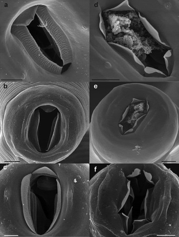
Male. Oral opening hexagonal surrounded by six smooth cuticular membranes: two subdorsal, two lateral and two subventral (fig. 2d–f). Subdorsal and subventral membranes of equal size and shape, triangular with rounded outer edges, while lateral membranes elongate, each with two rounded edges. Three intricate cuticular structures present at the base of buccal capsule: one dorsal and two subventral (figs 1c, d, 2f). Each structure comprises a complex of two cuticular plates arranged one after the other (fig. 1e). Inner plates, facing towards the lumen, apically divided by vertical groove into two parts that can bend to outside. Outer plates simple, bearing no groves or folds, can be slightly curved to outside. The dorsal pair of cuticular plates is narrower than subventral ones.
SEM micrographs of Ozolaimus linstowi n. sp., adults: (a) female oral opening, en face view; (b) female anterior end, apical view; (c) female oral opening with cuticular plates distinguishable inside the buccal capsule; (d) male oral opening, en face view; (e) male anterior end, subventral view; (f) male oral opening with cuticular plates distinguishable inside the buccal capsule. All scale bars are 10 μm.

Genital tract monorchic, vas deferens clearly divided into three parts: curved anterior part with flexure 1150–1200 μm posterior to basal bulb, middle part filled with small rounded cells with granular content and posterior part with walls composed of large polygonal cells (fig. 1a, h).
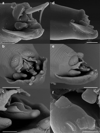
Copulatory papillae represented by two prominent, mammiform precloacal papillae situated ventrally above the cloacal aperture and two postcloacal pairs, of which anteriormost located on either side of genital cone that occupies the central position, and posterior one is situated subventrally close to tail tip (figs 1k, 3b, e). Precloacal papillae are followed by bifurcate membranous formations overhanging the cloaca. Cuticularized median duct often described as midventral papilla on the precloacal lip is absent. Cloacal aperture is surrounded by two lateral inflations joined with genital cone by thin folded cuticle, reaching its middle point and forming a kind of a groove (fig. 3c). The body of the genital cone is supported by a complicated funnelform structure that, in longitudinal section, can be described as Y-shaped (fig. 1l). The conical part of the structure is inserted into the genital cone, whereas the rest is immersed inside the body (fig. 1m). Two narrow, closely adpressed ducts pass through genital cone inside the conical part of the cuticular structure and open as small papilla-like pores at the cone's distal tip (fig. 3f). Surrounding tissues and thickness of cuticular structure prevent tracing of the way of ducts inside the body. The inner part of the cuticular structure resembles a funnel edged with four appendages.
SEM micrographs of Ozolaimus linstowi n. sp., male: (a) posterior end with protruding spicule; (b) cloaca region, ventral view; (c) genital cone, lateral view; (d) posterior end, lateral view; (e) posterior end, ventro-lateral view; (f) tail and genital cone distal tips. Scale bars: a, b, d, e = 30 μm; c, f = 10 μm.

Spicule very long, twisted along the entire length with slightly curved pointed distal tip (fig. 1h, i, j). Longitudinal striations on its surface clearly visible under scanning electron microscope (fig. 3a). Spicule head dorsally curved. Tail short, ventrally curved with well-developed caudal alae of rugose cuticle (fig. 3d). Pore-like openings of caudal glands discernible at the tail tip laterally of posteriormost papillae pair (fig. 1k).
Female. Oral opening rather triangular with strongly reduced dorsal sector and rounded opposite side, surrounded by three rugose cuticular membranes: one dorsal and two subventral, almost parallel to each other (fig. 2a–c). Dorsal membrane short, triangular in shape with rounded outer edge. Subventral membranes elongate, extended along the sides of the oral opening with rounded free edges. Dorsal membrane overlaps dorsal edges of subventral membranes when relaxed. Three simple cuticular plates, unequal in size, are situated at the base of buccal capsule, one dorsal and two subventral, of which the dorsal is twice as narrow. Plates are rounded, bent to the outside along the clear fold line (figs 2c).
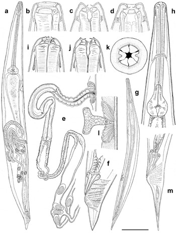
Ozolaimus linstowi n. sp. (a–f) Female: (a) entire view; (b–d) optical section through buccal capsule (longitudinal and transverse sections through middle and dorsal parts, respectively); (e) anterior part of genital tract; (f) tail. (g–m) J3 juvenile: (g) entire view; (h) anterior end, laterally; (i, j) optical sections through buccal capsule (longitudinal and transverse sections, respectively); (k) anterior end, apical view; (l) gonadal primordium, lateral view; (m) tail. Scale bars: a = 650 μm; b–d, l = 45 μm; e = 250 μm; f = 170 μm; g = 400 μm; h = 10 μm; i, j = 25 μm; k = 15 μm; m = 100 μm.

Vulva not salient, slit-like, located 1920 ± 276 (1357–2230) μm anterior to anus and covered by hanging anterior vulval lip (fig. 4a). Ring-like muscular sphincter well developed. Genital tract didelphic, prodelphic. Vagina vera muscular, long, anteriorly directed, followed by vagina uterina that immediately turns posterior, forming a loop. The cavity of vagina vera is filled with fibrous copulatory plug (fig. 4e). Uterus very long, meandering, then divided into two anteriorly directed uteri, each ending with its own ovary. Ovaries long, both extending anteriorly for a short distance then turning towards anus. Two seminal receptacles present at the boundary of each of the ovaries and uterus. Eggs not numerous, non-embryonated, oval, 117 ± 2.4 (114–120) × 70 ± 3.6 (65–75) μm in size with smooth, thin shells. Tail long, conical (fig. 4f).
Juveniles
Six juvenile specimens were found during the dissection of the host, which were identified as J3 stage females according to the development of gonad and vulva primordia.
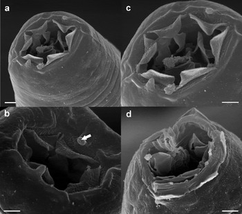
The body is spindle-shaped, reaching the maximum width in its middle (fig. 4g). Body cuticle well annulated, 2–3 μm apart. Cephalic capsule rounded, apically blunt, covered with smooth cuticle. Four cephalic papillae present. Two peg-like cephalic projections situated laterally close to oral opening (fig. 5a–c). Distal tips of projections provided with slit-like apertures (white arrow in fig. 5b). Oral opening of regular hexagonal shape surrounded with six rugose cuticular membranes of equal size and shape, and arranged as: two subdorsal, two lateral and two subventral (fig. 4k). Membranes triangular in shape with rounded free edges. Ring of rugose cuticle surrounds the oral opening prior to cuticular membranes. Buccal capsule rounded, 7–10 μm long, supported by 22–24 cuticular processes with fused bases (fig. 5d). Three intricate cuticular structures morphologically identical to those found in males, but of equal size, present at the base of buccal capsule and arranged as: one dorsal and two subventral (fig. 4i, j). Pharyngeal corpus long, slender, cylindrical along all its length; isthmus short, 40–60 μm in length and of same width as pharyngeal corpus; basal bulb well developed, slightly elongate with three short cardial lobes projecting into a gut lumen (fig. 4h). Nerve ring situated at the mid-level of pharyngeal corpus. Excretory pore located in first third of the body length, significantly posterior to basal bulb level. Narrow excretory duct surrounded by valve-like structure widens into funnel-like expansion. Intestine initially narrow, straight, filled with granular content. Rectal valve at its junction with rectum surrounded by well-developed muscular circular sphincter (fig. 4m). Rectum 100–110 μm long.
SEM micrographs of Ozolaimus linstowi n. sp., J3 juveniles: (a) anterior end, subventral view; (b) oral opening (white arrow indicates a slit-like aperture in the cephalic projection); (c) oral opening with cuticular plates distinguishable inside the buccal capsule; (d) damaged anterior end with prominent cuticular structures of the buccal capsule. All scale bars are 3 μm.

Vulval primordium 783 ± 64.3 (670–850) μm anterior to anus. Gonadal primordium well developed, comprising a T-shaped group of cells, 37–40 μm along the body axis, associated with vulval primordium (fig. 4l). Distal tip cells with dense granular content present on each proximal tip of gonadal primordium. Tail long, conical.
Diagnosis and relationships
Four species of Ozolaimus have been described from large iguanid lizards (Iguana, Cyclura, Ctenosaura) and reported from Central and South America (Brazil, Colombia, Costa Rica, Mexico, Venezuela) and Islands of the Caribbean Basin (Haiti, Cuba, Lesser Antilles). Ozolaimus linstowi n. sp. described herein was found in I. iguana which was reported previously as a host for O. megatyphlon and O. cirratus (Linstow, Reference Linstow1906; Leussink, Reference Leussink1958; Munakata et al., Reference Munakata, Inoue and Shirai1999; Leukopoulos et al., Reference Leukopoulos, Komnenou, Papadopoulos and Psychas2007; Breves et al., Reference Breves, Porto, Pissinatti, Luz and Menezes2011). The presence of a slender, cylindrical pharynx not divided into sections in O. linstowi n. sp. means that the new species most closely resembles O. monhystera from Cyclura cornuta (Haiti) and O. ctenosauri from Ctenosaura acanthura (Mexico).
Ozolaimus linstowi n. sp. can be distinguished from O. monhystera (according to the original description) by its shorter spicule length (1–1.3 vs. 1.97–2 mm) and smaller body size (4 vs. 7.11 mm in males and 5.8 vs. 7.6 mm in females). In spite of the fact that only average values of body size were given in the original description of O. monhystera, the body size variations in O. linstowi n. sp. are outside the specified values in O. monhystera. Males of O. linstowi n. sp. are similar to those of O. ctenosauri in spicule length and its common morphology, while females differ greatly by the markedly smaller body size (4.7–6.4 vs. 8–11 mm in O. ctenosauri), form of oral aperture (more elongate in O. linstowi n. sp.) and shape of the surrounding subventral cuticular membranes (almost rectangular in O. linstowi n. sp. and triangular in O. ctenosauri).
Phylogenetic relationships
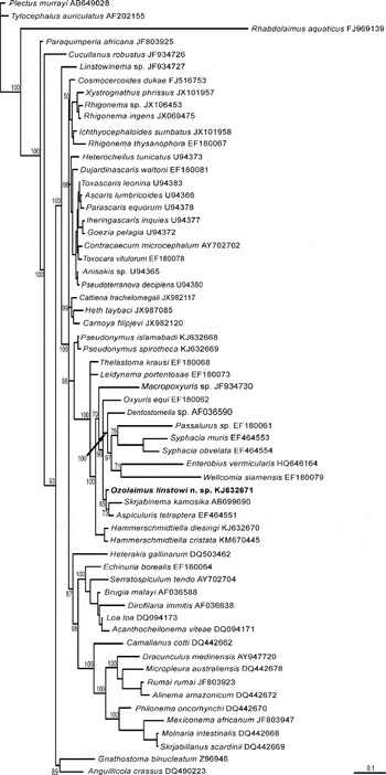
In the trees obtained with all four (MP, NJ, ML and MrB) methods of phylogenetic analysis, O. linstowi n. sp. is a part of a clade consisting of the representatives of several oxyurid genera: Oxyuris, Aspiculuris Schulz, 1924, Syphacia Seurat 1916, Skrjabinema Wereschtschagin, 1926, Dentostomella Schulz & Krepkogorskaja, 1932, Enterobius Leach, 1853, Wellcomia Sambon, 1907 and Passalurus Dujardin, Reference Dujardin1845. The position of O. linstowi n. sp. in this clade is variable from the basal position up to the terminal position. Bayesian analysis also gave significant support for the clade comprised all studied Oxyuridomorpha, i.e. oxyurids of vertebrates and Thelastomatoidea of invertebrates (fig. 6). Unlike the Bayesian analysis, the ML, MP and NJ trees did not support the relationships of Pseudonymus nematodes from water beetles with all remaining Oxyuridomorpha. An analysis of nucleotide differences in the partial SSU rDNA sequences compared with other Oxyuridomorpha nematodes shows that Ozolaimus differs from the vertebrate parasitic Skrjabinema sp. (EF180060) in 23 bp, while for insect-parasitic Leidynema Schwenk, 1929 and Hammerschmidtiella Chitwood, 1932 this difference is 26 and 27 bp, respectively. Differences with all other Oxyuridomorpha are higher (30 bp or more). The inference of the phylogenetic position of O. linstowi n. sp. among the Oxyuridomorpha based on D2–D3 LSU rDNA data was hampered because very few equivalent Pharyngodonidae sequences are available in the NCBI GenBank. These fragmentary data indicate that O. linstowi n. sp. is closest to Parapharyngodon Chatterji, 1933 and Thelandros Wedl, 1862 genera, with differences of 110 bp and 102 bp, respectively.
Phylogenetic relationships of Ozolaimus linstowi n. sp. inferred from Bayesian analysis of partial sequences of SSU rDNA. Total number of characters = 1334; evolution model GTR+G+I; burn-in = 400,000; scale bar = 0.1 expected changes per site.

Discussion
Despite the presence of numerous papers on Ozolaimus representatives, some aspects of their morphology (particularly the head-end structures) still remain understudied, and adequate illustrations are lacking. The first SEM images of the anterior extremity were obtained for O. ctenosauri, but, unfortunately, only female specimens were examined and sexual dimorphism in the head-end cuticular structures was not discovered (Moravec et al., Reference Moravec, Salgado-Maldonado and Mayen-Peña1996).
The current SEM studies revealed distinct sexual dimorphism in O. linstowi n. sp., in which the female oral opening was surrounded by three lip-like elevations framed by three cuticular membranous structures, whereas in males, the oral opening was hexagonal and surrounded by six cuticular membranous structures. The presence of similar sexual dimorphism of oral cuticular structures has been described for several other Pharyngodonidae: Parapharyngodon osteopili Adamson, Reference Adamson1981; Alaeuris vogelsangi Lent & Freitas, 1948 and some Trachygonetria species from South Africa (Petter, Reference Petter1969; Adamson, Reference Adamson1981; Bouamer & Morand, Reference Bouamer and Morand2004).
To date, at least two types of pharyngeal corpus are distinguished within the genus, but the diagnostic value of this character is undermined by the contradictory information given by different authors. Inglis et al. (Reference Inglis, Díaz Ungría and Coles1960) assumed that differences in pharynx morphology were mostly artefacts caused by fixation, while Leussink (Reference Leussink1958) illustrated pronounced differences in the pharynx shape, even for the morphologically close O. megatyphlon and O. cirratus, often found parasitizing the same host specimen. The recent communication of Breves et al. (Reference Breves, Porto, Pissinatti, Luz and Menezes2011) clearly illustrates the presence of two unequivocally different morphological shapes of the pharynx in O. cirratus and O. megatyphlon from I. iguana (Brazil). Light microscope photographs show the presence of a narrow pharynx with anterior fusiform swelling in O. cirratus and a two-part pharynx of O. megatyphlon, with an anterior sac-like part and posterior slender one; parts differ in optical density, unlike in O. cirratus. It is also worth noting that the illustrations of pharynx morphology for O. cirratus and O. megatyphlon found in Leussink (Reference Leussink1958) and Inglis et al. (Reference Inglis, Díaz Ungría and Coles1960) contradict that of Breves et al. (Reference Breves, Porto, Pissinatti, Luz and Menezes2011).
Ozolaimus linstowi n. sp. possesses a narrow, slender pharynx; however, minor changes in its width caused by muscle contracture due to fixation have been observed in some specimens. In the author's opinion, pharynx morphology is indeed an important diagnostic feature, and morphological differences shown by Breves et al. (Reference Breves, Porto, Pissinatti, Luz and Menezes2011) indicate the existence of two (or three?) taxa of generic level combined into one genus. However, morphological data based on light microscopy alone are insufficient to justify major taxonomic revisions.
Another distinguishing feature of the Ozolaimus genus and some other representatives of the Pharyngodonidae is the presence of a complex, strongly cuticularized structure supporting the genital cone. The sclerotized structure of O. linstowi n. sp., projecting into a genital cone, is funnel-shaped with an aperture through its distal end. Chitwood (Reference Chitwood1931) noted that a cone-shaped organ of O. monhystera ( = M. monhystera sensu Chitwood) did not look sensory as no cuticle thinning or sensory cells were observed in its vicinity. In O. linstowi n. sp. two narrow ducts can be distinguished passing through the genital cone and opening as small papilla-like pores at its distal tip. While the contact of the sclerotized structure with the spicule was not observed, the presence of thin, folded cuticle forming a groove on the genital cone surface suggests that it may have a guidance role for the spicule. The function of the papilla-like pores at the distal tip of the cone is unknown, but possibly they are associated with glands involved in the formation of copulatory plugs found in fertilized females. The author inclines to Chitwood's view on the gubernacular origin of a sclerotized structure, but further study with the use of transmission or confocal microscopy is needed.
No morphological data on Ozolaimus juvenile morphology has been published before this research. The pharyngeal corpus of J3-stage juveniles is slender and not divided into sections, although the isthmus, often not present in adults, is well defined. Unlike adults, the heads of J3-stage juveniles possess a pair of peg-like projections with slit-like apertures (white arrow in fig. 5b). The morphology of these projections resembles the pedunculate amphids described for adult stages of Thaparia macrospicum Ortlepp, 1933 from Testudo verreauxi Smith, 1839, Cithariniella khalili Petter, Vassiliades & Troncy, Reference Petter, Vassiliades and Troncy1972 and Synodontisia thelastomoides Petter, Vassiliades & Troncy, Reference Petter, Vassiliades and Troncy1972 from African fishes (Ortlepp, Reference Ortlepp1933b; Petter et al., Reference Petter, Vassiliades and Troncy1972). The SEM study of the anterior of female juveniles has shown that the mouth structure and organization of cuticular armature of the buccal capsule in adult males retain some features of juvenile morphology.
The phylogenetic analysis of the obtained rDNA sequences gives limited information about the taxonomic affiliation of Ozolaimus. There are few closely related SSU rDNA sequences in GenBank and even fewer LSU rDNA sequences available for analysis. The level of nucleotide differences indicates that Ozolaimus is equally close to both vertebrate- and invertebrate-parasitic Oxyuridomorpha. Parasitic oxyurids of the genus Pseudonymus found in water beetles are basal for the entire infraorder, and Hammerschmidtiella from cockroaches occupies the basal position for the Oxyuroidea family (i.e. oxyurids of vertebrates). Earlier studies using LSU rDNA also suggested the basal position of Pseudonymus towards Thelastomatoidea (Guzeeva & Spiridonov, Reference Guzeeva and Spiridonov2013). Different genera of Oxyuridomorpha link with Ozolaimus in all four methods of analysis applied. According to the contemporary classification of Oxyuridomorpha (Petter & Quentin, Reference Petter, Quentin, Anderson, Chabaud and Willmott2009; Gibbons, Reference Gibbons2010) these genera belong to different families of the Oxyuroidea: Oxyuris, Enterobius, Passalurus, Wellcomia, Syphacia and Skrjabinema to the Oxyuridae; Dentostomella and Aspiculuris to the Heteroxynematidae; and Ozolaimus itself to the Pharyngodonidae. These discrepancies likely result from the artificial nature of modern Oxyuridomorpha classification, which needs to be reconstructed on the basis of more extensive molecular phylogenetic analysis.
Iguana iguana is widely used as food in some countries of Central and South America and some Caribbean Islands, and is also becoming popular as an exotic pet in some countries. Recent publications about lethal infections in iguanas caused by representatives of Ozolaimus from Japan and Greece (Munakata et al., Reference Munakata, Inoue and Shirai1999; Leukopoulos et al., Reference Leukopoulos, Komnenou, Papadopoulos and Psychas2007) give reasons to revaluate the significance of these parasites in terms of veterinary control. Therefore further taxonomic and ecological studies are warranted to determine the true influence of Ozolaimus on host animals.
Acknowledgements
The author is grateful to Anna Prudanova of Moscow Zoo for providing the host material; to Dr S.E. Spiridonov and Dr E.A. Guzeeva from the Laboratory of Taxonomy and Evolution of Parasites (IPEE RAS) for their help with phylogenetic analysis and providing the oxyurid specimens used for comparative purposes; to Dr M. Pedram from the Department of Plant Pathology of Agricultural College at Tarbiat Modares University for his assistance with finding rare literature sources; and to the Laboratory of Electron Microscopy of the Moscow State University and, in particular, A.G. Bogdanov for technical assistance.
Financial support
This research was supported by grants 14-04-31561 mol_a from the Russian Foundation of Basic Research and MK-3636.2015.4 from the President of the Russian Federation to S.V.M.
Conflict of interest
None.