INTRODUCTION
During the interaction of a high intensity laser beam with a solid, collective absorption mechanisms transfer part of the laser energy into hot electrons, which are accelerated to multi-keV energies (see, e.g., Gibbon, Reference Gibbon2005) and penetrate into the cold solid behind the surface plasma, where they generate characteristic X-rays via K-shell ionization. The characteristic X-ray radiation arising at 2p-1s transitions is used, for example, for diagnostics of the dense plasmas with picosecond temporal resolution (Reich et al., Reference Reich, Gibbon, Uschmann and Förster2000; Glenzer & Redmer, Reference Glenzer and Redmer2009). Efficiency of the laser energy conversion to K α-radiation depends on the mechanism of hot electrons production, which is determined by conditions of the laser-target interaction.
In the case of subpicosecond p-polarized laser pulses of nonrelativistic or weekly relativistic intensities, hot electrons generation can be governed basically by two collisionless mechanisms, namely, vacuum heating (Brunel, Reference Brunel1987) and resonance absorption (Ginzburg, Reference Ginzburg1973). If the plasma scale length in the vicinity of critical electron density is considerably smaller than amplitude of electron oscillations, hot electrons can be accelerated by vacuum driving electric field, normal to the target surface. It was shown that in the definite range of laser pulse parameters K α yield from a massive iron target, irradiated by femtosecond laser pulses with wavelength 1.24 µm (Agranat et al., Reference Agranat, Andreev, Ashitkov, Ovchinnikov, Sitnikov, Fortov and Shevel'ko2006), is described by the model of vacuum (Brunel, Reference Brunel1987) heating of fast electrons (Kostenko & Andreev, Reference Kostenko and Andreev2011a). In the case of vacuum heating, growth of K α yield can be achieved via controlling laser pulse duration and modifying temporal intensity profile (Chen et al., Reference Chen, Kando, Xu, Li, Koga, Chen, Xu, Yuan, Dong, Sheng, Bulanov, Kato, Zhang and Tajima2008), or due to optimization of the size of spherical clusters introduced on flat surface of a solid (Sumeruk et al., Reference Sumeruk, Kneip, Symes, Churina, Belolipetski, Donelly and Ditmire2007; Kostenko & Andreev, Reference Kostenko and Andreev2010). Nishikawa et al. (Reference Nishikawa, Suzuki, Watanabe, Zhou and Nakano2004) demonstrated an enhancement of short pulse K α X-ray emission from a substrate covered with nano-tubes. The multiwalled carbon nano-tubes with high aspect ratio: the diameter around 30 nm and the length about 12 µm, were deposited on a Si substrate and irradiated by 100 fs pulses of titanium-sapphire laser with wavelength λ = 0.79 µm. A growth of the electron temperature was shown in experiments with nano-structured targets for short laser pulses over an intensity range 1015–1017 W/cm2 (Bagchi et al., Reference Bagchi, Kiran, Bhuyan, Bose, Ayyub, Krishnamurthy and Kumar2008).
The purpose of this paper is experimental investigation of K α yield from a metal foil covered with submicron sized metal rods with low aspect ratio. The research is motivated by the fact that, in the case of spherical clusters, peak in X-ray K α yield from Si wafer was observed when spheres with the diameter slightly larger than half the laser wavelength λ = 0.4 µm were employed (Sumeruk et al., Reference Sumeruk, Kneip, Symes, Churina, Belolipetski, Donelly and Ditmire2007). In accordance with these measurements, the analysis, which takes into account electric field structure at the spherical cluster surface and its depletion due to laser power absorption by fast electrons generated according to the vacuum heating mechanism, showed strong dependence of X-ray production on the sphere size (Kostenko & Andreev, Reference Kostenko and Andreev2010; Kostenko & Andreev, Reference Kostenko and Andreev2011b). We applied this model for interpretation of experimental results reported below.
EXPERIMENT
Experimental Setup
A femtosecond terawatt laser system (Coherent Inc.) was used for experimental research at Joint Institute for High Temperatures RAS. An optical scheme of the facility consists of standard components used for powerful femtosecond laser pulses production by CPA technique (Strikland & Mourou, Reference Strikland and Mourou1985): a seed oscillator (Mira Seed), a stretcher, a regenerative amplifier (Legend), a multipass amplifier (MPA), and a time compressor assembled in a vacuum chamber.
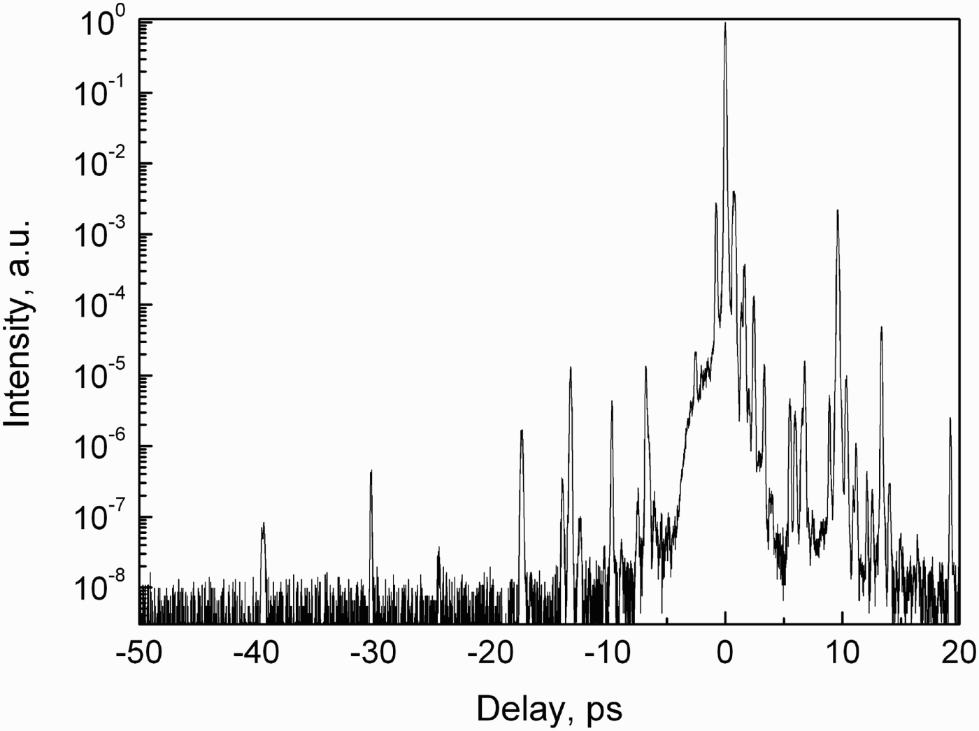
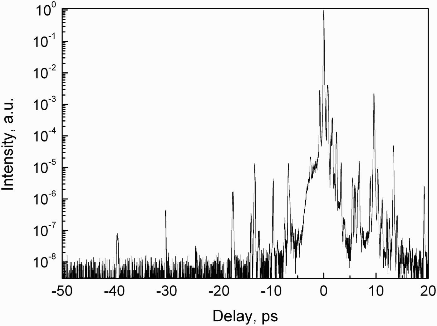
The laser facility produces a train of pulses with time duration of 40 ± 5 fs (full width at half maximum), energy up to 250 mJ and 10 Hz repetition rate. Pulse duration measurements were made by second order autocorellator. The Gaussian function was used to fit the temporal intensity profile. The pulse contrast is an important parameter of femtosecond laser interaction with a solid at intensity of about 1017 W/cm2, because prepulses can lead to significant modification of a target surface. A pulse temporal profile was measured using a third-order autocorrelator. Temporal pulse shape with prepulses in a time interval of 50 ps preceding the main pulse is presented in Figure 1. There are also prepulses on the order of 50 fs duration at the time delays—150 ps and —10 ns with intensity of 10−6 and 10−7, respectively, relative to the main pulse peak intensity. Amplified spontaneous emission intensity in the nano-second time domain does not exceed 10−8.
Third order cross-correlation function of the laser pulse.
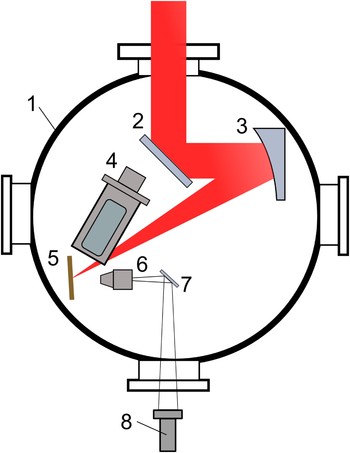
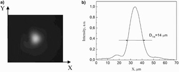
The schematic of experimental setup in use is shown in Figure 2. The experiments were conducted in a vacuum chamber evacuated to 10−4 mbar and connected to the vacuum chamber of time compressor. Laser beam of 30 mm diameter was focused on a target using off-axis parabolic mirror with focal length f′ = 254 mm. Laser spot size on a target was about 14 µm on 1/е level relative to the maximum intensity. Focal spot intensity distribution is presented in Figure 3.
(Color online) Experimental setup. (1) vacuum chamber, (2) deflecting mirror, (3) off-axis parabolic mirror, (4) Von Hamos spectrometer, (5) target, (6) microscope objective, (7) mirror, (8) CCD camera.

Spatial distribution of the laser pulse intensity in the focal plane of parabolic mirror: (а) two dimensional intensity distribution; (b) the intensity vs. X-coordinate.

The laser intensity spatial distribution in the focal plane was monitored by a microscope objective. It formed an image of the laser spot on a charge-coupled device (CCD)-array with 20× magnification. Targets (foils with a flat surface, and also covered with submicron sized metal rods, see below) were mounted on a three-dimensional motorized translation stages. One axis was used to set a target in a focal plane of parabolic mirror, while the rest were used to provide a fresh area on a target from shot to shot. A target position relative to the focal plane was controlled by observation system that consisted of a microscope objective and a CCD-camera. Accuracy of the target focal position was about ±30 µm, with the Rayleigh length 2z R = 385 µm for Gaussian beam waist radius w 0 = 7 µm.
To enable a single shot laser operation, an electro-optical modulator (DKDP crystal) with a synchronization unit was set at MPA output. This single pulse pick-out system was governed by a half-wavelength electric pulse of 10 ns time duration. The distance between electro-optical modulator and a target was about 5 m or about 15 ns for a laser pulse to travel. So the single pulse pick-out system also secured MPA optical elements (mirrors and active media) from back-reflected laser radiation. A thin quartz plate mounted right after MPA was used to reflect 2% of pulse energy onto a control photodiode. The photodiode calibration was carried out by means of laser calorimeter Sigma (Coherent). To vary laser pulse energy a polarized laser attenuator was applied. It consisted of a half-wave plate and a Glan prism. The energy fluctuations were about ±7%.
The angle between а target surface normal and a p-polarized laser beam was set to be 45 ± 2°. X-ray characteristic spectra were investigated using absolutely calibrated focusing crystal von Hamos spectrometer (Shevelko et al., Reference Shevelko, Kasyanov, Yakushev and Knight2002). In the von Hamos scheme, a mica crystal (2d = 19.84 Å) is bent into a cylindrical surface with R = 20 mm radius. The commercial CCD-array (Toshiba model TCD 1304AP) was used for X-ray detection. The angle of inclination of the spectrometer axis to the target surface normal was 30°.
Experimental Results
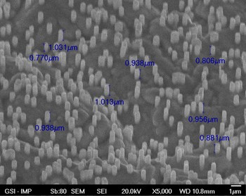
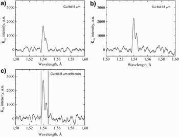
An absolute yield of X-ray characteristic K α-radiation for p-polarized laser pulses was investigated experimentally depending on a target type. There were three target types used: Cu foils of 8 and 31 µm thicknesses and a target with Cu rods. The Cu nano-rods target (Fig. 4) consists of about 108 rods/cm2, each of 500 nm in diameter and about 1 µm of height (~20% of the solid density). The nano-rods were supported by the 8 µm thick Cu layer, obtained by means of electrochemical deposition. This layer serves as a bulk material for production of K α-radiation by hot electrons generated in the interaction of ultrashort laser pulses with the nano-structured surface.
(Color online) Electron-microscope image of the target with Cu rods (GSI, No. 712).

The nano-structured targets were fabricated at GSI (the Gesellschaft für Schwerionenforschung, Germany). For these productions, polycarbonate membranes of 30 µm thickness were irradiated with heavy ion beams of 108 ion/cm2 fluence. The ion beam fluence can be varied depending on the desired nano-rod density from 1 to 109 ions/cm2. The distribution of the ion impacts is stochastic. Coulomb interaction of highly charged uranium ions of 11.4 MeV/u energy with target leads to material ion tracks along the ion stopping path. Via chemical etching process, one can remove the track area, and form cylindrical nano-channels of high aspect ratio (length to radius). The diameter of nano-channels depends on the etching time and is from 0.02 µm up to some micrometers.
The next step in the nano-rods fabrication is the electrochemical deposition of the desired material as Cu, Ag, and Au, etc. inside nano-channels (Liu et al., Reference Liu, Duan, Toimil-Molares, Karim, Cornelius, Dobrev, Yao, Sun, Hou, Mo, Wang and Neumann2006; Duan et al., Reference Duan, Liu, Mo, Yao, Maaz, Chen, Sun, Hou, Qu, Zhang and Chen2010). The material deposition rate and deposition time define the length of nano-rods. In order to obtain stable vertical structure of the nano-rods, the aspect ratio should not be higher than 20. After deposition process, the polycarbonate membrane is solved in the dichloromethane.
The typical X-ray spectra measured for various targets are presented in Figure 5. A laser energy value in the experiments was 9.2 ± 0.5 mJ. With pulse duration of 40 fs and laser spot diameter of 14 µm, it results in the maximum laser intensity on a target surface of 1.4 × 10¹⁷ W/cm2. All the recorded spectra were obtained by signal accumulation over 10 laser shots. The measurements for each target were repeated five times. Measured K α yield within given spectral interval Δλ = 0.01 Å (marked by vertical lines in Fig. 5c) is shown in Table 1 together with the results of modeling described in the next section. The errors are defined as a standard deviation from a mean value for five measurements.
X-ray spectra for different targets: (a) polished Cu foil of 8 µm thickness, (b) polished Cu foil of 31 µm thickness, (c) Cu foil of 8 µm thickness with nano-rods (GSI, No. 712).

Results of the measurements and calculations of K α yield from the different targets

MODELLING AND DISCUSSION
In spite of the high contrast laser pulses used in the discussed experiments, pilot modeling of the surface modifications under the action of the nano-second prepulse showed some smearing of the initial inhomogeneity of the target surface covered with micron sized rods. At the same time, a surface roughness with the typical scale on the order of micron remains, keeping sharp density gradients. To estimate the influence of a clustered surface on K α generation, we considered the model of characteristic X-ray emission from a foil covered with half-spherical clusters (as an approximate model of the surface with rods of low aspect ratio modified by pre-pulses) taking into account given laser pulse and target parameters.
In view of a time and radial dependence of the laser pulse intensity, the total number of K α photons, generated in a foil by fast electrons and emitted from the foil front side, is given by

under the assumption of compact arrangement of the clusters (Kostenko & Andreev Reference Kostenko and Andreev2010; Kostenko & Andreev, Reference Kostenko and Andreev2011b). Here, θ is the laser pulse angle of incidence; χ is the angle between the direction of fast electron propagation and the normal to the foil surface, φ is the azimuth angle in the surface plane; S h is the averaged over laser cycle electron flux directed to the cluster centre; N g(E) is the number of photons into 2π solid angle generated in the foil by an electron of incidence energy E, and f e(E) is the fraction of these photons that escape from the front the side of the foil. It is convenient to use Maxwellian distribution of hot electrons to approximate results of measurements and modeling of K α yield (Bastiani et al., Reference Bastiani, Audebert, Geindre, Schlegel and Gauthier1999; Reich et al., Reference Reich, Gibbon, Uschmann and Förster2000; Gibbon, Reference Gibbon2005). Electron energy distribution was fitted by a one-dimensional Maxwellian function f h(E) with the temperature related to the incident electron energy, averaged over a laser cycle. The fast electron parameters were evaluated according to the Brunel (Reference Brunel1987) analytic model of vacuum heating. To facilitate numerical integration, we generalized the simplified model of K α generation in a solid (Reich et al., Reference Reich, Gibbon, Uschmann and Förster2000) in case of an oblique incidence of an electron into the foil.
In case of the normal incidence of an electron with energy E into a solid target with plane surface, emission factor was approximated by a step function (Reich et al., Reference Reich, Gibbon, Uschmann and Förster2000)

with ɛ = E/E k, where E k is the K-shell ionization energy, ɛ1 = 20, U(x) = 1 for x ≥ 0, and 0 otherwise. According to Figure 1b in Reich et al. (Reference Reich, Gibbon, Uschmann and Förster2000), the mean depth of K α generation is a monotonous function of ɛ, and we approximated this function for copper by expression
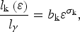
with b k = 4 × 10⁻³ and σk = 1.77 under ɛ > ɛ0 ≈ 3.3. Absorption length for self-emitted K α radiation in copper is l γ = 22.32 µm (Henke et al., Reference Henke, Gullikson and Davis1993). Eq. (2) can be rewritten as
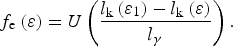
Physical interpretation of this expression is that most of the photons generated at the depth less than l k(ɛ1) = 0.8 l γ can escape from the target (cf. Reich et al., Reference Reich, Gibbon, Uschmann and Förster2000). Hence, in case of the oblique incidence of an electron at angle χ, we obtain

In the same way, the number of photons generated by an electron incident upon a foil of thickness d is

where N g0 = 4 × 10⁻³Z −1.67(E/keV)3/2U(ɛ – 1) is the number of photons into 2π solid angle generated in a solid target, and Z is the atomic number (Green & Cosslett, Reference Green and Cosslett1968; Reich et al., Reference Reich, Gibbon, Uschmann and Förster2000). Then, the number of photons emitted by one electron from the front side of the foil, on the assumption of d > l k(ɛ0) ≈ 0.67 µm, is given by
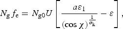
with  .
In accordance with experimental conditions, Gaussian laser pulse with wavelength λ = 0.8 µm, energy 9.2 mJ and duration 40 fs (full width at half maximum) was considered to be focused up to peak intensity 1.4 × 10¹⁷ W/cm2, with focal spot diameter 14 µm (at e −1 level of the maximum intensity) and θ = 45°. Calculations carried out for polished copper foils of 8 µm and 31 µm thickness (with Z = 29 and E k = 8.98 keV) according to analogous model (Kostenko & Andreev, Reference Kostenko and Andreev2011b), with Maxwellian distribution of hot electrons, give K α yields of 4.4 × 10⁸ and 6 × 10⁸ photons, respectively. Taking into account isotropic emission of X-rays in the context of considered model, these numbers (recalculated per steradian) are in reasonable agreement with measured K α yields and reproduce well the relative increase of K α emission with increase of the foil thickness (see Table 1). Note that according to Eq. (7), growth of K α yield with increase of the foil thickness ceases for d ≥ l k(ɛ1) ≈ 18 µm.
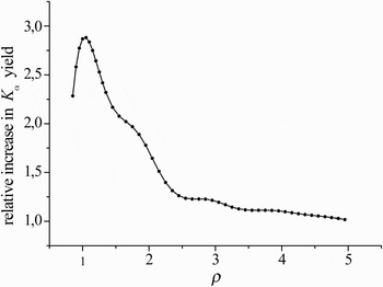
For a foil, as in case of a solid substrate (Kostenko & Andreev, Reference Kostenko and Andreev2010), dependence of K α yield on the cluster size shows a sharp maximum at ρ = πD/λ ≈ 1, with D being the cluster diameter (Fig. 6) (Kostenko & Andreev, Reference Kostenko and Andreev2011b). Measured relative increase in X-ray yield from the foil of 8 µm thickness with clustered surface is best fitted by the clusters of 0.5 µm diameter at ρ = 2 (see Table 1). This increase is caused by enhancement of accelerating electric field at the cluster surface (Sumeruk et al., Reference Sumeruk, Kneip, Symes, Churina, Belolipetski, Donelly and Ditmire2007; Kostenko & Andreev, Reference Kostenko and Andreev2010).
Relative increase in K α yield from a copper foil of 8 µm thickness versus ρ.

Obtained results indicate an applicability of the used model, based on the vacuum heating of electrons, under conditions of carried out experiments and show an availability of nano-structured targets for increasing of K α radiation under the action of high contrast femtosecond laser pulses. Further experiments could be concerned with optimization of the rods sizes, their surface density and the laser pulse contrast.
ACKNOWLEDGMENTS
This work was supported in part by the Programs on fundamental research of the Russian Academy of Sciences and Russian Foundation for Basic Research, RFBR-PICS project No. 11-02-91058.