Introduction
Vertebral artery dissection is an uncommon antecedent and cause of stroke. It accounts for between 0.13 and 2.5 per cent of all strokes.Reference Biller, Hingtgen, Adams, Smoker, Godersky and Toffol1, Reference Bogousslavsky and Regli2 Vertebral artery dissection is more common in younger adults, representing 16–20 per cent of strokes in patients below the age of 44 years.Reference Bogousslavsky and Regli2, Reference Kristensen, Malm, Carlberg, Stegmayr, Backman and Fagerlund3
Direct laryngoscopy is a common diagnostic and therapeutic procedure performed by ENT surgeons. In Australia, over 9000 cases were performed between January 2007 and December 2007. There have been reports of spinal cord injury following direct laryngoscopy in patients with cervical spine disease.Reference McLeod and Calder4 There are, however, no previously reported cases of vertebral artery dissection occurring following direct laryngoscopy, with only a single case noted in any post-operative patient.Reference Ringrose and Thompson5
We report for the first time a case of vertebral artery dissection in a woman who had undergone direct laryngoscopy. Symptoms of this dissection developed 48 hours following the procedure, which is consistent with the pathogenesis of vertebral artery dissection. We propose that this complication is under-reported, given the delay between the procedure and presentation and the self-limiting nature of vertebral artery dissection.
Case report
A 71-year-old woman with idiopathic subglottic stenosis was admitted for elective dilatation of the stenosed segment. She had previously been treated with steroids, which had lead to impaired glucose tolerance. She had a history of cervical spondylosis associated with many years of chronic neck pain (see Figure 1) and a limited range of motion in the cervical spine. She also had a history of hypertension. There was no history of cerebrovascular disease, and the patient had no other risk factors for arteriopathy.
Lateral cervical spine radiograph demonstrating cervical spondylosis.

The patient had previously undergone flexible rather than rigid bronchoscopy, due to concern regarding her cervical spine disease. However, this procedure had failed to identify the area of stenosis. When her symptoms worsened and exercise tolerance was limited to walking 10 metres, we decided to undertake direct laryngoscopy and dilatation, having obtained informed consent.
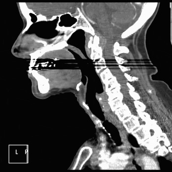
A pre-operative computed tomography (CT) scan of the patient's airway demonstrated a short segment of stenosis in the subglottis (see Figure 2).
Computed tomography neck scan (sagittal reconstruction) demonstrating a web-like stenosis in the subglottis.

The direct laryngoscopy procedure took 43 minutes and was performed under spontaneous ventilation without endotracheal intubation. A Dedo laryngoscope with gallows suspension was used, and visualisation of the glottis was optimal with minimal suspension. Manipulation of the cervical spine was very limited. Carbon dioxide laser radial incisions of the stenosis and CRT™ 15 mm balloon dilatation (Boston Scientific, Boston, Massachusetts, USA) were performed without any adverse consequences. There were no anaesthetic complications, and the patient's blood pressure remained stable throughout the procedure. The patient recovered rapidly but remained an in-patient for two days for social reasons.
Forty-eight hours after her procedure, the patient experienced a sudden onset of vertigo with nausea and vomiting. She complained of neck and occipital pain, which differed from her usual chronic neck pain, and was unable to mobilise.
Clinical examination revealed mild cerebellar signs, including past-pointing. Gait could not be assessed due to the severity of the patient's vertigo. There was no focal neurological deficit or nystagmus.
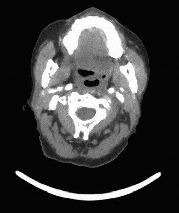
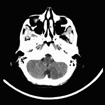
An urgent enhanced arterial-phase CT of the brain demonstrated occlusion of the left vertebral artery above the level of C2, with an associated wedge hypodensity of the medial left cerebellar lobe (see Figures 3 and 4). Further imaging studies were not performed.
Axial computed tomography angiogram demonstrating occlusion of the left vertebral artery within the foramen transversarium of the C2 vertebra.

Axial computed tomography head scan demonstrating a wedge opacity of the medial left cerebellar lobe.

The patient was subsequently transferred to the stroke unit. Her balance progressively recovered and her vertigo and nausea resolved.
The patient was discharged home on post-operative day seven. At three months' follow up, she had fully recovered and her respiratory function had improved.
Discussion
Vertebral artery dissection is an uncommon condition and has an estimated annual incidence of 0.97–1.12 cases per 100 000 population.Reference Lee, Brown, Mandrekar and Mokri6 Studies indicate a mortality of 1–2 per cent at six months post-event.Reference Arnold, Bousser, Fahrni, Fischer, Georgadis and Gandjour7, Reference Dziewas, Konrad, Drager, Evers, Besselmann and Ludemann8 Vertebral artery dissection is often spontaneous; however, reports have linked it to various risk factors.
Anatomy
The vertebral artery consists of four defined segments. It arises from the subclavian artery and ascends (V1) to enter the foramen transversarium of the C6 vertebra. It then climbs through the foramina of successive cervical vertebrae, forming the V2 portion. Subsequently, the artery reaches C2 and forms a lax posterior loop (V3) which then lies on the posterolateral arch of C1 and passes between the atlas and occiput, encased in the atlanto-occipital membrane. The V3 segment gives rise to the small meningeal branch. The artery ascends through the foramen magnum then pierces the dura (V4) before giving the posterior spinal, anterior spinal and posterior inferior cerebellar branches. The artery then joins its contralateral number on the clivus to form the basilar artery. The location of the proximal extent of the dissection is commonly in the V2 and V3 segments.Reference Arnold, Bousser, Fahrni, Fischer, Georgadis and Gandjour7 In the present case, occlusion of the vertebral artery was noted at C2 level (V3), at which level both rotation and extension of the cervical spine occur.
Risk factors
Connective tissue disorders (including Marfan's syndrome, Ehlers–Danlos syndrome and fibromuscular dysplasia) are present in 10–15 per cent of patients with vertebral artery dissection.Reference Schievink, Mokri and O'Fallon9 Arnold et al. Reference Arnold, Bousser, Fahrni, Fischer, Georgadis and Gandjour7 report that 11 per cent of patients with vertebral artery dissection display evidence of fibromuscular dysplasia on angiography. More recently, it has been suggested that 55–57 per cent of patients with cervical artery dissection demonstrate ultrastructural connective tissue aberrations.Reference Brandt, Orberk, Weber, Werner, Busse and Muller10, Reference Hausser, Muller, Engelter, Lyrer, Pezzini and Padovani11 However, there is no demonstrable clinical phenotype associated with such findings,Reference Schievink, Mokri and O'Fallon9 and patients lack the specific point mutations that are seen in connective tissue diseases.Reference Brandt, Orberk, Weber, Werner, Busse and Muller10–Reference Dittrich, Heidbreder, Rohsbach, Schmalhorst, Nassenstein and Maintz12 Attempts to demonstrate a consistent arteriopathic phenotype associated with connective tissue changes in vessel walls have been fruitless.Reference Dittrich, Heidbreder, Rohsbach, Schmalhorst, Nassenstein and Maintz12–Reference Calvet, Boutouyrie, Touze, Laloux, Mas and Laurent15 Vertebral artery dissection has also been linked to chronic vascular risk factors, such as diabetes, smoking and hypertension, which increase the risk of intimal damage. A recent systematic review found no definitive risk factor for vertebral artery dissection,Reference Rubinstein, Peerdeman, van Tulder, Riphagen and Haldeman16 with bias confounding most studies.
Trauma to the cervical spine, involving prolonged or forceful extension and/or rotation, and therapeutic spinal manipulation are commonly implicated as causes of vertebral artery dissection. However, attempts to quantify this risk and to demonstrate causality have been hampered by low case numbers and bias in study designs.Reference Haldeman, Kohlbeck and McGregor17–Reference Smith, Johnston, Skalabrin, Weaver, Azari and Albers20 It is suggested that risks associated with movement are increased in patients with cervical spine disease, including rheumatoid arthritis and spondylotic disease.Reference Howell and Molyneux21, Reference Gaikwad, Suri, Garg, Kanodia, Gupta and Mishralf22
There are also many minor traumatic events which have been associated with vertebral artery dissection, including: sports (tennis, golf and yoga), gardening, painting, giving birth, riding roller-coasters, massage, having a hair cut (the so-called ‘beauty parlour stroke syndrome’), reversing a car and minor whiplash injuries.Reference Dziewas, Konrad, Drager, Evers, Besselmann and Ludemann8, Reference Haldeman, Kohlbeck and McGregor19, Reference Smith, Johnston, Skalabrin, Weaver, Azari and Albers20, Reference Mas, Bousser, Hasboun and Laplane23–Reference Maroon, Gardner, Abla, El-Kadi and Bost26
Pathogenesis
It is known that vertebral artery dissection involves an intimal tear and disruption of the media due to the shearing forces of turbulent blood flow, with formation of a pseudolumen which may or may not have a re-entry point to the true lumen. Re-entry appears to be less common and is associated with lower morbidity than non-re-entry. Mizutani et al. Reference Mizutani, Kojima, Asamoto and Miki27 reported that disruption of the internal elastic lamina occurs longitudinally rather than axially. It is also noted that medial degeneration was not detected, suggesting that atherosclerotic plaques are not involved in the pathogenesis of vertebral artery dissection.Reference Mizutani, Kojima, Asamoto and Miki27 The plane of dissection may result in both internal and external compressive effects,Reference Mizutani, Kojima, Asamoto and Miki27, Reference Engelter, Brandt, Debette, Caso, Lichy and Pezzini28 and sub-arachnoid haemorrhage may result from dissection in a sub-adventitial plane.
Association between vertebral artery dissection and cervical spine movement
In the case described, and in many cases in the literature, the movements associated with vertebral artery dissection involve extension and/or rotation of the head With rotation of the head, the vertebral artery is stretched by the downward and forward motion of the contralateral atlantoaxial joint.Reference Maroon, Gardner, Abla, El-Kadi and Bost26 Evidence suggests that such a mechanical risk factor may be greater in patients with cervical spine disease. There are reports of patients with rheumatoid arthritis who demonstrated angiographically proven occlusion of the vertebral artery on extension of the head.Reference Howell and Molyneux21, Reference Gaikwad, Suri, Garg, Kanodia, Gupta and Mishralf22
However, in a recent systematic review of risk factors, Rubinstein et al. Reference Rubinstein, Peerdeman, van Tulder, Riphagen and Haldeman16 concluded that neck movements are not independent risk factors for vertebral artery dissection. It is suggested that manipulation of the cervical spine is the final insult that precipitates an ischaemic event, and that vertebral artery dissection occurs as a random and unpredictable sequel to manipulation.Reference Haldeman, Kohlbeck and McGregor18, Reference Haldeman, Kohlbeck and McGregor19
Clinical presentation
Vertebral artery dissection is most common in patients aged below 50 years, and has a well demonstrated accompanying symptom cluster and a typical history. Nearly 80 per cent of patients will present with ischaemic symptoms, and 88 per cent experience pain.Reference Arnold, Bousser, Fahrni, Fischer, Georgadis and Gandjour7 Pain will be ipsilateral to a unilateral dissection,Reference Sturzenegger29 and is occipital or cervical in location. The pain is sudden, but may be temporally dissociated from the onset of ischaemic symptoms. The majority of strokes will occur within 24 hours and before 10 days,Reference Blunt and Galton30 with 82 per cent of patients presenting within seven days of the onset of pain.Reference Biousse, D'Anglejan-Chatillon, Touboul, Amarenco and Bousser25 However, one to two months may elapse between onset of pain and occurrence of ischaemic symptoms.Reference Biousse, D'Anglejan-Chatillon, Touboul, Amarenco and Bousser25, Reference Nazir and Muir31
Ischaemic symptoms result from occlusion of the posterior cerebral circulation and may also be followed by lateral medullary infarction (Wallenberg's syndrome).Reference Arnold, Bousser, Fahrni, Fischer, Georgadis and Gandjour7, Reference Schievink, Mokri and O'Fallon9, Reference Mas, Bousser, Hasboun and Laplane23, Reference Biousse, D'Anglejan-Chatillon, Touboul, Amarenco and Bousser25, Reference Nazir and Muir31 Dissection may also be associated with subarachnoid haemorrhage in cases of intracerebral extension or origin of the dissection.
Investigations
The ‘gold standard’ in confirming the diagnosis is digital subtraction angiography, but magnetic resonance angiography and Doppler ultrasonography have superseded this invasive investigation. Vertebral artery dissection is commonly diagnosed on CT angiography, as this test is more readily available and commonly performed in the routine investigation of stroke; however, it is not the investigation of choice to demonstrate the precise location of dissection.
Treatment
The treatment of vertebral artery dissection is largely conservative. There is little evidence to support the use of antiplatelet agents or anticoagulants. Whilst a randomised, controlled trial is needed, it is unlikely to accrue sufficient cases.Reference Engelter, Brandt, Debette, Caso, Lichy and Pezzini28
Prognosis
The outcome following vertebral artery dissection is good. Youth and high baseline function are independent predictors of a favourable outcome. Between 70 and 82 per cent of patients have a modified Rankin scale score of less than one (i.e. no symptoms or no functional limitation despite symptoms) at early follow up.Reference Arnold, Bousser, Fahrni, Fischer, Georgadis and Gandjour7, Reference Dziewas, Konrad, Drager, Evers, Besselmann and Ludemann8 The most commonly reported residual symptom is loss of coordination. It is reported that, at follow-up angiography, 63 per cent of dissected arteries display resolution and a further 26 per cent display improvement.Reference Mas, Bousser, Hasboun and Laplane23 It is suggested that 8 per cent of patients will suffer recurrent dissection, with the risk of recurrence after one month being approximately 1 per cent per year.Reference Schievink, Mokri and O'Fallon9
Conclusion
We have presented a case of vertebral artery dissection following direct laryngoscopy. It is accepted that vertebral artery dissection is often under-diagnosed by virtue of an absence of robust clinical signs and a confounding temporal relationship between onset of symptoms and signs. It must be recognised that vertebral artery dissection is a potential complication of direct laryngoscopy, regardless of whether gallows suspension is used. Patients with cervical spine disease must be warned of their increased risk of this unpredictable condition, even with minimal cervical manipulation. They should be advised to seek immediate treatment in the event of heralding symptoms, which may occur several days (or weeks) after the procedure.
Vertebral artery dissection associated with neck movement accounts for a small percentage of an uncommon condition.Reference Rubinstein, Peerdeman, van Tulder, Riphagen and Haldeman16 During the consent procedure, the ENT surgeon should formally inform all patients, particularly those with cervical spine disease, of the possibility of vertebral artery dissection after direct laryngoscopy.