The scimitar syndrome accounts for a variable combination of anomalous connections of some or all of the right pulmonary veins to the inferior caval vein, hypoplasia of the right lung, hypoplasia and abnormal development of the right pulmonary artery, anomalous systemic arterial supply to the lower lobe of the right lung, and horseshoe or crossover lung.Reference Neill, Ferencz and Sabiston1, Reference Freedom, Yoo, Goo, Mikailian and Anderson2 The condition is very rare, occurring in no more than 1 to 3 cases in each 100,000 live births. When present, patients typically present either as infants,Reference Dupuis, Charaf and Brevière3 or as children or adults.Reference Dupuis, Charaf and Brevière4 Those presenting as infants tend to have by a higher incidence and severity of associated defects, cardiac failure, pulmonary hypertension, and higher ratios of pulmonary-to-systemic flow ratio, with correspondingly increased morbidity and mortality.Reference Najm, Williams, Coles and Rebeyka5 Stenosis of the scimitar vein is found in one-tenth of those with the infantile form.Reference Brown, Ruzmetov and Minnich6 We report here an infant with scimitar syndrome in whom the scimitar vein was completely obstructed.
Case report
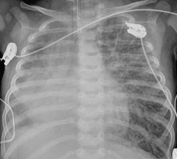
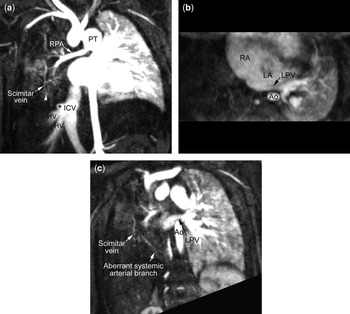
A female infant, born at 40 weeks gestation, presented at birth with tachypnoea. The chest radiograph revealed haziness of the right lung, with displacement of the heart to the right (Fig. 1). An echocardiogram, performed to investigate a murmur, demonstrated hypoplasia of the right pulmonary artery, but failed to identify right pulmonary veins. The infant required supplemental oxygen for 9 days, and was discharged home on the 13th day of life. At  months, she was readmitted because of poor feeding and tachypnoea. Her saturation of oxygen measured in room air was 81%. Her weight had dropped from an initial position on the 50th to the 3rd centile. She was intubated because of the acute respiratory distress and transferred to our hospital. Transthoracic and transesophageal echocardiography demonstrated very dilated right atrial and ventricular chambers, suprasystemic right ventricular systolic pressures, decreased right ventricular function, an atrial septum defect in the oval fossa measured at 13 millimetres, 2 small apical ventricular septal defects, and a common left pulmonary vein. Magnetic resonance imaging, with contrast-enhanced angiography,Reference Kramer, Domberger and Fenchel7 showed a severely hypoplastic vascular structure that was vertically oriented in the right lower lung, and aligned with an out-pouching from the right lateral aspect of the inferior caval vein above the right hepatic venous confluence, consistent with an occluded scimitar vein (Fig. 2a). The common left pulmonary vein was compressed between the enlarged left atrium and the spine (Fig. 2b). Both lungs showed severe pulmonary oedema. Phase-contrast velocity mapping revealed that the right lung received only 2% of the total flow to the lungs. The ratio of pulmonary to systemic flows was 2.2 to 1. A tiny aberrant systemic artery arose from the abdominal aorta and supplied the right lower lung (Fig. 2c). Subsequent cardiac catheterization confirmed the findings at magnetic resonance imaging, and additionally revealed the presence of a small persistently patent arterial duct.
The chest radiograph shows haziness of the right lung, with displacement of the heart to the right.

The reformatted contrast-enhanced magnetic resonance angiogram in an oblique coronary plane through the scimitar vein (a) shows the vein to be very hypoplastic, with complete occlusion in its lower part (arrow head). A round out-pouching (*) is seen to arise from the inferior caval vein (ICV) in the direction toward the atretic scimitar vein. The reformatted image in an axial plane seen from below (b) shows that the common left pulmonary vein (LPV) is compressed between the enlarged heart and the descending aorta (Ao). RA = right atrium, LA = left atrium. The reformatted image in an oblique coronal plane (c) shows a tiny aberrant systemic arterial branch in the right lower lung. The scimitar vein is also seen. HV = hepatic vein, PT = pulmonary trunk, RPA = right pulmonary artery.

Immediate surgery was delayed because of growth of Staphylococcal aureus from broncheoalveolar lavage. Subsequent interventions were necessitated by the ongoing need for mechanical ventilatory support associated with pulmonary oedema. They consisted of surgical closure of the atrial septal defect at 4 months, occlusion of the arterial duct at 5 months using a coil, and so-called sutureless repair of the common left pulmonary vein at 5 1/2 months.
Cardiac catheterization performed prior to the repair of the pulmonary vein demonstrated a left pulmonary arterial wedge pressure of 18 mmHg, with a simultaneous left ventricular pressure of 76/10 mmHg, and a mean left atrial pressure of 7 mmHg. The pulmonary arterial pressure was half that in the systemic circuit.
Post-operatively, there was significant improvement in her ventilatory state. Extubation was impeded, however, by compromise of the upper airways, and she was not extubated until 1 month later. She was discharged home at 7 months of age, requiring supplemental oxygen. The echocardiogram at discharge showed unobstructed flow from the left lung, with good biventricular systolic function.
Discussion
To the best of our knowledge, complete congenital obstruction of a scimitar vein has not previously been described. When the right lung shows unexplained hypoplasia, the scimitar syndrome should figure in any list of differential diagnoses. In our patient, the large deficiency of the oval fossa, and the obstructed venous return from the left lung, compounded the severity of the clinical course.