Implications
Essentially all commercially reared pigs are given chelated iron compounds to prevent anemia, a management practice that has been followed for several decades. Because iron is a highly reactive, potentially toxic metal, there still remain questions about whether such iron treatment may indeed be associated with increases in deleterious oxidative metabolism. The results reported here suggest that iron dextran, one of the most common iron-containing therapeutic compounds, when given as recommended, is not associated with oxidation of circulating proteins. Further, administration of treatment either directly following birth or 3 days later does not influence the serum protein oxidation process.
Introduction
Intracellular and circulating proteins are subject to many post-translational modifications that influence their turnover rate, function and activity, and can have marked effects on metabolism. Although many modifications are the result of direct enzymatic action, oxidation of proteins occurs following exposure to oxygen radicals that appear as non-reversible carbonyl modifications of specific amino acids, typically proline, arginine, threonine and lysine (Nyström, Reference Nyström2005). In addition, protein carbonyl formation also occurs as a result of metal-catalyzed oxidation. Irrespective of how they are derived, increased carbonyls are often associated with oxidative stress and many disease conditions and processes (Stadtman and Levine, Reference Stadtman and Levine2000; Adams et al., Reference Adams, Green, Claxton, Simcox, Williams, Walsh and Leeuwenburgh2001).
In modern confinement reared production facilities, essentially all pigs are treated with supplemental iron shortly after birth, usually in the form of iron dextran, to prevent the development of anemia in these rapidly growing animals (Zimmerman, Reference Zimmerman1980). Pigs in outdoor production environments also benefit from iron treatment for optimal performance and health (Szabo and Bilkei, Reference Szabo and Bilkei2002). Because iron is a potentially toxic trace element (Zimmerman, Reference Zimmerman1980, Lipiński et al., Reference Lipiński, Starzyński, Canonne-Hergaux, Tudek, Oliński, Kowalczyk, Dziaman, Thibaudeau, Gralak, Smuda, Woliński, Usińska and Zabielski2010) that can catalyze the formation of protein carbonyls (Robinson et al., Reference Robinson, Keshavarzian, Pasco, Frommel, Winship and Holmes1999; Yoo and Regnier, Reference Yoo and Regnier2004) it was our intention to evaluate the potential role of iron dextran in the induction of protein oxidation in the serum of neonatal pigs. Specifically, recommended dosages of iron dextran have recently been shown to induce DNA oxidation in piglet liver (Lipiński et al., Reference Lipiński, Starzyński, Canonne-Hergaux, Tudek, Oliński, Kowalczyk, Dziaman, Thibaudeau, Gralak, Smuda, Woliński, Usińska and Zabielski2010), the site of major cellular accumulation of injected iron dextran (Caperna et al., Reference Caperna, Failla, Steele and Richards1987) and the production of serum proteins. In addition, a single high dose of iron dextran has been shown to induce hepatic mitochondrial oxidation in rats (Pardo Andreu et al., Reference Pardo Andreu, Inada, Vercesi and Curti2009). We have recently quantified and identified protein carbonyls in serum of fetal and neonatal pigs, but did not investigate whether iron dextran treatment may impact serum protein oxidation (Caperna et al., Reference Caperna, Shannon, Blomberg, Garrett and Ramsay2010). Here we show that the standard treatment for the prevention of anemia in newborn pigs has little influence on the status or metabolism of circulating oxidized proteins.
Material and methods
Reagents
Chemicals for electrophoresis were purchased from Bio-Rad (Hercules, CA, USA) and biochemicals including biotin hydrazide, 2,4-dinitrophenylhydrazine (DNPH), sodium cyanoborohydride and acetonitrile were from Sigma-Aldrich (St. Louis, MO, USA). Urea and avidin-fluorescein (avidin-FITC) were from Pierce (Rockford, IL, USA). Methanol and acetic acid were from Fisher (Suwanee, GA, USA); trifluoroacetic acid was from J.T. Baker (Phillipsburg, NJ, USA).
Collection of serum samples
Care and treatment of all pigs in this study were approved by the Institutional Animal Care and Use Committee of the US Department of Agriculture. Seven litters (from first or second parity gilts and sows) of crossbred pigs (Landrace × York × Poland China) were obtained on day 1 (D1; within 24 h) and evaluated for use in the study. Each litter utilized, contained at least eight piglets. Five healthy non-runt piglets of the same sex were chosen from each litter, such that each treatment group contained three gilts and four boars. D1 controls (D1-Fe) were immediately bled and euthanized. On day 1, two piglets were administered 1.0 ml of iron dextran solution (100 mg Fe/ml; Butler Co., Columbus OH, USA) intramuscularly and were bled on day 3 (D3 + FeD1) or day 5 (D5 + FeD1); another control non-iron-treated piglet was bled and euthanized on D3 (D3-Fe). The final piglet was administered the iron dextran solution on D3 and bled on D5 (D5 + FeD3). All piglets except D1-Fe were also given a 1-ml bolus of vitamin B complex solution (Vedco Inc., St. Joseph, MO, USA) intramuscularly on D1. Blood samples were collected by venous puncture and allowed to clot on ice. Serum was harvested by two successive centrifugation steps (3500 × g in a swinging bucket rotor) and frozen in aliquots at −80°C until analyzed. Protein content in whole serum and serum extracts was determined by the method of Lowry following precipitation in 7% trichloroacetic acid (TCA) and 0.07% Triton-X 100 (final concentrations) respectively, and re-solubilized in 1 N NaOH (Nerurkar et al., Reference Nerurkar, Marino and Adams1981). Bovine serum albumin was used as a standard.
Quantification of protein carbonyls
To quantify the amount of carbonyls in serum samples, the spectrophotemetric method of Levine et al. (Reference Levine, Garland, Oliver, Amici, Climent, Lenz, Ahn, Shaltiel and Stadtman1990), and Yoo and Regnier (Reference Yoo and Regnier2004) as described by Caperna et al. (Reference Caperna, Shannon, Blomberg, Garrett and Ramsay2010) was used. Protein carbonyls in serum were converted to dinitrophenyl (DNP) derivatives with DNP-hydrazine, precipitated with TCA and extracted with ethyl acetate : ethanol (1 : 1). The final pellets were solubilized with 6 M guanidine hydrochloride in 20 mM KH2PO4/10 mM H3PO4, pH 2.3, and the absorbance at 362 nm was determined. Protein content of each dissolved sample was also determined and the extinction coefficient for DNP-conjugated carbonyls was considered to be 22 000 M/cm (Levine et al., Reference Levine, Garland, Oliver, Amici, Climent, Lenz, Ahn, Shaltiel and Stadtman1990). The background absorbance of each sample was determined by preparing a set of control duplicates by addition of 2 NHCl (without the DNPH) to the serum samples, which were treated as described above. Background values for each sample were calculated (nmoles carbonyls/mg protein) and subtracted from each DNPH experimental determination.
Identification of protein carbonyls: biotin conjugation
To separate, visualize and identify carbonylated proteins, biotin hydrazide at neutral pH was used as a carbonyl coupling reagent (Yoo and Regnier, Reference Yoo and Regnier2004) as described by Caperna et al. (Reference Caperna, Shannon, Blomberg, Garrett and Ramsay2010). Serum samples were precipitated with TCA and incubated for 2 h at room temperature in 5 mM biotin hydrazide with end-over-end mixing. Following reduction with sodium cyanoborohydride, and precipitation with TCA they were extracted with ethyl acetate : ethanol. The final precipitates were vacuum dried and solubilized with modified two-dimensional electrophoresis re-hydration buffer without dithiothreitol (7.55 M urea, 4% 3-[(3-cholamidopropyl)dimethylammonio] 1-propanesulfonate (CHAPS), 0.5% Bio-Lytes and 0.001% bromophenol blue (BPB)). Total protein content was determined as described above. Following separation by two-dimensional polyacrylamide gel electrophoresis (2D-PAGE), the presence of biotin was confirmed by directly staining the gel with avidin-FITC (Nakamura et al., Reference Nakamura, Tsumoto, Ishimura and Kumagai2002; Yoo and Regnier, Reference Yoo and Regnier2004) as described previously (Caperna et al., Reference Caperna, Shannon, Blomberg, Garrett and Ramsay2010). Biotin–avidin-FITC conjugates were visualized by fluorescence image scanning (488 nm excitation/520BP40 filter; Typhoon, GE Healthcare, Piscataway, NJ, USA). Each gel was then fixed and stained with a standard colloidal Coomassie blue (CCB) stain (Caperna et al., Reference Caperna, Shannon and Garrett2008) and scanned (Image Scanner III; GE Healthcare) to visualize total proteins.
2D-PAGE
The first dimension isoelectric focusing of biotin-conjugated serum proteins was performed using 7 cm immobilized pH gradient (IPG) gel strips (pH 3 to 10; Bio-Rad) in the IPGphor II system (GE Healthcare) essentially as described previously (Caperna et al., Reference Caperna, Shannon and Garrett2008 and Reference Caperna, Shannon, Blomberg, Garrett and Ramsay2010). Focused strips were rinsed in dH20 and stored at −80°C. Thawed strips were equilibrated, treated with iodoacetamide and the two-dimensional electrophoretic separation (10% acrylamide) was performed as described previously (Caperna et al., Reference Caperna, Shannon, Blomberg, Garrett and Ramsay2010) using a mini-Protean II (Bio-Rad) electrophoresis system. Following electrophoresis, gels were fixed and stained with avidin-FITC and CCB as described above.
Gel image analysis
Following fluorescence and optical scanning, all raw image files (.gel files) were converted to .tif files with IQTL software (GE Healthcare) and evaluated with Progenesis SameSpots software version 3.1 (Nonlinear Dynamics USA Inc., Durham, NC, USA). To determine individual spot volumes, the gel regions to be incorporated in the analysis area were sized uniformly, and gels were aligned to a single reference gel for each experimental grouping. Individual spot volumes were determined for avidin-stained gels and then the corresponding spots from the CCB-stained gels were identified and quantified, separately as described previously (Caperna et al., Reference Caperna, Shannon, Blomberg, Garrett and Ramsay2010). For quantitative spot analysis, equal amounts of biotinylated protein from the seven individual animals were pooled and each pooled sample was separated by 2D-PAGE and analyzed three separate times. Once protein spots were identified by mass spectrometry (MS), some proteins that were represented by multiple spots or regions were grouped and labled as single proteins on the 2D-PAGE map image.
Serum iron analysis
Confirmation of iron dextran treatment was achieved by analyzing ferrozine-reactive iron in serum samples using a total serum iron reagent kit (Pointe Scientific Inc., Canton, MI, USA). Serum samples were assayed according to the manufacturer's directions; however, the volumes were modified so that a 96-well format could be utilized. All samples were analyzed in duplicate and compared with an iron standard curve performed on the same plate.
MS and protein identification
Coomassie-stained protein spots corresponding to FITC-avidin-stained proteins were excised from the gel and placed in a 96-well V bottom plate (no. 3363; Costar, Corning, NY, USA) in 200 μl dH20 and stored at −80°C. Samples obtained from gels were thawed, digested and prepared for MS as described previously (Caperna et al., Reference Caperna, Shannon and Garrett2008 and Reference Caperna, Shannon, Blomberg, Garrett and Ramsay2010). A matrix-assisted laser desorption/ionization-time of flight mass spectrometer (Voyager DE-STR MALDI-TOF-MS, Applied Biosystems, Framingham, MA, USA) operated in positive ion reflector mode was used to analyze tryptic peptides by peptide mass fingerprinting (PMF). Spectra were acquired with 75 shots of a 337-nm nitrogen laser operating at 20 Hz and were internally calibrated using the porcine trypsin autolysis peaks at m/z 842.51 and 2211.10 as internal standards. Samples that were not initially identified by PMF were vacuum-dried, re-suspended in 20 μl 5% acetonitrile, 0.1% formic acid and subjected to MS/MS analysis (ThermoFisher Scientific LTQ Orbitrap XL hybrid linear ion trap, mass spectrometer, San Jose, CA, USA). Peptides were separated by reverse phase chromatography on a 100 × 0.18 mm BioBasic-18 column using a 30 min linear gradient from 5% to 40% acetonitrile in 0.1% formic acid at a flow rate of 3 μl/min. The instrument was operated in data dependent mode with a duty cycle that acquired and recorded MS/MS spectra in the linear ion trap of the five most abundant ions determined by a high-resolution survey scan in the Orbitrap (r = 30 000 at m/z 400) over the range of 400 to 1600 m/z. High mass accuracy of the survey scans was achieved and maintained by calibration in real time using the lock mass option. The polydimethylcyclosiloxane ion (Si(CH3)2O)6 (protonated m/z 445.120025), which is generated in the electrospray process from ambient air (Schlosser and Volkmer-Engert, Reference Schlosser and Volkmer-Engert2003), was used for the calibration lock mass. Dynamic exclusion was used to prevent the continuous analysis of the same ions. Once two MS/MS spectra had been acquired from any given ion, the parent mass was placed on an exclusion list for the duration of 3 min. Electrospray voltage was set at 3.5 kV and desolvation was assisted with 10 units of sheath gas; the capillary transfer tube temperature was set to 200°C. The minimum ion count required to trigger an MS/MS spectrum was set to 5000 and normalized collision energy was set at 30%. Mascot Distiller version 2.1.1 (www.matrixscience.com) was used to prepare searchable peak lists for both PMF data and LC (liquid chromatography)-MS/MS data.
Protein identification by PMF or MS/MS was performed by searching the National Center for Biotechnology Information non-redundant and SwissProt primary databases using the Mascot search engine that uses a probability-based scoring system (Perkins et al., Reference Perkins, Pappin, Creasy and Cottrell1999). The following parameters were used for PMF database searches: monoisotopic mass, 25 ppm mass accuracy, trypsin as digesting enzyme with 1 missed cleavage allowed, fixed modification of carbamidomethylation of cysteine, while oxidation of methionine, N-terminal pyroglutamic acid from glutamic acid or glutamine were set as variable modifications; taxonomy was limited to Mammalia or other Mammalia. For MS/MS data, monoisotopic mass was chosen, peptide tolerance was set to 5 ppm, MS/MS tolerance to 1 Da and peptide charges set to 1+, 2+ and 3+. Cleavage enzyme, allowed missed cleavages and fixed and variable modifications were the same as for PMF searches. To qualify the MALDI-TOF-MS data as a positive identification, a protein's score (MOWSE) was equal to or exceeded the minimum significant score. Positive identification of proteins by MS/MS analysis required a minimum of two unique peptides, both with significant ion scores and with an apparent protein molecular weight within approximately 10 kD of the expected mass of each protein. In cases in which multiple proteins were potentially present in a single spot, the protein with the highest score and/or the greatest peptide representation was considered to be the identified protein of record.
Statistical analysis
Spot volume values obtained from differentially stained 2D gels (n = 3) were analyzed by GLM ANOVA (NCSS 2000, Kaysville, UT, USA) using a one-way factorial model to evaluate differences among individual protein spots. Values of P<0.05 were considered different and means were separated by Fisher's LSD post hoc analysis. When proteins were represented by multiple spots, volumes for the individual spots were obtained and summed before statistical analysis. Levels of protein carbonyls and iron in serum were also analyzed by GLM ANOVA (n = 7) with five treatment groups, and means in which significant differences were observed, were separated by Fisher's LSD.
Results
Quantitative analysis of DNP-conjugated protein carbonyls in serum of experimental pigs is presented in Figure 1a. On D1 (D1-Fe), the carbonyl levels were 0.87 + 0.07 nmole/mg serum protein. On D3 (D3-Fe) carbonyl levels were similar to those of newborn piglets. Injection with iron dextran on D1 had no apparent influence on serum protein oxidation at 48 or 96 h following injection, D3 + FeD1 and D5 + FeD1, respectively. In addition, waiting until the third day of life for the application of iron dextran also had no apparent influence on protein carbonyl content of piglets when analyzed 48 h following treatment (D5 + FeD3; P > 0.05). The presence of elevated circulating iron in iron dextran-treated piglets was confirmed by measurement of ferrozine-reactive iron in serum of piglets (Figure 1b). Treatment with iron dextran on D1 resulted in a fourfold increase (P < 0.05) in serum iron 48 h following injection (D3 + FeD1) and levels returned to nearly pre-treatment levels by 96 h (D5 + FeD1). Iron dextran treatment on D3 resulted in a similar increase in ferrozine-reactive iron 48 h after treatment (D5 + FeD3 v. D3 + FeD1).
Protein carbonyls (a) and iron (b) in serum of control and iron dextran-treated piglets. Values are means ± s.e.m. (n = 7 pigs/group). The experimental groups were: D1-Fe (day 1 untreated controls); D3-Fe (day 3 untreated controls); D3 + FeD1 (day 3 treated with iron dextran on day 1); D5 + FeD1 (day 5 treated with iron dextran on day 1) and D5 + FeD3 (day 5 treated with iron dextran on day 3). Data were analyzed by GLM ANOVA and means were separated by Fisher's LSD post hoc test.

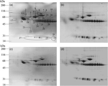
Representative avidin-FITC and Coomassie-stained gels of biotin-conjugated serum samples from the D3 ± iron dextran treatment groups are shown in Figure 2. Essentially all of the major serum proteins, as observed by Coomassie staining (Figure 2b and d), had detectable levels of carbonyls (avidin-FITC-stained; Figure 2a and c) in either control or iron-treated piglets. With few exceptions (this section), the overall molecular distribution of carbonylated proteins was similar for all groups. In addition, although individual proteins varied widely in their fluorescence staining, comparison of carbonyl staining intensity of specific individual proteins across the experimental groups of piglets whether they were treated with iron dextran was quite similar.
Avidin-fluorescein (avidin-FITC) and colloidal Coomassie-stained two-dimensional polyacrylamide gel electrophoresis (2D-PAGE) of pooled piglet serum samples labeled with biotin hydrazide. (a) avidin-FITC-stained pooled D3 + FeD1 (day 3 treated with iron dextran on day 1) serum; (b) colloidal Coomassie-stained pooled D3 + FeD1 serum; (c) avidin-FITC-stained pooled D3-Fe (day 3 untreated controls) serum; and (d) colloidal Coomassie-stained pooled D3-Fe serum. Numbered protein spots were identified by peptide mass fingerprinting (PMF) or LC-MS/MS. In each gel pair, the same gel was stained with avidin-FITC and colloidal Coomassie blue.

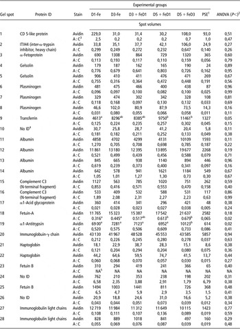
The corresponding identities of the major avidin-stained proteins and quantitative spot analyses are given in Table 1. Proteins that showed high levels of carbonyl staining were immunoglobulin heavy and light chains, albumin, fetuin A, transferrin and α1-antitrypsin; other identified less-prominent oxidized proteins were complement component 3, CD 5-like protein, fetuin B, α-fetoprotein, plasminogen, gelsolin, haptoglobin, α1-acid glycoprotein and inter-α-trypsin inhibitor 4.
Protein identification, relative distribution of carbonylated (avidin-stained) proteins and the ratio of avidin to total Coomassie-stained proteins (A : C) in serum of pigs treated with iron dextran and untreated controls

Gel spot numbers correspond to those identified in Figure 2a. Spot volumes for each spot or group of spots were determined as outlined in Material and methods section. All listed proteins met the strict criteria for significant positive identifications as described in Materials and Methods. All represent primary matches directly to Sus scrofa except fetuin B, which was identified by searching the EST (expressed sequence tag)-mammals database and a significant match to a porcine EST submission was obtained; the peptide sequence was subsequently BLASTED to the non-redundant sequence database and identified using the bovine sequence.
1PSE (pooled standard error) was determined using ANOVA (n = 3).
2Significance values based on one-way ANOVA.
3Ratio of the spot volumes for avidin-stained spots/spot volume from the same spots stained with Coomassie blue.
4A specific protein could not be identified for this spot.
5NA = not applicable as no corresponding Coomassie-stained spot was detected.
a,bMeans within a row with different superscript letters are significantly different at P < 0.05 (Fisher's LSD post hoc analysis) .
Statistical analysis (ANOVA) of avidin-FITC-stained proteins revealed that transferrin (Figure 2a; spot 9) was altered by treatment (P < 0.05); means separation (Fisher's LSD) indicated that the increase in carbonyl content of transferrin was due to the age of the piglet and not to the iron-dextran treatment. Further, the ratio of carbonylation to total transferrin protein (A : C ratio) was not different (P = 0.15), suggesting that while the level of transferrin increased over time, the specific carbonylation also increased coordinately. Oxidation of α1-antitrypsin (Figure 2a; spot 19) was influenced by iron treatment (P < 0.02). When means were separated by post hoc analysis, carbonylation of this protein was higher in the D5 + FeD3 group compared with the other groups, while A : C ratio was similar among all five groups, indicating an increase in both carbonylation and total protein. The total avidin-staining intensity of fetuin-A (Figure 2a; spot 18) was not influenced by iron dextran treatment; however, the A : C ratio within the spot was significantly elevated in the D5 iron dextran-treated groups compared with untreated D1 and D3 piglets.
Discussion
In this study, a combined analytical and proteomic approach was used to both quantify and identify specific oxidized proteins in serum of young piglets as might be influenced by iron treatment. High levels of iron are known to be toxic and may potentially lead to enhanced susceptibility to bacterial infection (Knight et al., Reference Knight, Klasing and Forsyth1983). However, the levels that are recommended and are given to piglets at birth in the form of iron dextran are thought to be safe and effective at providing additional iron to meet the demand for rapid growth during the first few weeks of life (Zimmerman, Reference Zimmerman1980; Caperna et al., Reference Caperna, Failla, Steele and Richards1987; Egeli and Framstad, Reference Egeli and Framstad1999).
In our preliminary evaluations, it initially appeared that iron dextran treatment was associated with elevated serum protein carbonyls. However, we subsequently determined that all of the increases in absorbance values obtained were due to background, and most likely associated with circulating levels of iron complexes and not DNP-conjugated carbonyls. In our previous study (Caperna et al., Reference Caperna, Shannon, Blomberg, Garrett and Ramsay2010), individual background values for each sample were also determined for the calculation of total serum carbonyls, although background absorbance values were relatively similar among the developmental groups investigated. For the data presented here the simultaneous determination of a background value for each sample was essential to obtain analytical integrity.
Quantification of total carbonyls by direct spectroscopic analysis revealed that iron dextran treatment had very little influence on serum protein oxidation. This was further confirmed when the distribution of oxidized proteins as analyzed by 2D-PAGE of biotin conjugated serum proteins was found to be similar among the five groups of piglets. With few noted exceptions (viz., α1-antitrypsin and fetuin-A), analysis of biotin-labeled spot volumes by quantitative fluorescence imaging of the proteins separated individually indicated that there were no consistent or major effects attributable to iron dextran. In the experimental 2D-PAGE analysis, identical amounts of protein were loaded on each gel and protein spots were quantified if they occurred in all gels. This allows for direct comparison of all recognized spots for both fluorescently stained and Coomassie-stained gels. Overall, differences in the distribution or in the intensity of individual proteins (i.e. Coomassie-stained protein spots) were not observed. Taken together, this would indicate that, at least for the major serum proteins, iron dextran treatment had minimal influence on the distribution of oxidized proteins and did not appear to induce carbonyl formation. This observation does not negate the possibility that iron dextran could induce oxidative damage in other tissues or classes of molecules. Indeed, treatment of neonatal pigs with similar levels of iron dextran (40 or 100 mg Fe/injection) induced significant amounts of hepatic 8-oxo-7,8 dihydro-2’-deoxyguanosine, a marker of DNA oxidation (Lipiński et al., Reference Lipiński, Starzyński, Canonne-Hergaux, Tudek, Oliński, Kowalczyk, Dziaman, Thibaudeau, Gralak, Smuda, Woliński, Usińska and Zabielski2010). Whether intracellular carbonyl formation is induced in specific hepatic cell populations where iron accumulates (Caperna et al., Reference Caperna, Failla, Steele and Richards1987) or in other tissues is a topic for further investigation.
The data presented here suggest a specific increase in carbonylated α1-antitrypsin 48 h after treatment with iron dextran on D3 with no change in the A:C ratio, indicating a coordinate increase in both carbonyls and circulating levels of this protein. It has been recently suggested that in human serum, α1-antitrypsin binds to prohepcidin, a circulating form of the iron regulatory hormone, hepcidin (Pandur et al., Reference Pandur, Nagy, Poór, Sarnyai, Huszár, Miseta and Sipos2009). Interestingly, Lipiński et al. (Reference Lipiński, Starzyński, Canonne-Hergaux, Tudek, Oliński, Kowalczyk, Dziaman, Thibaudeau, Gralak, Smuda, Woliński, Usińska and Zabielski2010) have recently demonstrated that iron dextran induces the expression of hepatic hepcidin in neonatal piglets. Thus, these data could implicate a specific role for α1-antitrypsin in the regulation of iron metabolism via its potential interaction with prohepcidin in the neonatal pig. Circulating levels of hepcidin or prohepcidin have not been reported in piglets and the potential impact of carbonylation on the binding characteristics of α1-antitrypsin to prohepcidin has not been investigated.
In this study, we observed an increase in the level of oxidation (A:C ratio) of fetuin-A in the serum of iron-treated D5 piglets. Owing to the experimental design, the confounding influence of age cannot be ruled out, as all the D5 piglets were treated with iron dextran. However, the carbonyl levels in both D5 groups were similar to the iron-treated D3 piglets but higher than the untreated pigs at D1 or D3. In addition, except for immunoglobulin heavy chains and albumin, fetuin-A represents the richest source of protein carbonyls. Fetuin-A is a serum glycoprotein and primarily functions as a mineral chaperone for the transport of calcium phosphate (Jahnen-Dechent et al., Reference Jahnen-Dechent, Schäfer, Ketteler and McKee2008) and has been shown to regulate ectopic and vascular tissue calcification (Koos et al., Reference Koos, Brandenburg, Mahnken, Mühlenbruch, Stanzel, Günther, Floege, Jahnen-Dechent, Kelm and Kühl2009). In addition, fetuin-A is a negative acute phase protein that has been proposed to serve as a general carrier of a variety of molecules, similar to the function of serum albumin (Moe and Chen, Reference Moe and Chen2005). Although evidence suggests that high levels of Fetuin-A are associated with reversal of vascular oxidative inflammatory disease (Moe and Chen, Reference Moe and Chen2005), the capacity of carbonylated fetuin-A to attenuate the disease process has not been addressed. Further, the association of fetuin-A with iron transport or metabolism has not been investigated previously.
In conclusion, we have shown that iron dextran treatment markedly elevates the level of serum iron approximately fourfold at 48 h post-injection. The maximum level of circulating iron was probably much higher as in a similar study, where the peak concentrations of serum iron were observed for 8 to 16 h following treatment of piglets with 100 mg iron dextran (Knight et al., Reference Knight, Klasing and Forsyth1983). Despite the large increase in iron availability, carbonyls remained relatively constant throughout the 5-day period reported here. In addition, in our previous study, serum carbonyl levels were similar between pigs at 1 day of age (without iron treatment) and on day 7, that is, 6 days following iron dextran treatment (Caperna et al., Reference Caperna, Shannon, Blomberg, Garrett and Ramsay2010). From the standpoint of defining serum carbonyls as a biomarker of oxidative stress, iron dextran, when given at recommended levels either on D1 or D3, does not appear to induce protein oxidation in the neonatal pig.
Acknowledgements
We thank Dr Neil C. Talbot for critical review of the manuscript. We also thank Margo Stoll for animal handling, sample collection and technical help. Jenile Tapscott, James Woods and Clayton Wieben are thanked for assistance in maintaining and handling experimental animals.