Introduction
After the emergence of the human immunodeficiency virus (HIV) in the 1980s, there has been a considerable resurgence of interest in human parasitic protozoan infections in general, with many cases of Cystoisospora belli infections being reported in patients with HIV. In addition, frequent C. belli infections were reported in immunocompetent patients that were given immunosuppressive therapy for organ transplants or those treated for tumours worldwide. Having reviewed many of those reports, it became clear that there are many uncertainties concerning the biology of C. belli, including its endogenous stages, misdiagnoses, and mechanism(s) for persistent infections in humans. Many details of the development of C. belli stages are lacking. Additionally, non-protozoal structures in the gallbladders of patients that were originally identified as C. belli are now considered artefacts. Although, trimethoprim sulfamethoxazole (TMP/SMX or cotrimoxazole), is the drug most commonly used for cystoisosporiasis, relapses with the clinical disease are frequent in immunosuppressed patients and many people are allergic to sulfonamides. Here, we review all aspects of the biology of C. belli.
History
Isospora-like intestinal coccidia have been known for two centuries to parasitize humans, dogs and cats (Levine, Reference Levine1973). Connal (Reference Connal1922) reviewed more than 150 cases of intestinal coccidiosis in humans worldwide, but he was uncertain of the species involved. Wenyon (Reference Wenyon1923) proposed the name Isospora belli for the large, bell-shaped 25–30 × 12–15 µm oocysts in human feces. Despite the characteristic shape of its oocysts, I. belli continued to be misdiagnosed as Isospora hominis (Elsdon-Dew and Freedman, Reference Elsdon-Dew and Freedman1953; Jarpa Gana, Reference Jarpa Gana1966; Smitskamp and Oey-Muller, Reference Smitskamp and Oey-Muller1966). Until the 1970s, Isospora species were considered non-host-specific and to have a direct fecal–oral cycle (Dubey, Reference Dubey2018). When the life cycle of Sarcocystis was discovered in 1972, it became clear that the parasite I. hominis was a mixture of more than two species of Sarcocystis with an obligatory two-host cycle involving cattle and pigs as intermediate hosts and humans as definitive hosts (reviewed in Dubey et al., Reference Dubey, Calero-Bernal, Rosenthal, Speer and Fayer2016). Additionally, Isospora spp. of cats were found to have a tissue cyst stage in extra-intestinal organs of paratenic hosts as well as in the definitive host (Dubey and Frenkel, Reference Dubey and Frenkel1972; Frenkel and Dubey, Reference Frenkel and Dubey1972), leading to the creation of a new genus, Cystoisospora (Frenkel, Reference Frenkel1977) for Isospora species with a tissue cyst stage. In 1987, the tissue cyst stage of I. belli was discovered (Restrepo et al., Reference Restrepo, Macher and Radany1987). Based on morphologic and phylogenetic relationships with feline and canine Isospora, I. belli was transferred to the genus Cystoisospora (Carreno et al., Reference Carreno, Schnitzler, Jeffries, Tenter, Johnson and Barta1998; Franzen et al., Reference Franzen, Müller, Bialek, Diehl, Salzberger and Fätkenheuer2000; Barta et al., Reference Barta, Schrenzel, Carreno and Rideout2005). Thus, the correct designation for I. belli is Cystoisospora belli (Wenyon, Reference Wenyon1923) Frenkel, Reference Frenkel1977.
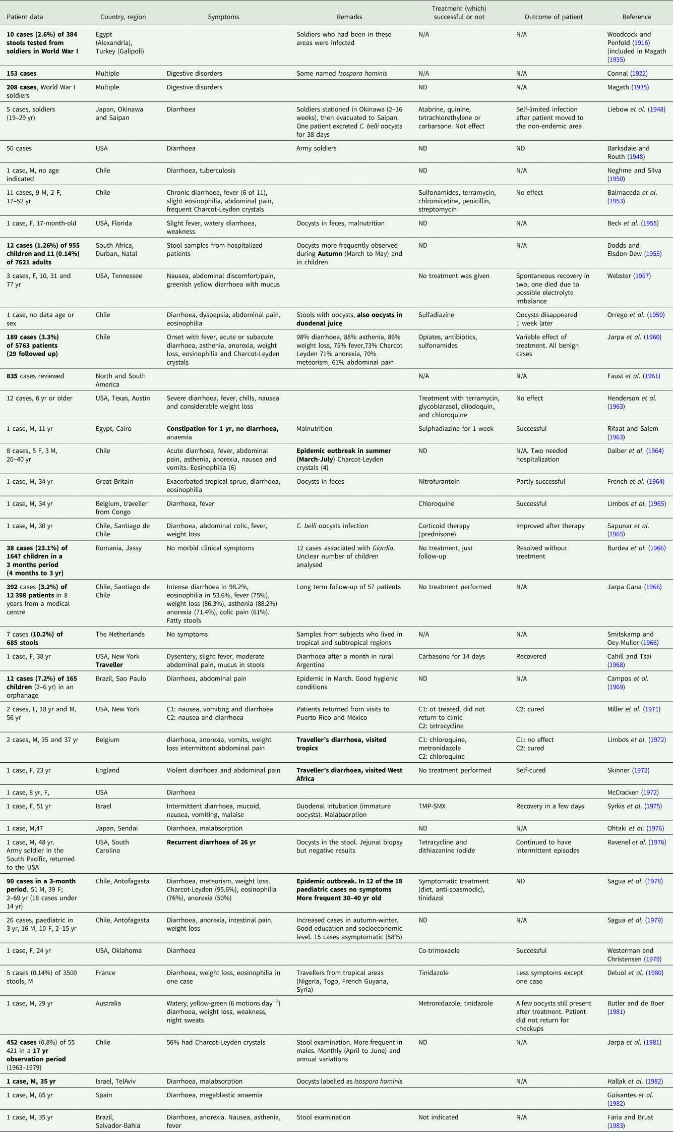
Although there are several previous reviews on C. belli (e.g. Knight and Wright, Reference Knight and Wright1978; Soave, Reference Soave1988; Curry et al., Reference Curry, Turner and Lucas1991; Pape and Johnson, Reference Pape and Johnson1991; Gellin and Soave, Reference Gellin and Soave1992; Marcial-Seoane and Serrano-Olmo, Reference Marcial-Seoane and Serrano-Olmo1995; Goodgame, Reference Goodgame1996; Heyworth, Reference Heyworth1996, Marshall et al., Reference Marshall, Naumovitz, Ortega and Sterling1997; Lindsay et al., Reference Lindsay, Dubey and Blagburn1997a; Field, Reference Field2002; Arora and Arora, Reference Arora and Arora2009; Chawla and Ichhpujani, Reference Chawla and Ichhpujani2011; Kotloff et al., Reference Kotloff, Blackwelder, Nasrin, Nataro, Farag, van Eijk, Adegbola, Alonso, Breiman, Faruque, Saha, Sow, Sur, Zaidi, Biswas, Panchalingam, Clemens, Cohen, Glass, Mintz, Sommerfelt and Levine2012; Legua and Seas, Reference Legua and Seas2013; Nhampossa et al., Reference Nhampossa, Mandomando, Acacio, Quintó, Vubil, Ruiz, Nhalungo, Sacoor, Nhabanga, Nhacolo, Aide, Machevo, Sigaúque, Nhama, Kotloff, Farag, Nasrin, Bassat, Macete, Levine and Alonso2015; Ortega and Cama, Reference Ortega, Cama, Ortega and Sterling2018), they are either more than two decades-old or are not comprehensive. After the emergence of the HIV, many cases of C. belli infections were reported in patients with HIV, and in immunocompetent patients that were given immunosuppressive therapy for organ transplants or those treated for tumours (Tables 1–3).
Early reports (1935–1983, before the emergence of AIDS) of intestinal C. belli infections in humans based only on stool examination, presented in chronological order

N/A, not applicable; ND, not done or o data; yr, age in years; M, male; F, female; TMP-SMX, trimethoprim sulfamethoxazole or cotrimoxazole.
Important findings are in bold.
Summary of positive reports of intestinal C. belli infections in humans after the emergence of AIDS, based only on stool examination, presented in chronological order

M, male; F, female; yr, age in years; ND, not done or o data; IC, immunocompetent; TMP-SMX, trimethoprim sulfamethoxazole or cotrimoxazole; Oo, oocysts; neg, negative; pos, positive; N/A, not applicable; ID, Immunosuppressed.
Important/rare findings are in bold.
Summary of reports of C. belli infections in humans with histologic findings presented in chronological order, including stages found, treatments and outcome

SC, schizonts; ND, not done or o data; F, female; yr, age in years; Oo, oocysts; GA, gamonts; M, male; IC, immunocompetent; TEM, transmission electron microscopy; TMP-SMX, trimethoprim sulfamethoxazole or cotrimoxazole; ME, merozoites; pos, positive; N/A, Not applicable; TC, tissue cysts; HARRT, highly active antiretroviral therapy.
Important/rare findings are in bold.
Morphology and life cycle
Oocysts are excreted in an unsporulated stage in freshly passed human feces. Wenyon (Reference Wenyon1923) named and illustrated the organism; however, he failed to provide a description of the oocysts. The following authors have contributed to the description of C. belli oocysts: Barksdale and Routh (Reference Barksdale and Routh1948); Jeffery (Reference Jeffery1956); Ferreira et al. (Reference Ferreira, Coutinho, Argento and da Silva1962); Henderson et al. (Reference Henderson, Gillepsie, Kaplan and Steber1963); French et al. (Reference French, Whitby and Whitfield1964); Jarpa Gana (Reference Jarpa Gana1966); Cahill and Tsai (Reference Cahill and Tsai1968); Zaman (Reference Zaman1968); Levine (Reference Levine1973); Guisantes et al. (Reference Guisantes, Rull, Rubio, Tobalina and de Landazuri1982); Morakote et al. (Reference Morakote, Muangyimpong, Somboon and Khamboonruang1987); Jongwutiwes et al. (Reference Jongwutiwes, Putaporntip, Charoenkorn, Iwasaki and Endo2007); Resende et al. (Reference Resende, Pedrosa, Correia, Cabrine-Santos, Lages-Silva, Meira and Oliveira-Silva2011); Ros Die and Nogueira Coito (Reference Ros Die and Nogueira Coito2018). Oocysts are 17–37 × 8–21 µm and colourless; the oocyst wall is very thin and smooth. The oocysts are not always bell-shaped and have one, both, or no pointed ends (Lindsay et al., Reference Lindsay, Dubey and Blagburn1997a; Jongwutiwes et al., Reference Jongwutiwes, Putaporntip, Charoenkorn, Iwasaki and Endo2007). The length to width ratio is >1.2 (usually 2.1). Usually the sporulated oocysts contain two sporocysts and four sporozoites in each sporocyst, but occasionally Caryospora-like oocysts (oocysts with one sporocyst containing 8 sporozoites) are present (Jongwutiwes et al., Reference Jongwutiwes, Putaporntip, Charoenkorn, Iwasaki and Endo2007). A micropyle and polar granule are present in some oocysts. The sporocysts are 9–14 × 7–12 µm, spherical to ellipsoidal, and without a Stieda body. Sporulation occurs in the environment but apparently, in some cases, oocysts sporulate in the gut lumen (Brandborg et al., Reference Brandborg, Goldberg and Breidenbach1970, Neira and Villaon, Reference Neira and Villalón1989; Nateghi Rostami et al., Reference Nateghi Rostami, Nikmanesh, Haghi-Ashtiani, Monajemzadeh, Douraghi, Ghalavand and Kashi2014) and partially and fully sporulated oocysts have been seen in freshly excreted feces. Sporulation time varies depending on the environmental conditions from 24 h to 10 days. Oocysts are excreted intermittently or continuously up to 50 days (Jeffery, Reference Jeffery1956). The specific gravity of C. belli oocysts is between 1.045–1.090 (Dodds and Elsdon-Dew, Reference Dodds and Elsdon-Dew1955). Oocysts can be stored in 0.5% formaldehyde in water and proceed to maturation, while storing them in anaerobic conditions will stop maturation (Dodds and Elsdon-Dew, Reference Dodds and Elsdon-Dew1955).
Monozoic tissue cysts are present in extra-intestinal organs of immunosuppressed humans but a paratenic host has not been demonstrated. Tissue cysts were found in the spleen, liver, mediastinal, trachea-bronchial, and mesenteric lymph nodes, and in the intestinal lamina propria (Brandborg et al., Reference Brandborg, Goldberg and Breidenbach1970; Restrepo et al., Reference Restrepo, Macher and Radany1987, Comin and Santucci, Reference Comin and Santucci1994; Michiels et al., Reference Michiels, Hofman, Bernard, Saint Paul, Boissy, Mondain, Lefichoux and Loubiere1994; Lindsay et al., Reference Lindsay, Dubey and Blagburn1997a; Reference Lindsay, Dubey, Toivio-Kinnucan, Michiels and Blagburn1997b; Velásquez et al., Reference Velásquez, Carnevale, Mariano, Kuo, Caballero, Chertcoff, Ibáñez and Bozzini2001; Frenkel et al., Reference Frenkel, de Oliveira Silva, Saldanha, de Silva, Correia Filho, Barata, Lages, Ramirez and Prata2003; Meamar et al., Reference Meamar, Rezaian, Mirzaei, Zahabiun, Faghihi, Oormazdi and Kia2009; Velásquez et al., Reference Velásquez, Osvaldo, Di Risio, Etchart, Chertcoff, Perissé and Carnevale2011; Lindsay et al., Reference Lindsay, Houk, Mitchell and Dubey2014; Velásquez et al., Reference Velásquez, di Risio, Etchart, Chertcoff, Nigro, Pantano, Ledesma, Vittar and Carnevale2016; Swanson et al., Reference Swanson, March, Clayton, Couturier, Arcega, Smith and Evason2018).
Based on morphological evaluation of 1000 tissue cysts in sections of spleen and lymph nodes, tissue cysts were monozoic, with a 1.5–4.0 µm thick cyst wall (Lindsay et al., Reference Lindsay, Dubey, Toivio-Kinnucan, Michiels and Blagburn1997b). The zoites were 10.0–14.0 × 1.5–4.0 µm and mostly crescent-shaped. A pale eosinophilic staining was present distal to the nucleus. Ultrastructurally, the tissue cyst consisted of a parasitophorous vacuolar membrane enclosing the thick granular cyst wall, and intravacuolar tubules. The zoite inside the cyst has the structure of coccidian sporozoite, including numerous micronemes, a few rhoptries, dense granules, polysaccharide granules, and a crystalloid body. Occasionally two tissue cysts were seen in one vacuole.
Attempts to infect non-human primates, livestock, rodents, and other animals with C. belli were unsuccessful (Jeffery, Reference Jeffery1956). Experimental infections in human volunteers (Matsubayashi and Nozawa, Reference Matsubayashi and Nozawa1948; Ferreira et al., Reference Ferreira, Coutinho, Argento and da Silva1962) confirmed the direct fecal–oral transmission cycle of C. belli. Human volunteers who ingested sporulated oocysts became infected and excreted unsporulated C. belli oocysts 9 to 17 days later. In the first experiment conducted in Japan, two healthy adults ingested C. belli oocysts. On September 22, 1946, one of the authors of the paper, a medical student volunteer, ingested 3000 C. belli sporulated oocysts, obtained from feces of a Japanese soldier who returned from China (inoculum #1). He developed diarrhoea, fever, headache, poor appetite, lassitude, muscular pain and oedema of the face between day 8 and 17 after oocyst ingestion. Oocysts were first detected in feces on the 9th day of infection and were seen occasionally for 30 days. On December 5, 1946 (day 74 after the first infection), he ingested 2500 C. belli oocysts a second time (inoculum #2). He remained symptomless, thus suggesting the development of protective immunity. The second volunteer, a 19-year-old, ingested 2500 oocysts (inoculum #2) on 5 December 1946 developed diarrhoea on the 9th day. Oocysts were first detected on day 11 and were present in his feces for 38 days. Based on this experiment the prepatent period was between nine and 11 days (Matsubayashi and Nozawa, Reference Matsubayashi and Nozawa1948).
The second experiment was conducted in Brazil (Ferreira et al., Reference Ferreira, Coutinho, Argento and da Silva1962). Five, 20–25 years old males each ingested 6000 C. belli oocysts (Ferreira et al., Reference Ferreira, Coutinho, Argento and da Silva1962). Infection became evident in four of the subjects; the fifth person did not excrete oocysts and remained healthy. Oocysts were first detected in feces of three volunteers on day 15 and in one volunteer on day 17 and oocysts were excreted for 14 days in one person, 28 days in one, 29 days in one and 34 days in another. The incubation period (first day of symptoms) was 7–10 days. The following symptoms were recorded for the four individuals: fever in three, nausea in two, vomiting and headache in one, and asthenia and abdominal pain in all four. The gastrointestinal symptoms included meteorism (excess gas), fetid flatus, diarrhoea (as many as 13 motions daily) and voluminous stool. All four volunteers recovered; the experiment was terminated on day 48 (Ferreira et al., Reference Ferreira, Coutinho, Argento and da Silva1962).
Infections thought to be laboratory-acquired have also been reported (Connal, Reference Connal1922; Jeffery, Reference Jeffery1956; Henderson et al., Reference Henderson, Gillepsie, Kaplan and Steber1963; McCracken, Reference McCracken1972). A 28-year-old male technician from the laboratory where C. belli oocysts were processed for animal experimentation (Jeffery, Reference Jeffery1956), developed a fever and intermittent diarrhoea for 2 months; oocysts were found in his feces for 50 days (Jeffery, Reference Jeffery1956). A 26-year-old male laboratory technician got infected analysing positive C. belli fecal samples from an 8-year-old girl in Texas. The technician had mild abdominal discomfort, malaise, low grade fever and diarrhoea that subsided after 5 days, although C. belli continued to be excreted for a further 10 days (McCracken, Reference McCracken1972).
Two healthy scientists apparently ingested C. belli oocysts while attempting to infect rabbits (Henderson et al., Reference Henderson, Gillepsie, Kaplan and Steber1963). Both developed gastrointestinal symptoms 11 or 12 days later. The symptoms included nausea, fever, vomits, headaches, weight loss, and abdominal distention. In the first worker, nausea persisted for 30 days, becoming severe at nightfall. In the second worker, diarrhoea was severe with as many as 40 stools daily. Oocysts were first detected on day 16 in one and on day 23 in the other scientist after accidental ingestion (Henderson et al., Reference Henderson, Gillepsie, Kaplan and Steber1963). In the one case reported from Nigeria, a scientist apparently became infected by accidently ingesting sporulated C. belli oocysts (Connal, Reference Connal1922). He developed diarrhoea on the 6th day but oocysts were not detected in his feces until day 28 after the accidental ingestion.
Fragmentary information on endogenous stages have been obtained by examination of human biopsy tissues from the small intestine (Table 3) (Niedmann, Reference Niedmann1963; French et al., Reference French, Whitby and Whitfield1964; Brandborg et al., Reference Brandborg, Goldberg and Breidenbach1970; Henry et al., Reference Henry, Bird and Doe1974; Trier et al., Reference Trier, Moxey, Schimmel and Robles1974; Liebman et al., Reference Liebman, Thaler, DeLorimier, Brandborg and Goodman1980; Veldhuyzen van Zanten et al., Reference Veldhuyzen van Zanten, Lange, Sauerwein, Rijpstra, Laarman, Rietra and Danner1984; Modigliani et al., Reference Modigliani, Bories, LeCharpentier, Salmeron, Messing, Galian, Rambaud, Lavergne, Cochand-Priollet and Desportes1985; Restrepo et al., Reference Restrepo, Macher and Radany1987; Peng and Tsai, Reference Peng and Tsai1991; Benator et al., Reference Benator, French, Beaudet, Levy and Orenstein1994; Comin and Santucci, Reference Comin and Santucci1994; Michiels et al., Reference Michiels, Hofman, Bernard, Saint Paul, Boissy, Mondain, Lefichoux and Loubiere1994; Hamour et al., Reference Hamour, Curry, Ridge, Baily, Wilson and Mandal1997; Bialek et al., Reference Bialek, Overkamp, Rettig and Knobloch2001; Field, Reference Field2002; Jongwutiwes et al., Reference Jongwutiwes, Sampatanukul and Putaporntip2002; Sasaki et al., Reference Sasaki, Tanaka, Nishimura, Tsujikawa, Andoh, Ishizuka, Koyama, Fujiyama and Kushima2004; Murphy et al., Reference Murphy, Hoogestraat, Sengupta, Prentice, Chakrapani and Cookson2011; Rao et al., Reference Rao, Geetha, Ranjini, Vidhyalakshmi and Rupashree2012; Kim et al., Reference Kim, Kim, Jung, Chai and Chai2013; Agholi et al., Reference Agholi, Shahabadi, Motazedian and Hatam2016b; Swanson et al., Reference Swanson, March, Clayton, Couturier, Arcega, Smith and Evason2018) and bile ducts/or gallbladder (Benator et al., Reference Benator, French, Beaudet, Levy and Orenstein1994; Frenkel et al., Reference Frenkel, de Oliveira Silva, Saldanha, de Silva, Correia Filho, Barata, Lages, Ramirez and Prata2003; Lagrange-Xélot et al., Reference Lagrange-Xélot, Porcher, Sarfati, de Castro, Carel, Magnier, Delcey and Molina2008; Walther and Topazian, Reference Walther and Topazian2009; Agholi et al., Reference Agholi, Aliabadi and Hatam2016a).
Schizonts, gamonts and unsporulated oocysts were seen in the enterocytes of the small intestine and in the biliary epithelium. Histopathologic examination of tissues obtained in post-mortem studies confirmed that asexual and sexual stages of C. belli are confined to the small intestine, bile ducts and gallbladder epithelium (Brandborg et al., Reference Brandborg, Goldberg and Breidenbach1970; Restrepo et al., Reference Restrepo, Macher and Radany1987; Benator et al., Reference Benator, French, Beaudet, Levy and Orenstein1994; Michiels et al., Reference Michiels, Hofman, Bernard, Saint Paul, Boissy, Mondain, Lefichoux and Loubiere1994; Frenkel et al., Reference Frenkel, de Oliveira Silva, Saldanha, de Silva, Correia Filho, Barata, Lages, Ramirez and Prata2003; Agholi et al., Reference Agholi, Aliabadi and Hatam2016a, among others) (Table 3). Restrepo et al. (Reference Restrepo, Macher and Radany1987) mentioned finding schizonts and gamonts also in the large intestine but did not provide any details. In another study, the parasite was also reported to be present in epithelial cells of the large intestine (in the colon) (Figueroa et al., Reference Figueroa, Palacios, Rivero, Oddo, Roa, Honeyman, Gatica and Acuna1985). Additionally, tissue cysts (thought to be encysted sporozoites) were found in the lamina propria of the small and large intestine, lymph nodes, spleen, and liver of immunosuppressed patients (Restrepo et al., Reference Restrepo, Macher and Radany1987; Comin and Santucci, Reference Comin and Santucci1994; Michiels et al., Reference Michiels, Hofman, Bernard, Saint Paul, Boissy, Mondain, Lefichoux and Loubiere1994; Lindsay et al., Reference Lindsay, Dubey and Blagburn1997a, Reference Lindsay, Dubey, Toivio-Kinnucan, Michiels and Blagburn1997b; Velásquez et al., Reference Velásquez, Carnevale, Mariano, Kuo, Caballero, Chertcoff, Ibáñez and Bozzini2001; Frenkel et al., Reference Frenkel, de Oliveira Silva, Saldanha, de Silva, Correia Filho, Barata, Lages, Ramirez and Prata2003; Meamar et al., Reference Meamar, Rezaian, Mirzaei, Zahabiun, Faghihi, Oormazdi and Kia2009; Velásquez et al., Reference Velásquez, Osvaldo, Di Risio, Etchart, Chertcoff, Perissé and Carnevale2011; Swanson et al., Reference Swanson, March, Clayton, Couturier, Arcega, Smith and Evason2018).
Partial development of schizonts containing 2–4 merozoites was seen in human cell lines seeded with C. belli sporozoites (Siripanth et al., Reference Siripanth, Punpoowong, Amarapal and Thima2004; Oliveira-Silva et al., Reference Oliveira-Silva, Lages-Silva, Resende, Prata, Ramirez and Frenkel2006).
In a severe case involving a 2-year-old child who had prolonged corticoid therapy due to systemic vasculitis, a high number of C. belli oocysts and the unusual presence of merozoites were observed in stools (Malik et al., Reference Malik, Samantaray, Bagga and Das2005). The authors proposed that the mucosal injury probably provided an environment more prone to invasion by C. belli and that the weak defences of the child allowed the infection of extra intestinal sites from which the merozoites would have been released (Malik et al., Reference Malik, Samantaray, Bagga and Das2005).
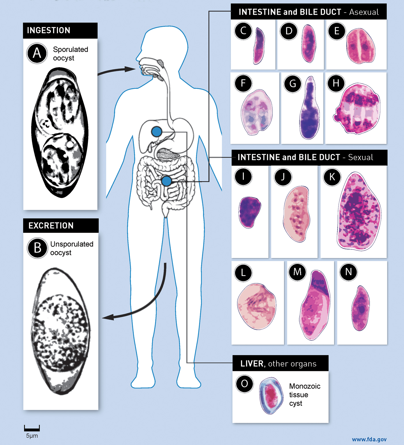
After a review of these reports, it became clear that details regarding C. belli developmental stages were lacking. Additionally, non-protozoal structures in the gallbladder of patients that were initially identified as C. belli (Lai et al., Reference Lai, Goyne, Hernandez-Gonzalo, Miller, Tuohy, Procop, Lamps and Patil2016; Martelli and Lee, Reference Martelli and Lee2016; Akateh et al., Reference Akateh, Arnold, Benissan-Messan, Michaels and Black2018; Noor et al., Reference Noor, Katzman, Huber, Findeis-Hosey, Whitney-Miller, Gonzalez, Zhou, N'kodia, Skonick, Abell, Saubermann, Lamps and Drage2019) are now considered artefacts. Therefore, an intensive search was made to identify the developmental stages of C. belli in the duodenum and common bile duct of two patients reported previously (Walther and Topazian, Reference Walther and Topazian2009; Swanson et al., Reference Swanson, March, Clayton, Couturier, Arcega, Smith and Evason2018). The study revealed that C. belli schizonts and gamonts were like other species of Cystoisospora in animals (Dubey et al., Reference Dubey, Evason and Walther2019). Schizonts were often crescent-shaped, up to 25 × 18 µm, and contained up to 10 merozoites that were 10–11 × 3–4 µm. The macrogamonts were elongated. The microgamonts contained as many as 30 nuclei, and 1–3 residual bodies. The microgametes are about 4 µm long (Dubey et al., Reference Dubey, Evason and Walther2019) and have flagella. The life cycle of C. belli is illustrated in Fig. 1.
Life cycle of Cystoisospora belli. Cystoisospora belli has a direct fecal–oral transmission cycle. If a susceptible human ingests a sporulated oocyst (A) with contaminated food or water, the sporozoites inside the sporocysts are released in the gut lumen and invade the epithelial cells of the intestine, bile ducts and gallbladder where the parasite multiplies asexually and sexually within a parasitophorous vacuole (pv) of the host. In the asexual reproduction (C–H; C: merozoite; D: schizont with dividing nucleus; E: paired organisms; F: paired crescent-shaped schizonts; G: schizont with a conical (conoidal) and a rounded non-conoidal end; H: schizont with mature merozoites). In the sexual reproduction two types of gamonts are formed: microgamonts (males) and macrogamonts (female) (I–N; I: multinucleated microgamont; J: an elongated microgamont with several peripheral nuclei with condensed chromatin; K: a microgamont with two residual bodies and several microgametes; L: microgametes; M: an elongated macrogamont with a large nucleus and small granules in the cytoplasm; N: an elongated macrogamont with a single nucleus and a large nucleolus). After a microgamete fertilizes a macrogamont, oocyst wall is formed around the zygote and unsporulated oocyst (B) is excreted in feces. Some sporozoites encyst in extra-intestinal organs (spleen, liver, mediastinal, trachea-bronchial, and mesenteric lymph nodes) and in intestinal lamina propria and produce monozoic tissue cysts (O). Oocysts sporulate in the environment and can survive in soil and water for months. All stages of the parasite are microscopic (<50 µm in maximum dimension). Fig. A and B are from unstained, unfixed oocysts, C–O are from histological sections of tissues. Bar applies to all parts.

Distribution and prevalence
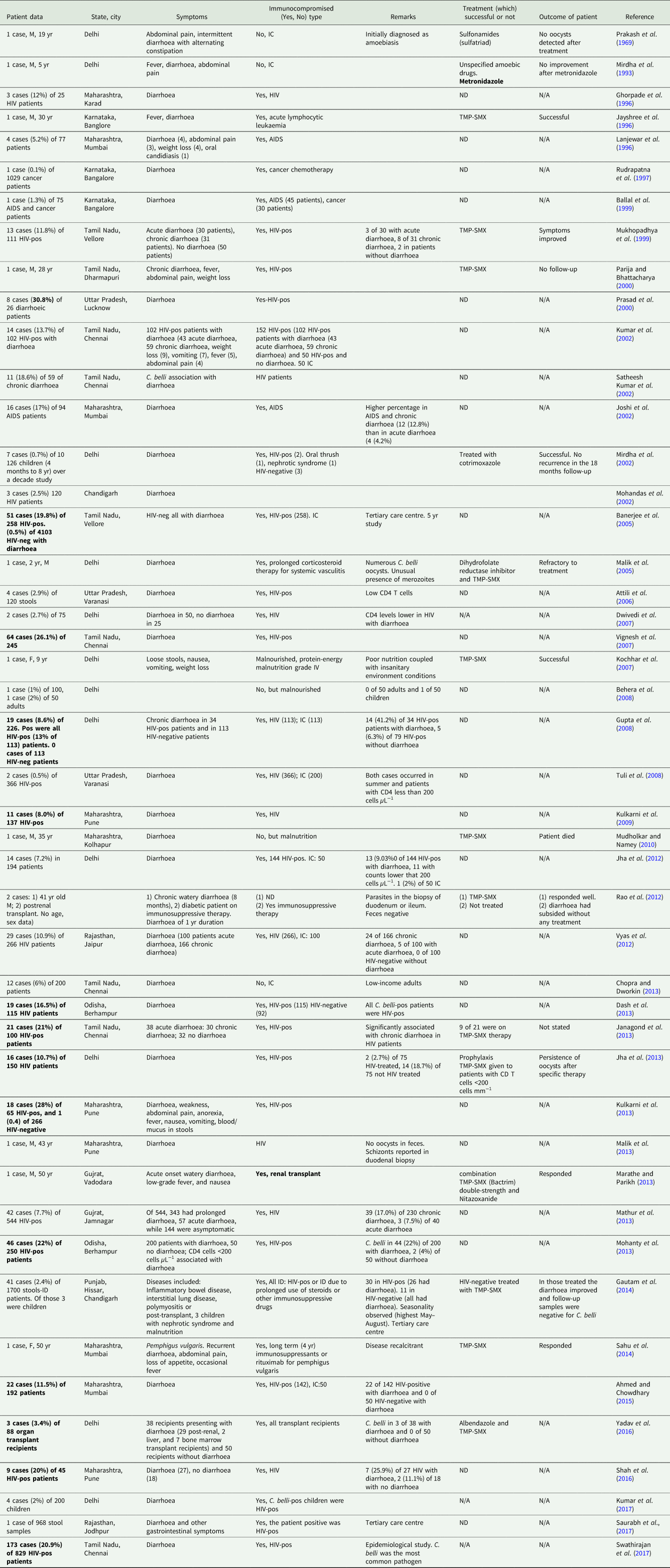
Cystoisospora belli is globally distributed, but infections are found mainly in tropical and subtropical areas of the world such as the Caribbean, Central and South America, Africa and South-East Asia (Tables 1–3). There are more reports of C. belli infections from India than other countries and these are summarized in Table 4. Additionally, many cases were recorded as Isospora homonis before the recognition of C. belli (Mukherjee, Reference Mukherjee1947).
Summary of positive reports of C. belli infections in humans in India, presented in chronological order

M, male; yr, age in years; IC, immunocompetent; ND, not done or o data; N/A, not applicable; pos, positive; TMP-SMX, trimethoprim sulfamethoxazole, cotrimoxazole; F, female; ID, Immunosuppressed; SC, schizonts; Oo, oocysts; ART, active antiretroviral therapy.
Important/rare findings are in bold.
Most C. belli infections have been reported in the last century. Faust et al. (Reference Faust, Giraldo, Caicedo and Bonfante1961) summarized more than 1500 cases from the Western Hemisphere. Most of these reports were based on finding C. belli oocysts in feces (Tables 1 and 2). A higher number of studies were reported after the emergence of HIV, and currently, the prevalence of C. belli is being reported worldwide (Table 2). Reports confirmed by autopsy or biopsy are summarized in Table 3. Epidemic outbreaks of cystoisosporiasis were reported in the 1960s and in the late 1970s in Chile and Brazil (Daiber et al., Reference Daiber, Vuillemin and Schleef1964; Campos et al., Reference Campos, Neto and Campos1969; Sagua et al., Reference Sagua, Soto, Delano, Fuentes and Becker1978). In the largest outbreak in Chile, 90 clinical cases were reported in a 3-month period during autumn–winter months (May–June 1977). Oocysts of C. belli were found in an industrial water treatment plant, which could have been used to water and contaminate vegetables (Sagua et al., Reference Sagua, Soto, Delano, Fuentes and Becker1978). The epidemic report in Brazil occurred in a children orphanage (Campos et al., Reference Campos, Neto and Campos1969).
Prevalence rates vary even in endemic areas. Consistently high prevalence of C. belli infection (12–15%) was observed in several studies in Haiti (DeHovitz et al., Reference DeHovitz, Pape, Boncy and Johnson1986; Pape et al., Reference Pape, Verdier and Johnson1989; Verdier et al., Reference Verdier, Fitzgerald, Johnson and Pape2000). In Africa, prevalence levels varied greatly among countries, ranging from 0.2% to 28% (Henry et al., Reference Henry, De Clercq, Lokombe, Kayembe, Kapita, Mamba, Mbendi and Mazebo1986; Kelly et al., Reference Kelly, Baboo, Wolff, Ngwenya, Luo and Farthing1996; Cranendonk et al., Reference Cranendonk, Kodde, Chipeta, Zijlstra and Sluiters2003; Olusegun et al., Reference Olusegun, Okaka and Machado2009; Vouking et al., Reference Vouking, Enoka, Tamo and Tadenfok2014; Sangaré et al., Reference Sangaré, Bamba, Cissé, Zida, Bamogo, Sirima, Yaméogo, Sanou, Drabo, Dabiré and Guiguemdé2015; Casmo et al., Reference Casmo, Lebbad, Maungate and Lindh2018). In South America prevalences ranged from 1.1% to 30.0% (Jarpa Gana, Reference Jarpa Gana1966; Sauda et al., Reference Sauda, Zamarioli, Ebner Filho and Mello1993; Certad et al., Reference Certad, Arenas-Pinto, Pocaterra, Ferrara, Castro, Bello and Núñez2003; Kaminsky et al., Reference Kaminsky, Stovall, Mayer, Martin, Bowers and Didier2007; Chincha et al., Reference Chincha, Bernabé-Ortiz, Samalvides, Soto, Gotuzzo and Terashima2009; Cardoso et al., Reference Cardoso, Galisteu, Schiesari Júnior, Chahla, da Silva Canille, Belloto, Franco, Maia, Rossit and Machado2011; Assis et al., Reference Assis, Resende, Cabrine-Santos, Correia and Oliveira-Silva2013; Barcelos et al., Reference Barcelos, de Freitas e Silva, Dias, de Menezes Filho and Rodrigues2018). Several studies were performed in Brazil where high variations in prevalence were also reported (1.1–13.0%) (Sauda et al., Reference Sauda, Zamarioli, Ebner Filho and Mello1993; Cardoso et al., Reference Cardoso, Galisteu, Schiesari Júnior, Chahla, da Silva Canille, Belloto, Franco, Maia, Rossit and Machado2011; Assis et al., Reference Assis, Resende, Cabrine-Santos, Correia and Oliveira-Silva2013, Barcelos et al., Reference Barcelos, de Freitas e Silva, Dias, de Menezes Filho and Rodrigues2018). In the Indian subcontinent, high prevalence levels have also been reported, ranging between 20.9 and 30.8% (Prasad et al., Reference Prasad, Nag, Dhole and Ayyagari2000; Vignesh et al., Reference Vignesh, Balakrishnan, Shankar, Murugavel, Hanas, Cecelia, Thyagarajan, Solomon and Kumarasamy2007; Kulkarni et al., Reference Kulkarni, Patsute, Sane, Chandane, Vidhate and Risbud2013; Swathirajan et al., Reference Swathirajan, Vignesh, Pradeep, Solomon, Solomon and Balakrishnan2017), but also the prevalences in the same area varied from report to report (from 1.2 to 31%) (reviewed by Mohanty et al., Reference Mohanty, Panda, Sahu, Dash, Narasimham, Padhi and Parida2013). Analysis of 88 transplant recipients in India, revealed that 3 (3.4%) had the parasite (Yadav et al., Reference Yadav, Khalil and Mirdha2016). There are many reports from India and these are presented separately in Table 4.
In non-endemic areas, such as Europe, the USA and Australia, prevalences of C. belli are low. In Europe, prevalence ranges from <0.01 to 5% (Smitskamp and Oey-Muller, Reference Smitskamp and Oey-Muller1966; Ros et al., Reference Ros, Fueyo, Llach, Moreno and Latorre1987; Lagrange-Xélot et al., Reference Lagrange-Xélot, Porcher, Sarfati, de Castro, Carel, Magnier, Delcey and Molina2008). A high prevalence (25.5%) was observed in a study in Germany but samples were selected for comparison of staining techniques (Bialek et al., Reference Bialek, Binder, Dietz, Knobloch and Zelck2002). In the USA, an old study found C. belli oocysts in 1% (127/16 351) of fecal samples of patients with HIV infection (Sorvillo et al., Reference Sorvillo, Lieb, Seidel, Kerndt, Turner and Ash1995). An average prevalence of 0.2% (61/30 632) in AIDS patients was observed in another study in the USA (Selik et al., Reference Selik, Starcher and Curran1987). In that study, Haitian-born adults had 13-times higher cystoisosporiasis (1.7%) that all other AIDS patients in the USA. Cystoisospora belli oocysts were not found in 315 HIV-positive patients with diarrhoea in a more recent study (Ribes et al., Reference Ribes, Seabolt and Overman2004).
Cases initially reported in Europe, the USA and Australia were mostly in people who came from endemic areas or who had travelled to tropical and subtropical countries, such as Haiti, South America, India or Africa. In fact, in the USA, one of the risk factors for infection was immigration from or travel to Latin America (Sorvillo et al., Reference Sorvillo, Lieb, Seidel, Kerndt, Turner and Ash1995). Clinical cases related to traveller's diarrhoea have been reported in Great Britain (Skinner, Reference Skinner1972), France (Junod, Reference Junod1988; Agnamey et al., Reference Agnamey, Djeddi, Oukachbi, Totet and Raccurt2010), Belgium (Limbos et al., Reference Limbos, Van Ros and De Muynck1972), Spain, (Pérez-Ayala et al., Reference Pérez-Ayala, Monge-Maíllo, Díaz-Menéndez, Norman, Pérez-Molina and López-Vélez2011), the USA (Pitchenik et al., Reference Pitchenik, Fischl, Dickinson, Becker, Fournier, O'Connell, Colton and Spira1983; Ma et al., Reference Ma, Kaufman and Montana1984; Whiteside et al., Reference Whiteside, Barkin, May, Weiss, Fischl and MacLeod1984; Godiwala and Yaeger, Reference Godiwala and Yaeger1987; Peters et al., Reference Peters, Kathpalia, Chittom-Swiatlo, Kallick, Sherer and Kocka1987; Greenberg et al., Reference Greenberg, Davey, Zierdt and Waldmann1988; Weiss et al., Reference Weiss, Perlman, Sherman, Tanowitz and Wittner1988; Sorvillo et al., Reference Sorvillo, Lieb, Seidel, Kerndt, Turner and Ash1995; Ud din et al., Reference Ud Din, Torka, Hutchison, Riddell, Wright and Gajra2012; Hasan et al., Reference Hasan, Bishburg and Nagarakanti2017) and Australia (Butler and de Boer, Reference Butler and de Boer1981).
Cystoisospora belli oocysts were found in vegetables and fruits (Bekele et al., Reference Bekele, Tefera, Biresaw and Yohannes2017) as well as in surface and recreational water (Elshazly et al., Reference Elshazly, Elsheikha, Soltan, Mohammad and Morsy2007; Bilung et al., Reference Bilung, Tahar, Yunos, Apun, Lim, Nillian and Hashim2017) in endemic areas. Eleven (3.1%) of 360 fruit and vegetable samples (avocado, lettuce, cabbage, carrot, tomato, banana and mango) collected in Southern Ethiopian markets were positive for the presence of this parasite (Bekele et al., Reference Bekele, Tefera, Biresaw and Yohannes2017), although it is unclear which individual commodities were contaminated. C. belli oocysts were also found in 4 of 12 water samples used for recreational activities in Malaysia (Bilung et al., Reference Bilung, Tahar, Yunos, Apun, Lim, Nillian and Hashim2017), and in Egypt 0.47% positive samples of 840 treated potable water samples tested positive for the presence of C. belli (Elshazly et al., Reference Elshazly, Elsheikha, Soltan, Mohammad and Morsy2007).
Since the 1990s, many studies have been reported on C. belli infection in immunocompromised patients worldwide. The prevalence of infection to C. belli is significantly higher in HIV-infected patients compared to HIV-negative patients in different geographical areas: 0% (0/81) in HIV-negative vs 9.9% (13/131) in HIV-positive individuals in Brazil (Sauda et al., Reference Sauda, Zamarioli, Ebner Filho and Mello1993); 0.4% (1/266) in HIV-negative patients vs 28% (18/65) in HIV-positive patients in India (Kulkarni et al., Reference Kulkarni, Patsute, Sane, Chandane, Vidhate and Risbud2013); 0% (0/164) in HIV-negative patients vs 12.2% (26/214) in HIV-positive patients in Ethiopia (Assefa et al., Reference Assefa, Erko, Medhin, Assefa and Shimelis2009); and 2.9% (50/1722) in HIV-negative patients compared to 8.4% (28/334) in HIV-positive patients in Peru (Chincha et al., Reference Chincha, Bernabé-Ortiz, Samalvides, Soto, Gotuzzo and Terashima2009). In HIV-infected persons, C. belli prevalence rates varied from 0.4% to 28% (DeHovitz et al., Reference DeHovitz, Pape, Boncy and Johnson1986; Ros et al., Reference Ros, Fueyo, Llach, Moreno and Latorre1987; Sauda et al., Reference Sauda, Zamarioli, Ebner Filho and Mello1993; Lagrange-Xélot et al., Reference Lagrange-Xélot, Porcher, Sarfati, de Castro, Carel, Magnier, Delcey and Molina2008; Chincha et al., Reference Chincha, Bernabé-Ortiz, Samalvides, Soto, Gotuzzo and Terashima2009; Kulkarni et al., Reference Kulkarni, Patsute, Sane, Chandane, Vidhate and Risbud2013; Swathirajan et al., Reference Swathirajan, Vignesh, Pradeep, Solomon, Solomon and Balakrishnan2017; Barcelos et al., Reference Barcelos, de Freitas e Silva, Dias, de Menezes Filho and Rodrigues2018; Casmo et al., Reference Casmo, Lebbad, Maungate and Lindh2018) (Table 2).
Pathogenesis
Cystoisospora belli is an intracellular parasite. It multiplies in enterocytes that are replaced every few days during the normal host cycle. Judging from the reports of examination of biopsy specimens from patients, the severity of symptoms does not seem to correlate with parasite density. In a typical coccidian parasite, such as Eimeria infections in livestock, coccidiosis is self-limiting. Cystoisospora belli infections, however, can persist for months (Trier et al., Reference Trier, Moxey, Schimmel and Robles1974). One hypothesis for persistency of infection is that oocyst can sporulate in the host and cause reinfection. Another hypothesis is that tissue cysts formed by C. belli in extra-intestinal organs or in the lamina propria of the intestine can initiate a relapse. However, there is no report of histological evidence for tissue cyst rupture. It remains uncertain that the high prevalence of C. belli in immunosuppressed patients is due to relapse of subclinical infections or recently acquired infections. There are no data pertaining to the infectious dose of C. belli. The occasional findings of schizonts and gamonts with the associated disease in the bile duct and gallbladder are probably due to the spread of intestinal infection by reverse intestinal peristalsis.
A study in Nigeria suggests that the parasite could also be mechanically transmitted by arthropods, since the parasite, together with many other pathogens, were found in cockroaches collected in houses (Tatfeng et al., Reference Tatfeng, Usuanlele, Orukpe, Digban and Okodua2005).
Clinical symptoms
Common symptoms of C. belli infection include profuse diarrhoea (chronic or intermittent), abdominal discomfort, fever, nausea, anorexia, and considerable weight loss (Henderson et al., Reference Henderson, Gillepsie, Kaplan and Steber1963; Faria and Brust, Reference Faria and Brust1983; Ma et al., Reference Ma, Kaufman and Montana1984; Whiteside et al., Reference Whiteside, Barkin, May, Weiss, Fischl and MacLeod1984; DeHovitz et al., Reference DeHovitz, Pape, Boncy and Johnson1986; Hasan et al., Reference Hasan, Bishburg and Nagarakanti2017). In a study of 57 patients sampled for 8 years (Jarpa Gana, Reference Jarpa Gana1966), the author noted intense watery diarrhoea in 98.2%, eosinophilia in 53.6%, fever in 75%, weight loss in 86.3%, asthenia in 88.2%, anorexia in 71.4% and colic pain in 61% of the patients. Fatty stools and the presence of Charcot-Leydel crystals were common and observed in more than 75% of these cases (Jarpa Gana, Reference Jarpa Gana1966).
Immunocompromised people can experience more severe disease with extreme diarrhoea, anorexia and weight loss. Clinical illness due to C. belli infection is more severe and of longer duration in HIV-positive than in HIV-negative patients (Certad et al., Reference Certad, Arenas-Pinto, Pocaterra, Ferrara, Castro, Bello and Núñez2003). HIV-positive individuals with diarrhoea have a higher prevalence of C. belli (Ravera et al., Reference Ravera, Reggiori and Riccioni1996; Cardoso et al., Reference Cardoso, Galisteu, Schiesari Júnior, Chahla, da Silva Canille, Belloto, Franco, Maia, Rossit and Machado2011, Mohanty et al., Reference Mohanty, Panda, Sahu, Dash, Narasimham, Padhi and Parida2013) and the risk for clinical disease is even higher in the HIV-positive individuals with low CD4+ lymphocyte counts (usually <200 cells µL−1) (DeHovitz et al., Reference DeHovitz, Pape, Boncy and Johnson1986; Pape et al., Reference Pape, Verdier and Johnson1989; Certad et al., Reference Certad, Arenas-Pinto, Pocaterra, Ferrara, Castro, Bello and Núñez2003; Assefa et al., Reference Assefa, Erko, Medhin, Assefa and Shimelis2009; Olusegun et al., Reference Olusegun, Okaka and Machado2009; Assis et al., Reference Assis, Resende, Cabrine-Santos, Correia and Oliveira-Silva2013; Mohanty et al., Reference Mohanty, Panda, Sahu, Dash, Narasimham, Padhi and Parida2013). In fact, some studies showed that among other parasites found in HIV-infected individuals with diarrhoea C. belli was the most common (Sauda et al., Reference Sauda, Zamarioli, Ebner Filho and Mello1993; Kulkarni et al., Reference Kulkarni, Patsute, Sane, Chandane, Vidhate and Risbud2013). Reactivation and migration of zoites from monozoic tissue cysts present in the intestinal lamina propria in HIV-patients was proposed to cause disseminated cystoisosporosis and parasitemia (Velásquez et al., Reference Velásquez, di Risio, Etchart, Chertcoff, Nigro, Pantano, Ledesma, Vittar and Carnevale2016). Infection can be chronic and if not treated can result in death.
Cystoisospora belli infections can be persistent; symptoms can last from weeks to months (Hallak et al., Reference Hallak, Yust, Ratan and Adar1982; Whiteside et al., Reference Whiteside, Barkin, May, Weiss, Fischl and MacLeod1984). The case in point is a C. belli infection in a 58-year-old male US veteran who had intermittent episodes of diarrhoea for 20 years since his discharge from the army in 1947 (Trier et al., Reference Trier, Moxey, Schimmel and Robles1974), and a similar case in another US veteran from the South Pacific with recurrent diarrhoea for 26 years (Ravenel et al., Reference Ravenel, Suggs and Legerton1976). Cystoisospora belli infection was continuously diagnosed by many intestinal biopsies over a 10-month period; C. belli endogenous stages were found in his intestinal epithelium. He was successfully treated with anticoccidials and the recovery was confirmed by several biopsies (Trier et al., Reference Trier, Moxey, Schimmel and Robles1974). Bloody diarrhoea can be occasionally recorded (Cahill and Tsai, Reference Cahill and Tsai1968).
Profound weight loss may be associated with chronic diarrhoea (Henry et al., Reference Henry, De Clercq, Lokombe, Kayembe, Kapita, Mamba, Mbendi and Mazebo1986; Ud Din et al., Reference Ud Din, Torka, Hutchison, Riddell, Wright and Gajra2012; Kim et al., Reference Kim, Kim, Jung, Chai and Chai2013). A man who was 75 kg in 1975 lost 23 kg in the 3 months before being admitted to the hospital in January 1979. The weight loss continued and in August 1979 he weighed 38 kg; he was also cachectic. After treatment with pyrimethamine-sulfadoxine, the diarrhoea subsided, and the patient started to progressively gain weight (Guisantes et al., Reference Guisantes, Rull, Rubio, Tobalina and de Landazuri1982).
Frequently, C. belli infection is associated with malabsorption (Trier et al., Reference Trier, Moxey, Schimmel and Robles1974; Hallak et al., Reference Hallak, Yust, Ratan and Adar1982; Michiels et al., Reference Michiels, Hofman, Bernard, Saint Paul, Boissy, Mondain, Lefichoux and Loubiere1994; Jongwutiwes et al., Reference Jongwutiwes, Sampatanukul and Putaporntip2002). The presence of Charcot-Leydel crystals in fecal samples (Jarpa Gana, Reference Jarpa Gana1966; Guisantes et al., Reference Guisantes, Rull, Rubio, Tobalina and de Landazuri1982; Veldhuyzen van Zanten et al., Reference Veldhuyzen van Zanten, Lange, Sauerwein, Rijpstra, Laarman, Rietra and Danner1984; Henry et al., Reference Henry, De Clercq, Lokombe, Kayembe, Kapita, Mamba, Mbendi and Mazebo1986; Junod, Reference Junod1988) and steatorrhea (high fat presence) have been found in a high percentage of patients (French et al., Reference French, Whitby and Whitfield1964; Brandborg et al., Reference Brandborg, Goldberg and Breidenbach1970; Guisantes et al., Reference Guisantes, Rull, Rubio, Tobalina and de Landazuri1982; Junod, Reference Junod1988).
Fever, mostly low grade, can occur (Niedmann, Reference Niedmann1963; Godiwala and Yaeger, Reference Godiwala and Yaeger1987; Junod, Reference Junod1988, Romeu et al., Reference Romeu, Clotet, Tural, Carles and Foz1989; Marathe and Parikh, Reference Marathe and Parikh2013; Post et al., Reference Post, Garnaud, Maubon, Pelloux, Mansard, Bosseray and Dard2018). However, fever is not always present. From 28 patients analysed only 7% presented fever, while diarrhoea was present in all (Lagrange-Xélot et al., Reference Lagrange-Xélot, Porcher, Sarfati, de Castro, Carel, Magnier, Delcey and Molina2008). Peripheral eosinophilia can also be a feature of C. belli infection (Niedmann, Reference Niedmann1963; Junod, Reference Junod1988; Certad et al., Reference Certad, Arenas-Pinto, Pocaterra, Ferrara, Castro, Bello and Núñez2003; Navaneethan et al., Reference Navaneethan, Venkatesh, Downs-Kelly and Shen2012; Kim et al., Reference Kim, Kim, Jung, Chai and Chai2013).
Immunosuppressed patients infected with C. belli have been reported in association with viral infections other than HIV, such as Human T-cell Leukaemia Virus type 1 (HTLV-1) (Greenberg et al., Reference Greenberg, Davey, Zierdt and Waldmann1988; Peng and Tsai, Reference Peng and Tsai1991; Ud Din et al., Reference Ud Din, Torka, Hutchison, Riddell, Wright and Gajra2012; Hasan et al., Reference Hasan, Bishburg and Nagarakanti2017). The parasite has also been reported for several individuals with cancers such as Hodgkin's lymphoma (Brandborg et al., Reference Brandborg, Goldberg and Breidenbach1970; Peng and Tsai, Reference Peng and Tsai1991; Resiere et al., Reference Resiere, Vantelon, Bouree, Chachaty, Nitenberg and Blot2003; Lagrange-Xélot et al., Reference Lagrange-Xélot, Porcher, Sarfati, de Castro, Carel, Magnier, Delcey and Molina2008), mediastinal thymoma (Meamar et al., Reference Meamar, Rezaian, Mirzaei, Zahabiun, Faghihi, Oormazdi and Kia2009), malignant lymphoma (Ohtaki et al., Reference Ohtaki, Michimata, Suzuki, Oikawa, Mikami, Onodera and Yoshinaga1976), acute leukaemia (Morakote et al., Reference Morakote, Muangyimpong, Somboon and Khamboonruang1987; Jayshree et al., Reference Jayshree, Acharya and Sridhar1996; Ud Din et al., Reference Ud Din, Torka, Hutchison, Riddell, Wright and Gajra2012), hypogammaglobulinemia (Desai et al., Reference Desai, Kurien, Simon, Dutta, Joseph and Chowdhury2014), or after organ transplant (bone marrow, intestine, liver, renal or cardiac) (Bialek et al., Reference Bialek, Binder, Dietz, Knobloch and Zelck2002; Yazar et al., Reference Yazar, Tokgöz, Yaman and Sahin2006; Atambay et al., Reference Atambay, Bayraktar, Kayabas, Yilmaz and Bayindir2007; Koru et al., Reference Koru, Araz, Yilmaz, Ergüven, Yenicesu, Pektas and Tanyüksel2007; Gruz et al., Reference Gruz, Fuxman, Errea, Tokumoto, Fernandez, Velasquez, Nagel, Ruf, Mauriño, Nachman, Rumbo and Gondolesi2010; Rao et al., Reference Rao, Geetha, Ranjini, Vidhyalakshmi and Rupashree2012; Usluca et al., Reference Usluca, Inceboz, Unek and Ü2012; Marathe and Parikh, Reference Marathe and Parikh2013; Sanches et al., Reference Sanches, Morgado, Carvalho and Anjos2015; Yadav et al., Reference Yadav, Khalil and Mirdha2016; Akateh et al., Reference Akateh, Arnold, Benissan-Messan, Michaels and Black2018). In some cases, a combination of several pathologies such as HTLV-1 positive with aggressive T-cell malignancy was observed in C. belli patients (Greenberg et al., Reference Greenberg, Davey, Zierdt and Waldmann1988; Peng and Tsai, Reference Peng and Tsai1991; Kawano et al., Reference Kawano, Nishida, Kurisaki, Tsukamoto, Satoh, Sanada, Shido, Obata, Kimura and Sasaki1992; Yamane et al., Reference Yamane, Takekawa, Tanaka, Hasuike, Hirai, Misu, Ota, Ohira, Nakao and Yasui1993; Ud Din et al., Reference Ud Din, Torka, Hutchison, Riddell, Wright and Gajra2012; Hasan et al., Reference Hasan, Bishburg and Nagarakanti2017).
Other disorders observed in persons infected with C. belli include alpha chain disease (disordered IgA) (Henry et al., Reference Henry, Bird and Doe1974), homozygous haemoglobin E (haemoglobinopathy) (Jongwutiwes et al., Reference Jongwutiwes, Sampatanukul and Putaporntip2002), ultrashort bowel syndrome with transplant (Gruz et al., Reference Gruz, Fuxman, Errea, Tokumoto, Fernandez, Velasquez, Nagel, Ruf, Mauriño, Nachman, Rumbo and Gondolesi2010), diabetes insipidus with or without immunosuppressive therapy (Sasaki et al., Reference Sasaki, Tanaka, Nishimura, Tsujikawa, Andoh, Ishizuka, Koyama, Fujiyama and Kushima2004; Rao et al., Reference Rao, Geetha, Ranjini, Vidhyalakshmi and Rupashree2012), eosinophilic gastroenteritis (Navaneethan et al., Reference Navaneethan, Venkatesh, Downs-Kelly and Shen2012), reactive arthritis (González-Dominguez et al., Reference González-Dominguez, Roldan, Villanueva, Kindelan, Jurado and Torre-Cisneros1994; Post et al., Reference Post, Garnaud, Maubon, Pelloux, Mansard, Bosseray and Dard2018), monoclonal gammopathy (Ros Die and Nogueira Coito, Reference Ros Die and Nogueira Coito2018), Lyell syndrome (Romeu et al., Reference Romeu, Clotet, Tural, Carles and Foz1989), active pulmonary tuberculosis (Hailu et al., Reference Hailu, G/Selassie, Merid, Gebru, Ayene and Asefa2015), Hyper-IgM syndrome (Cabral-Marques et al., Reference Cabral-Marques, Klaver, Schimke, ÉH, Khan, Pereira, Falcai, Vargas-Hernández, Santos-Argumedo, Bezrodnik, Moreira, Seminario, Di Giovanni, Raccio, Porras, Weber, Ferreira, Tavares, de Carvalho, Velente, Kuntze, Galicchio, King, Rosário-Filho, Grota, dos Santos Vilela, Di Gesu, Lima, de Souza Moura, Talesnik, Mansour, Roxo-Junior, Aldave, Goudouris, Pinto-Mariz, Berrón-Ruiz, Staines-Boone, Calderón, del Carmen Zarate-Hernández, Grunach, Sorensen, Durandy, Torgerson, Carvalho, Espinosa-Rosales, Ochs and Condino-Neto2014), hypogammaglobulinemia (Desai et al., Reference Desai, Kurien, Simon, Dutta, Joseph and Chowdhury2014), or Good's syndrome (Esvan et al., Reference Esvan, Suleková, Gabrielli, Biliotti, Palazzo, Spaziante and Taliani2018; Tavakol et al., Reference Tavakol, Mahdaviani, Ghaemi, Vaezi, Dorudinia, Jamaati and Velayati2018). Cystoisosporiasis was observed in two cases of ulcerative colitis: (1) in an HIV-positive patient (Alfandari et al., Reference Alfandari, Ajana, Senneville, Beuscart, Chidiac and Mouton1995) and (2) in another patient immunosuppressed during the treatment for ulcerative colitis with azathioprine in combination with infliximab (Stein et al., Reference Stein, Tannich and Hartmann2013). In addition, a patient suffering from pemphigus vulgaris treated with long-term immunosuppressants or rituximab also had a C. belli infection (Sahu et al., Reference Sahu, Koticha, Kuyare and Khopkar2014).
Clinically, gallbladder and biliary tract infection, mainly in immunosuppressed HIV patients, can also accompany cystoisosporiasis. Reports of biliary involvement associated with C. belli are summarized in Table 3. Recent cases of acalculous cholecystitis were observed in both HIV-positive patients (Lagrange-Xélot et al., Reference Lagrange-Xélot, Porcher, Sarfati, de Castro, Carel, Magnier, Delcey and Molina2008, Walther and Topazian, Reference Walther and Topazian2009; Agholi et al., Reference Agholi, Aliabadi and Hatam2016a) and in HIV-negative patients (Takahashi et al., Reference Takahashi, Falk, Cruise and Morris-Stiff2015; Lai et al., Reference Lai, Goyne, Hernandez-Gonzalo, Miller, Tuohy, Procop, Lamps and Patil2016; Martelli and Lee, Reference Martelli and Lee2016; Agholi et al., Reference Agholi, Aliabadi and Hatam2016a; Akateh et al., Reference Akateh, Arnold, Benissan-Messan, Michaels and Black2018). However, care must be taken when diagnosing C. belli in the gallbladder. A recent study analysed eight patients in which epithelial cytoplasmic vacuoles and inclusions within the gallbladder epithelium were previously diagnosed as having a C. belli infection. None of the patients could be confirmed by subsequent molecular methods (Swanson et al., Reference Swanson, March, Clayton, Couturier, Arcega, Smith and Evason2018). In a Vietnamese patient with 15 years of intermittent abdominal pain C. belli DNA was detected in the biliary fluid (Chiu et al., Reference Chiu, Chiou, Lu, Wu and Eng2016). No biopsies or analysis of the parasite were undertaken.
Some clinical cases have been also reported in children (Teschareon et al., Reference Teschareon, Jariya and Tipayadarapanich1983; Mirdha et al., Reference Mirdha, Kabra and Samantray2002; Malik et al., Reference Malik, Samantaray, Bagga and Das2005; Cengiz et al., Reference Cengiz, Yilmaz, Çiçek, Beyhan and Dülger2016), including a study in mentally handicapped children in Egypt (Shehata and Hassanein, Reference Shehata and Hassanein2015). A case of a 13-year-old with a heart transplant was reported (Sanches et al., Reference Sanches, Morgado, Carvalho and Anjos2015).
Immunocompetent individuals infected with C. belli can be asymptomatic, but more commonly diarrhoea, low grade fever and nausea are observed. These symptoms are often mild and if watery diarrhoea is present, it usually lasts 6–10 days, and infections are self-limiting and self-cure in 2–3 weeks (Miller et al., Reference Miller, Pizzuto and McCauley1971; Hamour et al., Reference Hamour, Curry, Ridge, Baily, Wilson and Mandal1997; Mirdha et al., Reference Mirdha, Kabra and Samantray2002; Pérez-Ayala et al., Reference Pérez-Ayala, Monge-Maíllo, Díaz-Menéndez, Norman, Pérez-Molina and López-Vélez2011; Lai et al., Reference Lai, Goyne, Hernandez-Gonzalo, Miller, Tuohy, Procop, Lamps and Patil2016). The sequential analysis of symptoms in immunocompetent individuals has been well described in healthy volunteers. Spontaneous C. belli recovery was observed even in a patient with acute leukaemia (Morakote et al., Reference Morakote, Muangyimpong, Somboon and Khamboonruang1987). However, severe cases may occur such as the one reported in an immunocompetent Myanmar refugee with chronic diarrhoea (Woon et al., Reference Woon, Yang, Ryan, Boan and Prentice2016).
Diagnosis
Oocysts of C. belli are morphologically distinctive and easy to diagnose by microscopic examination of fecal samples if large numbers are present. However, oocysts are excreted irregularly and in low numbers or are absent during acute infection. Therefore, repeated stool examinations and concentration procedures are recommended (Pacheco et al., Reference Pacheco, Silva, Martins, Oliveira, Alcântara-Neves, Silva, Soares and Teixeira2013). Although C. belli oocysts can be detected in direct fecal smears or by Lugol wet mounts, concentration methods such as Sheather's sugar flotation (Ma et al., Reference Ma, Kaufman and Montana1984) or sedimentation concentration followed by light microscopy are superior (Lindsay et al., Reference Lindsay, Dubey and Blagburn1997a; Bialek et al., Reference Bialek, Binder, Dietz, Knobloch and Zelck2002; Pacheco et al., Reference Pacheco, Silva, Martins, Oliveira, Alcântara-Neves, Silva, Soares and Teixeira2013). Confirmation is accomplished through morphometric characteristics of oocysts. In a comparison study of two sedimentation techniques for C. belli oocysts, sedimentation by centrifugation was found to be more efficient than formalin-ether acetate sedimentation; oocysts are lost in the fat ring interface using formalin-ether acetate sedimentation (Pacheco et al., Reference Pacheco, Silva, Martins, Oliveira, Alcântara-Neves, Silva, Soares and Teixeira2013).
Staining of fecal smears made from concentrated samples may improve detection (Ma et al., Reference Ma, Kaufman and Montana1984; Ng et al., Reference Ng, Markell, Fleming and Fried1984; Pape et al., Reference Pape, Verdier and Johnson1989; Lindsay et al., Reference Lindsay, Dubey and Blagburn1997a; Tresierra et al., Reference Tresierra, de León, Casanova, Granda and Mormontoy1998; Meurs et al., Reference Meurs, Polderman, Vinkeles Melchers, Brienen, Verweij, Groosjohan, Mendes, Mechendura, Hepp, Langenberg, Edelenbosch, Polman and van Lieshout2017). Among staining techniques, safranin, modified acid-fast staining, auramine-rhodamine and Giemsa staining can be used (Kobayashi et al., Reference Kobayashi, Kort, Berlin and Bruckner1985; Bush and Markus, Reference Bush and Markus1987; Sauda et al., Reference Sauda, Zamarioli, Ebner Filho and Mello1993; Lindsay et al., Reference Lindsay, Dubey and Blagburn1997a; Pacheco et al., Reference Pacheco, Silva, Martins, Oliveira, Alcântara-Neves, Silva, Soares and Teixeira2013; Cama and Mathison, Reference Cama and Mathison2015; Ros Die and Nogueira Coito, Reference Ros Die and Nogueira Coito2018). Better results were found with modified Zielh Neelsen or safranin staining compared to auramine which showed irregularity of fluorochrome uptake by C. belli (Varea et al., Reference Varea, Clavel, Doiz, Castillo, Rubio and Gómez-Lus1998; Pacheco et al., Reference Pacheco, Silva, Martins, Oliveira, Alcântara-Neves, Silva, Soares and Teixeira2013).
The use of fluorescent staining using 1% Unitex 2B was compared to the modified fast stain in stools and duodenal/bile juice and it was found to be a suitable method for diagnosis (Franzen et al., Reference Franzen, Müller, Salzberger, Hartmann, Diehl and Fätkenheuer1996). Epifluorescence using a 330–380 nm ultraviolet (UV) filter may also be used to detect autofluorescence of the oocysts (Berlin et al., Reference Berlin, Conteas and Sowerby1996; Bialek et al., Reference Bialek, Binder, Dietz, Knobloch and Zelck2002). Similar sensitivity was observed between the Kinyoun acid-fast stained smear and autofluorescence (Berlin et al., Reference Berlin, Conteas and Sowerby1996) but epifluorescence analysis was recommended because of its simple preparation (Berlin et al., Reference Berlin, Conteas and Sowerby1996). On the other hand, higher sensitivity was observed using epifluorescence method [95.7% (95% CI 85.2–99.5)] compared to iodine staining [48.4% (95% CI 37.7–59.1)] (Bialek et al., Reference Bialek, Binder, Dietz, Knobloch and Zelck2002).
Internal parasitic stages may also be identified in duodenal aspirates (Trier et al., Reference Trier, Moxey, Schimmel and Robles1974). However, the histological examination of intestinal biopsies of infected individuals, particularly in HIV-positive patients, are best. Although rare, as indicated earlier, intracellular stages have been found in other extra-intestinal locations. In older studies, the internal infection was analysed by using the duodenal string test, which consisted of swallowing a capsule containing a coiled string. The string was removed after 12 h and the mucus was collected and analysed (Pitchenik et al., Reference Pitchenik, Fischl, Dickinson, Becker, Fournier, O'Connell, Colton and Spira1983; Whiteside et al., Reference Whiteside, Barkin, May, Weiss, Fischl and MacLeod1984; Franzen et al., Reference Franzen, Müller, Salzberger, Hartmann, Diehl and Fätkenheuer1996; Tresierra et al., Reference Tresierra, de León, Casanova, Granda and Mormontoy1998).
There are currently several PCR methods that facilitate the identification of Cystoisospora spp. (Müller et al., Reference Müller, Bialek, Fätkenheuer, Salzberger, Diehl and Franzen2000; Lalonde and Gajadhar, Reference Lalonde and Gajadhar2011; Murphy et al., Reference Murphy, Hoogestraat, Sengupta, Prentice, Chakrapani and Cookson2011; ten Hove et al., Reference ten Hove, van Lieshout, Brienen, Perez and Verweij2008; Taniuchi et al., Reference Taniuchi, Verweij, Sethabutr, Bodhidatta, Garcia, Maro, Kumburu, Gratz, Kibiki and Houpt2011; Woon et al., Reference Woon, Yang, Ryan, Boan and Prentice2016). Molecular techniques have been used for identification of C. belli in clinical cases (Samarasinghe et al., Reference Samarasinghe, Johnson and Ryan2008; Woon et al., Reference Woon, Yang, Ryan, Boan and Prentice2016; Esvan et al., Reference Esvan, Suleková, Gabrielli, Biliotti, Palazzo, Spaziante and Taliani2018) and for molecular characterization of monozoic tissue cysts (Velásquez et al., Reference Velásquez, Osvaldo, Di Risio, Etchart, Chertcoff, Perissé and Carnevale2011). Cystoisospora belli oocysts were morphologically and molecularly characterized in a study using PCR analysis based on the small subunit ribosomal RNA (rRNA), 5.8S rRNA, internal transcribed spacer 1 (ITS-1), and ITS-2, 2 loci sequences, which showed that C. belli is highly conserved. This indicates that there were no cryptic species or extensive strain variation of the parasite (Jongwutiwes et al., Reference Jongwutiwes, Putaporntip, Charoenkorn, Iwasaki and Endo2007). However, the analysis of genetic profiles by restriction-fragment-length-polymorphism (RFLP) showed that the same patient with extra-intestinal infection may have been infected with several genotypes at the same time (Resende et al., Reference Resende, Pedrosa, Correia, Cabrine-Santos, Lages-Silva, Meira and Oliveira-Silva2011).
No serological tests have been described.
Treatment
Several anticoccidials have been evaluated for the treatment of C. belli infections. Among them, trimethoprim sulfamethoxazole (TMP-SMX or cotrimoxazole) is the drug of choice for cystoisosporiasis (Westerman and Christensen, Reference Westerman and Christensen1979; Comin and Santucci, Reference Comin and Santucci1994; Gruz et al., Reference Gruz, Fuxman, Errea, Tokumoto, Fernandez, Velasquez, Nagel, Ruf, Mauriño, Nachman, Rumbo and Gondolesi2010; Rao et al., Reference Rao, Geetha, Ranjini, Vidhyalakshmi and Rupashree2012; Kim et al., Reference Kim, Kim, Jung, Chai and Chai2013; Takahashi et al., Reference Takahashi, Falk, Cruise and Morris-Stiff2015; Woon et al., Reference Woon, Yang, Ryan, Boan and Prentice2016; Agholi et al., Reference Agholi, Shahabadi, Motazedian and Hatam2016b). A 7–10-day course of TMP-SMX usually provides a rapid clinical and parasitological cure (Pape et al., Reference Pape, Verdier and Johnson1989). However, it is common for immunosuppressed patients to relapse with clinical disease (Whiteside et al., Reference Whiteside, Barkin, May, Weiss, Fischl and MacLeod1984; Gaska et al., Reference Gaska, Tietze and Cosgrove1985; Godiwala and Yaeger, Reference Godiwala and Yaeger1987; Pape et al., Reference Pape, Verdier and Johnson1989; Peng and Tsai, Reference Peng and Tsai1991; Velásquez et al., Reference Velásquez, Carnevale, Mariano, Kuo, Caballero, Chertcoff, Ibáñez and Bozzini2001; Lagrange-Xélot et al., Reference Lagrange-Xélot, Porcher, Sarfati, de Castro, Carel, Magnier, Delcey and Molina2008; Boyles et al., Reference Boyles, Black, Meintjes and Mendelson2012). Up to 38% relapses (6/16 patients) were observed after treatment by Walther and Topazian (Reference Walther and Topazian2009). When recurrence occurs, patients may need to be treated longer and/or with higher daily doses to clear infections (Godiwala and Yaeger, Reference Godiwala and Yaeger1987; Pape et al., Reference Pape, Verdier and Johnson1989; Meamar et al., Reference Meamar, Rezaian, Mirzaei, Zahabiun, Faghihi, Oormazdi and Kia2009; Hasan et al., Reference Hasan, Bishburg and Nagarakanti2017). Secondary reactions and hypersensitivity to TMP-SMX have been reported including skin rash, Lyell syndrome or pancytopenia (decreasing white blood cells) (Romeu et al., Reference Romeu, Clotet, Tural, Carles and Foz1989; Dionisio et al., Reference Dionisio, Sterrantino, Meli, Leoncini, Orsi and Nicoletti1996; Hamour et al., Reference Hamour, Curry, Ridge, Baily, Wilson and Mandal1997; Esvan et al., Reference Esvan, Suleková, Gabrielli, Biliotti, Palazzo, Spaziante and Taliani2018) and in some cases TMP-SMX resistance or failed treatment were observed in C. belli infections (Peters et al., Reference Peters, Kathpalia, Chittom-Swiatlo, Kallick, Sherer and Kocka1987; Michiels et al., Reference Michiels, Hofman, Bernard, Saint Paul, Boissy, Mondain, Lefichoux and Loubiere1994; Musey et al., Reference Musey, Chidiac, Beaucaire, Houriez and Fourrier1988; Jongwutiwes et al., Reference Jongwutiwes, Sampatanukul and Putaporntip2002; Frenkel et al., Reference Frenkel, de Oliveira Silva, Saldanha, de Silva, Correia Filho, Barata, Lages, Ramirez and Prata2003).
Pyrimethamine and sulfadiazine or sulfadoxine treatment can be used as therapy in patients with failed TMP-SMX treatment or in patients who are allergic or intolerant to TMP-SMX (Trier et al., Reference Trier, Moxey, Schimmel and Robles1974; Guisantes et al., Reference Guisantes, Rull, Rubio, Tobalina and de Landazuri1982; DeHovitz et al., Reference DeHovitz, Pape, Boncy and Johnson1986; Weiss et al., Reference Weiss, Perlman, Sherman, Tanowitz and Wittner1988). Since pyrimethamine can suppress the bone marrow, patients should also take folinic acid.
Pyrimethamine can also produce side-effects such as pancytopenia or allergies (Veldhuyzen van Zanten et al., Reference Veldhuyzen van Zanten, Lange, Sauerwein, Rijpstra, Laarman, Rietra and Danner1984; Musey et al., Reference Musey, Chidiac, Beaucaire, Houriez and Fourrier1988; Weiss et al., Reference Weiss, Perlman, Sherman, Tanowitz and Wittner1988) and as with TMP-SMX, recurrences have also been observed in patients treated with pyrimethamine, therefore long-term treatment with pyrimethamine might be needed (Modigliani et al., Reference Modigliani, Bories, LeCharpentier, Salmeron, Messing, Galian, Rambaud, Lavergne, Cochand-Priollet and Desportes1985; Jongwutiwes et al., Reference Jongwutiwes, Sampatanukul and Putaporntip2002). In cases when an oral pyrimethamine formulation did not work, intravenous pyrimethamine was successfully used (Bialek et al., Reference Bialek, Overkamp, Rettig and Knobloch2001).
Ciprofloxacin is another alternative treatment for patients allergic or intolerant to TMP/SMX (Agnamey et al., Reference Agnamey, Djeddi, Oukachbi, Totet and Raccurt2010; Boyles et al., Reference Boyles, Black, Meintjes and Mendelson2012; Esvan et al., Reference Esvan, Suleková, Gabrielli, Biliotti, Palazzo, Spaziante and Taliani2018), although as with Cyclospora infection, it has been found to be less efficacious than TMP-SMX (Verdier et al., Reference Verdier, Fitzgerald, Johnson and Pape2000). Out of 22 patients treated, persistent elimination was observed in three of 12 patients treated with ciprofloxacin, and only in one of ten treated with TMP-SMX (Verdier et al., Reference Verdier, Fitzgerald, Johnson and Pape2000).
Additional treatments have been successful, when used alone or in combination with TMP-SMX, such as nitazoxanide (Marathe and Parikh, Reference Marathe and Parikh2013), diclofenac (González-Dominguez et al., Reference González-Dominguez, Roldan, Villanueva, Kindelan, Jurado and Torre-Cisneros1994), metronidazole (Forthal and Guest, Reference Forthal and Guest1984; Romeu et al., Reference Romeu, Clotet, Tural, Carles and Foz1989), albendazole and ornidazole (Dionisio et al., Reference Dionisio, Sterrantino, Meli, Leoncini, Orsi and Nicoletti1996), or diclazuril (Kayembe et al., Reference Kayembe, Desmet, Henry and Stoffels1989). Amprolium was successful after the failure of other treatments (Veldhuyzen van Zanten et al., Reference Veldhuyzen van Zanten, Lange, Sauerwein, Rijpstra, Laarman, Rietra and Danner1984). Nitrofurantoin was used with success in a patient (French et al., Reference French, Whitby and Whitfield1964); roxithromycin has also been used successfully in patients used after resistance to TMP-SMX and reactions to pyrimethamine and folinic acid (Musey et al., Reference Musey, Chidiac, Beaucaire, Houriez and Fourrier1988).
Metronidazole was unsuccessful or resulted in recurrence in some studies (Hallak et al., Reference Hallak, Yust, Ratan and Adar1982; Ng et al., Reference Ng, Markell, Fleming and Fried1984) and spiramycin was unsuccessful or partially successful in some others (Veldhuyzen van Zanten et al., Reference Veldhuyzen van Zanten, Lange, Sauerwein, Rijpstra, Laarman, Rietra and Danner1984; Gaska et al., Reference Gaska, Tietze and Cosgrove1985).
Some clinical cases were particularly difficult to treat, and treatment lasted for years. Two AIDS patients died of C. belli infections, despite immune recovery from AIDS based on evidence of reconstitution of CD4 T-cell count and suppression of HIV replication. Treatment included the use of single drug therapy or combinations of oral or intravenous cotrimoxazole, ciprofloxacin, albendazole and nitazoxanide in one patient and, cotrimoxazole, ciprofloxacin, paromomycin, pyrimethamine and nitazoxanide in the second patient (Boyles et al., Reference Boyles, Black, Meintjes and Mendelson2012).
There are no guidelines for patients with malignancies. The need of prophylaxis for these patients until their immunosuppression resolves has been suggested (Ud Din et al., Reference Ud Din, Torka, Hutchison, Riddell, Wright and Gajra2012).
Control and prevention
Infection of C. belli could be prevented by improved personal hygiene and sanitary conditions to eliminate possible fecal–oral transmission from contaminated food, water and possible environmental samples in endemic areas. The infection could also be prevented by avoiding consumption of raw fresh produce, particularly in endemic areas. Washing fruits and vegetables before consumption can reduce contamination. Using boiled water (after cooling) in endemic areas for all purposes (including drinking, and mouth washing) is one option to drinking bottled water. There are some exploratory methods to remove or inactivate oocysts in fresh fruits and raw vegetables. Studies conducted in other coccidia may shed light on the control and prevention of Cystoisospora. Using the poultry coccidian, Eimeria acervulina, γ-irradiation, at 1.0 KGy and higher, was effective in decontamination of raspberries (Lee and Lee, Reference Lee and Lee2001). Using the same surrogate, hydrostatic pressure (HPP) (550 MPa at 40° C in 2 min) in raspberries and basil appeared effective since broiler chickens that were fed the contaminated produce with treated oocysts were asymptomatic and did not excrete oocysts (Kniel et al., Reference Kniel, Shearer, Cascarino, Wilkins and Jenkins2007). Treatment of Cyclospora cayetanensis oocysts with magnesium oxide nanoparticles showed significant reductions in sporulation rates compared to untreated oocysts and could be used safely as a preventive agent in food and water disinfectant treatments for other similar parasites (Hussein et al., Reference Hussein, Ahmed, Mokhtar, Elzagawy, Yahi, Hussein and El-Tantawey2018). Water used for drinking, food preparation and washing of fresh produce to be eaten raw should be boiled or filtered. In general, coccidia are highly resistant to sanitizers and disinfectants.
Controlling sources of contamination in the field, in packinghouses and from farm workers is the key to the prevention of C. belli infection, particularly in endemic areas. Access to toilet facilities, thorough hand washing and proper disposal and treatment of human sewage are essential. Workers having any symptoms of gastro-enteritis should not be allowed to be in contact with vegetables or food. Other aspects of control and prevention used for other coccidian infections may also be applicable to C. belli. There is no vaccine for humans or livestock to protect against Cystoisospora infections (Gajadhar et al., Reference Gajadhar, Lalonde, Al-Adhami, Singh, Lobanov and Gajadher2015).
To avoid transmission for C. belli from potential paratenic hosts, the same recommendations as for Sarcocystis spp. for eating safe meat as should be considered.
Conclusion and future directions
After a review of the findings in many reports of C. belli worldwide, it has become evident that details of the development of C. belli stages are lacking. Additionally, non-protozoal structures, initially identified as C. belli in the gallbladder of patients, are now considered artefacts. There are many uncertainties concerning the biology of C. belli, including its endogenous stages, misdiagnosis and mechanism for persistent infections in humans. Further research needs to be conducted to understand many aspects of the life cycle and epidemiology of this coccidian parasite and whole genome sequencing of the parasite will be very useful to achieve that understanding.
Acknowledgements
We would like to thank Hediye Nese Cinar, Camila Cezar, Fernando Murata, Meghan Sadler and Oliver Kwok for their assistance in this review. This study is part of the Foodborne Parasitology Program of CFSAN, FDA and funding was obtained internally through US FDA appropriations (CARTS number IF01583).
Financial support
This research received no specific grant from any funding agency, commercial or not-for-profit sectors.
Conflict of interest
None.
Ethical standards
This study used archived material. No experiments were performed.