Introduction
Mammalian sperm have to undergo physiological modifications in order to fertilize in vivo and in vitro, a phenomenon called capacitation. Since the discovery of capacitation by Chang & Austin, many studies have been conducted to clarify the requirements for sperm capacitation in various mammalian species (reviewed in Yanagimachi, Reference Yanagimachi and 1994). In mice, the first in vitro capacitation was reported by Toyoda et al. (Reference Toyoda, Yokoyama and Hoshi1971) who used modified Krebs–Ringer's bicarbonate solutions (KRB) or TYH. The KRB-based media available currently for mice IVF include M16 (Sakkas et al., Reference Sakkas, Urner, Menezo and Leppens1993) and mWhitten (Hoppe & Pitt, Reference Hoppe and Pitts1973). Other media applied successfully to IVF are Tyrode's solutions, such as T6 (Szczygiel et al., Reference Szczygiel, Kusakabe, Yanagimachi and Whittingham2002) and modified Tyrode's (Fraser, Reference Fraser1993). Some researchers have also used complex tissue culture media such as TCM-199 and MEM with Earle's salt (Bleil, Reference Bleil1993; Ho et al., Reference Ho, Wigglesworth, Eppig and Schultz1995). Recently, KSOM, developed by Lawitts et al. (Lawitts & Biggers, Reference Lawitts and Biggers1993) by a simplex optimization computer program, with added glucose was used for mouse IVF (Summers et al., Reference Summers, Bhatnagar, Lawitts and Biggers1995). Although all these media are used successfully, they are applied for outbred strains only such as closed colonies and hybrid mice and there is little evidence that these media can be applied for all mouse strains, especially inbred mice.
In the last few decades, the number of newly developed gene-modified mice has increased explosively through advances in genetic engineering (Simpson et al., Reference Simpson, Linder, Sargent, Davisson, Mobraaten and Sharp1997). For efficient maintenance of newly developed gene-modified mice, assisted reproductive technologies (ARTs) that manipulate gametes and embryos in vitro have become essential techniques. For ARTs and other related techniques, a superovulation procedure followed by IVF plays an essential role in obtaining a sufficient number of embryos. However IVF procedure has not been tested for all inbred strains; poor fertilization (<50%) and is often encountered in major mouse strains important for biological research. Such strains include BALB/c and its descendant strains (BALB/c–nude, C.B-17 and C.B-17–scid mice), C3H and 129 (Thornton et al., Reference Thornton, Brown and Glenister1999; Choi et al., Reference Choi, Seng and Toyoda2000; Sztein et al., Reference Sztein, Farley and Mobraaten2000; Wu et al., Reference Wu, Chou, Lin and Huang2003; Byers et al., Reference Byers, Payson and Taft2006). Modification of IVF conditions for these strains have been attempted previously, but resulted in limited improvement (Wu et al., Reference Wu, Chou, Lin and Huang2003).
The objective of this study was to improve IVF for inbred BALB/c mouse strains, which are one of the most widely used strains, and to elucidate the requirements for IVF. In this paper we focus on requirements for capacitation of BALB/c sperm, because in our accompanying manuscript (this issue), we tried to optimize conditions for sperm : ova interaction, i.e. requirement for sperm to penetrate through the zona and male pronuclear formation. We showed previously in BALB/c mice that only modified human tubal fluid (mHTF) can support both in vitro capacitation and fertilization and that sperm preincubated in TYH penetrated through the zona pellucida faster than when using mHTF (Kito & Ohta, Reference Kito and Ohta2005). For the comparison of the compositions of mHTF and TYH with KSOM, which is incapable of supporting either capacitation or fertilization, major differences were found in osmolarity and concentrations of lactate and calcium chloride. Thus, we focused on the optimization of these components for sperm capacitation of BALB/c mice. KSOM was selected because it is the superior medium for in vitro culture of IVF embryos in many inbred strains (Kito et al., Reference Kito, Hayao, Noguchi-Kawasaki, Ohta, Hideki and Tateno2004) and, therefore, modification of this medium may be useful for reduction of stress on gametes and zygotes against changing environments during IVF and subsequent development in vitro.
Materials and Methods
Animals
Inbred BALB/cA mice were purchased from CLEA JAPAN, Inc. Animals were maintained at 22 ± 2°C under a light regimen of 12 L : 12 day (lights on from 07:00 to 19:00). All animals were treated according to the Recommendations for Handling of Laboratory Animals for Biomedical Research, compiled by the Safety and Ethical Handling Regulations Committee for Laboratory Animal Experiments, the National Institute of Radiological Sciences, Japan.
Medium preparation
All reagents were purchased from Nakalai Tesque Inc., unless stated otherwise. Glutamine (G5763), sodium pyruvate (P4562), sodium lactate(L7900) streptomycin sulphate (S1277), penicillin G (P4687) and trehalose (T0167) were obtained from Sigma–Aldrich Co. and EDTA (disodium salt) (no. 343–01861) from Wako Pure Chemical Industries.
The composition of modified human tubal fluid (mHTF) was indicated previously (Kito & Ohta, Reference Kito and Ohta2005), which had the following composition: NaCl 101.61 mM, KCl 4.69 mM, CaCl2 5.14 mM, KH2PO4 0.40 mM, mgSO4 0.20 mM, NaHCO3 25.00 mM, sodium pyruvate 0.30 mM, glucose 2.78 mM, sodium lactate 18.36 mM, penicillin 100 U/ml and streptomycin 0.05 mg/ml. This medium was used in all experiments as a positive control to manage the variability among males (Yanagimachi, Reference Yanagimachi1982). The basic composition of mKSOM was same as KSOM (Lawitts & Biggers, Reference Lawitts and Biggers1993) except for the glucose concentration, which was included at the same concentration as mHTF (2.78 mM), NaCl 95.0 mM, KCl 2.50 mM, CaCl2 1.71 mM, KH2PO4 0.35 mM, MgSO4 0.20 mM, NaHCO3 25.0 mM, sodium pyruvate 0.20 mM, glucose 2.78 mM, sodium lactate 10.0 mM, glutamine 1.00 mM, penicillin 100 U/ml and streptomycin 0.05 mg/ml. The osmolarities of media were measured using a freezing-point depression osmometer (OM802, Vogel). Media were stored without glutamine, pyruvate and bovine serum albumin (BSA) at 4 °C for no more than 1 week. All media used for capacitation and insemination contained 4 mg/ml BSA (Nakalai Tesque). The osmolarities of media were adjusted by changing the NaCl concentration except for experiments to distinguish the effects of NaCl and osmolarity. Complete media were equilibrated overnight at 37 °C under 5% CO2 in air with saturated humidity. When necessary, glutamine and BSA were added before equilibration and pyruvate was added immediately before gamete incubation.
Sperm preparation
Sperm were collected both from the distal cauda epididymides and the vas deferentis of mature males (11–16 weeks old) under mineral oil (M8410, Sigma–Aldrich). Males with sperm viability lower than 60% under dark-field dissecting microscope (Nikon) were excluded from experiments. Sperm incubation was performed at the concentration of 1–2 × 10⁷ cells/ml for 1.5 h under 5% CO2 at 37 °C.
Ovum collection and sperm insemination
The procedure of ovum collection was as previously described (Kito & Ohta, Reference Kito and Ohta2005). Briefly, mature females (10–15 weeks old) were injected with 5 IU pregnant mares serum gonadotropin (PMSG; Serotropin; Teikoku Hormone Mfg. Co., Ltd) and 5 IU human chorionic gonadotropin (hCG, gonadotropin; Teikoku Hormone) 46–50 h apart. The cumulus–oocyte complexes were collected 15–16 h post hCG injection. The cumulus cells were removed with 1 mg/ml bovine testis hyaluronidase (type I-S, H-3506, Sigma) containing 0.01 mg/ml soybean trypsin inhibitor (202–09221, Wako Pure Chemical Industries). In this study, cumulus-free ova were used instead of cumulus-intact ones in order to apply controlled pooling in which ova from individual animals were distributed equally into each treatment to control variability originated from individual animals.
Preincubated sperm from each experimental group were diluted 1 : 10 with mHTF to count sperm concentration under haemocytometer. The final sperm concentration during sperm : ova coincubation was adjusted to 1.5 × 10⁵ sperm/ml by dilution with mHTF (total of 1 : 50–100 dilution of preincubation suspension depending on sperm viability). Sperm preincubated in various conditions were inseminated into 100 μl drops of mHTF covered with mineral oil in 60 mm Petri dishes (No. 1007, Becton Dickinson) under 5% CO2 at 37 °C.
Evaluation of sperm penetration
After 2 h of sperm : ova coincubation, oocytes were washed a few times to remove attached but non-penetrating sperm and fixed in 2% formaldehyde and 2% glutaraldehyde (Kito & Ohta, Reference Kito and Ohta2005). Fixed ova were mounted on glass slides and overlaid with coverslips supported by a 3 : 1 paraffin wax–Vaseline mixture. Ova were stained with aceto-orcein and examined for penetration through the zona pellucida, sperm nuclear decondensation (SND) and the second meiotic division by Nomarski interference microscopy (Nikon). Ova were scored as penetrated if they had sperm in the perivitelline space or in the vitellus and exhibited the sperm tail. Ova with no sperm heads that had resumed second meiosis and ova that had only one pronucleus were scored as parthenogenotes and were excluded from the study because of their low incidence (<3%).
Chlortetracycline (CTC) staining of sperm
For the CTC staining, viable sperm were separated by Percoll gradient (Pharmacia), as described previously (Kito & Ohta, Reference Kito and Ohta2005). We used Percoll separation because our previous study showed that this method makes it possible to recover >90% motile sperm at a higher concentration than separation by Sephadex, as described by Fraser (Reference Fraser1993), which gave a six-fold reduction in sperm concentration compared with that of the original sperm suspension (Szczygiel et al., Reference Szczygiel, Kusakabe, Yanagimachi and Whittingham2002). Freshly collected sperm were used as a control and dispersed in PBS with 4 mg/ml BSA for 5 min at 37 °C before Percoll separation. Percoll concentration was adjusted by mixing water and 10× concentrate media without NaHCO3, which was added last from 1 M stock solutions. Media with Percoll were equilibrated at 5% CO2 for 2 h. Sperm were placed gently on top of 1 ml of 30% and 80% Percoll gradients in a conical tube (no. 2095, Becton Dickinson). The suspension was centrifuged at room temperature at 750 g for 10 min and the sperm pellet was transferred to 1 ml medium. After gentle mixing with fresh medium, sperm were washed by centrifugation 5 min at 750 g. Sperm pellets were transferred immediately to new tubes for staining with CTC (C4881, Sigma–Aldrich) as described elsewhere (Fraser, Reference Fraser1993). Sperm capacitation was assessed by fluorescence microscopy (Olympus) with a 405 nm band pass filter and a DM 455 nm dichroic mirror. A minimum of 200 sperm (Fraser & Herod, Reference Fraser and Herod1990) was scored in each replicated of experiment. Sperm were classified as uncapacitated (F pattern), capacitated (B pattern) or acrosome-reacted (AR pattern), depending on the staining pattern of sperm head region as described by Fraser (Fraser, Reference Fraser1993).
Experimental design and statistical analysis
Each experiment was replicated six times and each replication used one male. The CTC staining experiments were replicated four times. In all experiment, mHTF was included as a positive control. Sperm penetration and SND were recorded as a percentage data of the total oocytes inseminated and data of CTC staining pattern as a percentage of total sperm counted. All percentage data were transformed using arcsin to control unequal variance (Tukey–Freeman transformation) (Zar, Reference Zar1996). Data were analysed by analysis of variance (ANOVA) with a random block design using the SAS program (SAS Institute Inc.) with each male assigned as a block. Multiple comparisons were made using the Least Significant Difference (LSD) test. A probability of p < 0.05 was considered to be statistically significant.
Results
Experimental series 1: optimization of calcium and osmolarity in the presence of lactate
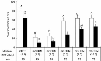
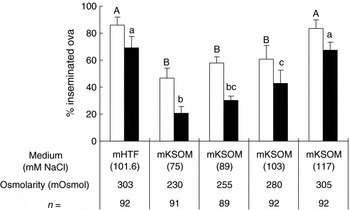
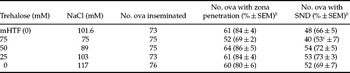
In the first experiment, the effect of various concentration of calcium (1.71, 2.5, 5, 7.5 and 10 mM) in mKSOM at 254 mOsmol on capacitation was examined. A calcium concentration at 5 mM or higher improved zona penetration and SND, but these percentages were still significantly lower than those of mHTF (Fig. 1). In the second experiment, the effect of osmolarity (230, 255, 280 and 305 mOsmol) was examined by changing the concentration of NaCl at 5.0 mM CaCl2 based on the first experiment. When sperm were preincubated at 305 mOsmol, both zona penetration (83 ± 6%) and SND (67 ± 6%) was the same level as with mHTF (86 ± 6% and 69 ± 9%, respectively; p > 0.05) (Fig. 2). In the third experiment, to distinguish the effects of NaCl from that of osmolarity, various amounts of NaCl corresponding 0, 25, 50 and 75 mOsmol were substituted with non-metabolizable sugar or trehalose. Substitution of NaCl with trehalose that corresponded to 50 mOsmol or lower osmolarity had no significant effects on both zona penetration and SND (Table 1). Substitution of NaCl corresponding to 75 mOsmol reduced SND significantly (53 ± 2%) compared with mHTF (66 ± 5%; p < 0.05). This condition was used in the second experimental series, because sperm preincubation at 5 mM CaCl2 and 305 mOsmol resulted in the same level of fertilization as with mHTF.
Sperm penetration through the zona pellucida (open bars) and SND (filled bars) of ova inseminated by sperm preincubated in mKSOM with various concentrations of calcium at 254 mOsmol in the presence of 10 mM lactate. Sperm were inseminated into mHTF. Ova were fixed 2 h after insemination and fertilization was scored. Error bars represent SEM. n = Total number of ova inseminated in six replicates. Within each fertilization process, noted with uppercase letters (zona penetration) and lowercase letters (SND), treatments with no common letter are significantly different (p < 0.05).

Sperm penetration through the zona pellucida (open bars) and SND (filled bars) of ova inseminated by sperm preincubated in mKSOM with various osmolarity adjusted by NaCl at 5 mM CaCl2 in the presence of 10 mM lactate. Sperm were inseminated into mHTF. Ova were fixed 2 h after insemination and fertilization was scored. Error bars represent SEM. n = Total number of ova inseminated in six replicates. Within each fertilization process, noted with uppercase letters (zona penetration) and lowercase letters (SND), treatments with no common letter are significantly different (p < 0.05).

Fertilization of BALB/c sperm capacitated in medium in which various concentrations of NaCl were substituted with corresponding osmolarity of trehalose without changing total osmolarity (305 mOsmol)a.

a Total of six replicates of experiments. Sperm were capacitated for 1.5 h at 1–2 × 10⁷ sperm/ml in various conditions and coincubated with cumulus-free ova at 1.5 × 10⁵ sperm/ml in mHTF. Zona penetration and sperm nuclear decondensation were examined at 2 h after coincubation. Osmolarities of all media were adjusted to 305 mOsmol.
b Percentage data were processed for arcsin transformation and analysed by ANOVA.
c Significantly different from the other treatments in the same column (p < 0.05).
Experimental series 2: effects of lactate on capacitation
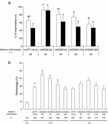
When sperm were preincubated with various concentrations of lactate (0, 5, 10, 20 mM), both percentages of zona penetration and SND by sperm preincubated at 0 mM (92±6% and 91±6%, respectively) were significantly higher than when using mHTF (65±5% and 46±8%, respectively; p < 0.05) (Fig. 3a). A similar tendency was found in the CTC staining pattern at 1.5 h incubation; the percentages of B-pattern (capacitated) sperm were significantly higher for sperm incubated in 0 and 5 mM lactate (45±3% and 40±1%, respectively) than for those sperm incubated in mHTF (28±2%; p < 0.05) (Fig. 3b). At 3 h incubation, the significant difference between the percentages of B-pattern sperm disappeared, although the percentages of B-pattern sperm in medium with 5 mM lactate was the highest among treatment (Fig. 3b). As removal of lactate was found to enhanced capacitation, the following experiments were conducted.
Fertilization (a) and capacitation (b) of sperm preincubated in mKSOM with various concentration of lactate with 5 mM CaCl2 at 305 mOsmol. (a) Sperm penetration through the zona pellucida (open bars) and SND (filled bars) of ova inseminated into mHTF. Fertilization was scored 2 h after sperm insemination. Error bars represent SEM. n = Total number of ova inseminated in six replicates. Within each fertilization process (noted with uppercase letters (zona penetration) and lowercase letters (SND)), treatments with no common letter are significantly different (p < 0.05). (b) Percentages of B-pattern sperm in CTC staining after 1.5 and 3 h incubation at various concentration of lactate. Error bars represent SEM. Treatments with no common letter are significantly different among the same incubation period (p < 0.05). n = Total number of sperm counted in four replicates.

Experimental series 3: optimization of osmolarity and calcium in the absence of lactate
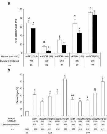
When sperm were preincubated in medium with varying osmolarities (230, 255, 280 and 305 mOsmol) adjusted by changing the NaCl concentration at 5 mM CaCl2, percentages of zona penetration and SND by sperm preincubated in 280 mOsmol (68±9% and 46±5%, respectively) were the same levels as those in mHTF (69±6% and 56±9%, respectively; p > 0.05) (Fig. 4a). At 305 mOsmol, both zona penetration (92±5%) and SND (88±5%) were significantly higher than when using mHTF (69±6% and 56±9%, respectively; p < 0.05) (Fig. 4a). The CTC staining pattern showed a similar tendency; percentages of B-pattern sperm incubated at 305 mOsmol for 1.5 (53±2%) and 3 h (50±5%) were higher than for the other treatment groups, including mHTF (21–33% and 21–33%, respectively; p < 0.05) (Fig. 4b).
Fertilization (a) and capacitation (b) of sperm preincubated in mKSOM at various osmolarity with 5 mM CaCl2 in the absence of lactate. (a) Sperm penetration through the zona pellucida (open bars) and SND (filled bars) of ova inseminated into mHTF. Fertilization was scored 2 h after sperm insemination. Error bars represent SEM. n = Total number of ova inseminated in six replicates. Within each fertilization process (noted with uppercase letters (zona penetration) and lowercase letters (SND)), treatments with no common letter are significantly different (p < 0.05). (b) Percentages of B-pattern sperm in CTC staining after 1.5 and 3 h incubation at various osmolarity. Error bars represent SEM. Within each incubation period, noted with lowercase letters (1.5 h) and uppercase letters (3 h), treatments with no common letter are significantly different (p < 0.05). n = Total number of sperm counted in four replicates.

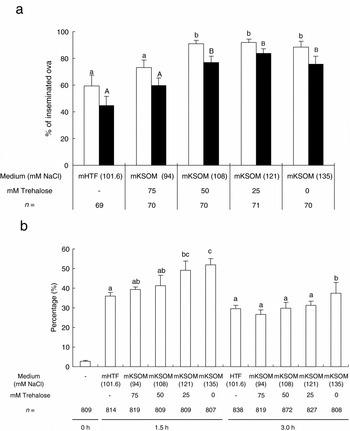
To clarify whether the improvement in fertilization was derived from an increased NaCl concentration or from a change in osmolarity, doses of NaCl corresponding to either 0, 25, 50 or 75 mOsmol were substituted with trehalose. Zona penetration and SND were significantly higher in sperm preincubated in media with an osmolarity of 50 mOsmol and a lower osmolarity of trehalose (95–97% and 82–84%, respectively) than those sperm in mHTF (76±8% and 56±3%, respectively; p < 0.05) (Fig. 5a). For the CTC staining pattern, percentages of B-pattern sperm incubated in media substituted with 0 and 25 mM trehalose (52±3% and 49±5%, respectively) were significantly increased compared with sperm in mHTF (36±2%; p < 0.05) (Fig 5b). When sucrose, choline chloride and sorbitol were used as osmotic reagent to substitute NaCl corresponding to 75 mOsmol, sucrose (84±4%) and choline chloride (81±4%), but not sorbitol (18±3%) significantly improved SND compared with mHTF (60±6%; p < 0.05). Because sperm viability was reduced to <10% in sorbitol, this reagent was determined to have detrimental effects on sperm in BALB/c mice.
Fertilization (a) and capacitation (b) of sperm preincubated in mKSOM in which NaCl was substituted with corresponding osmolarity of trehalose without changing total osmolarity (305 mOsmol) at 5 mM CaCl2 in the absence of lactate. (a) Sperm penetration through the zona pellucida (open bars) and SND (filled bars) of ova inseminated into mHTF. Fertilization was examined 1.5 h after sperm insemination. Error bars represent SEM. n = Total number of ova inseminated in six replicates. Within each fertilization process, noted with uppercase letters (zona penetration) and lowercase letters (SND), treatments with no common letter are significantly different (p < 0.05). (b) Percentages of B-pattern sperm in CTC staining after 1.5 and 3 h incubation. Error bars represent SEM. Within each incubation period, noted with lowercase letters (1.5 h) and uppercase letters (3 h), treatments with no common letter are significantly different (p < 0.05). n = Total number of sperm counted in four replicates.

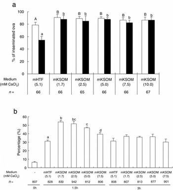
When various concentrations of calcium were tested in the absence of lactate at 305 mOsmol, zona penetration and SND was significantly higher at any concentration (87–91% and 82–88%, respectively) than those sperm in mHTF (79±4% and 54±7%; p < 0.05) and these finding were not significantly different among treatments groups (Fig. 6a). In CTC staining, the percentages of B-pattern sperm were decreased as calcium concentration increased from 1.7 to 7.5 mM CaCl2 at 1.5 h incubation, but this difference disappeared at 3 h incubation (Fig. 6b).
Fertilization (a) and capacitation (b) of sperm preincubated in mKSOM with various concentrations of calcium at 305 mOsmol in the absence of lactate. (a) Sperm penetration through the zona pellucida (open bars) and SND (filled bars) of ova inseminated into mHTF. Fertilization was examined 1.5 h after sperm insemination. Error bars represent SEM. n = Total number of ova inseminated in six replicates. Within each fertilization process, noted with uppercase letters (zona penetration) and lowercase letters (SND), treatments with no common letter are significantly different (p < 0.05). (b) Percentages of B-pattern sperm in CTC staining after 1.5 h and 3 h incubation. Error bars represent SEM. Treatments with no common letter are significantly different among the same incubation period (p < 0.05). n = Total number of sperm counted in four replicates.

Discussion
Inbred BALB/c strain and their descendant strains are one of the most widely used strains in biomedical research, including immunology, disease and toxicology, because of their clear genetic homozygosity. In spite of their importance, however, the in vitro manipulation of BALB/c gametes and embryos often encounters difficulties. These difficulties include poor in vivo and in vitro fertilizability after superovulation (Roudebush & Duralia, Reference Roudebush and Duralia1996; Choi et al., Reference Choi, Seng and Toyoda2000; Wu et al., Reference Wu, Chou, Lin and Huang2003), retardation of embryo development in vitro at 2-cell stage (2-cell block) (Sekirina et al., 1997), high sensitivity against freeze–thaw processes (Mobraaten, Reference Mobraaten1986) and high incidence of abnormal morphology of the sperm head (Burruel et al., Reference Burruel, Yanagimachi and Whitten1996). In this study, we attempted to clarify the requirements for in vitro capacitation of BALB/c sperm by optimizing lactate, calcium and osmolarity of medium. Our results showed that lactate and osmotic pressure are important factors for successful capacitation and subsequent fertilization in vitro and that the effect of calcium is dependent on the conditions employed.
The role of lactate during mammalian capacitation is somewhat equivocal. Miyamoto & Chang (Reference Miyamoto and Chang1973a, Reference Miyamoto and Changb) reported beneficial effects of lactate, pyruvate and BSA on mouse capacitation. In rats, Tsunoda & Chang (Reference Tsunoda and Chang1975) showed the importance of pyruvate, lactate and glucose. Conversely, lactate itself was shown to be non-essential for the in vitro capacitation of mice and hamsters (Hoppe, Reference Hoppe1976; Bavister & Yanagimachi, Reference Bavister and Yanagimachi1977; Fraser & Herod, Reference Fraser and Herod1990). Neil & Olds-Clarke (Reference Neill and Olds-Clarke1988) showed the inhibitory effects of lactate at a 5 mM or higher concentration on sperm capacitation using CF1 outbred mice. Our observations in BALB/c mice are consistent with the results of Neil & Olds-Clark (Reference Neill and Olds-Clarke1988). In BALB/c mice, lactate inhibited capacitation in a dose-dependent manner. Lowering lactate concentration during capacitation significantly increased zona penetration, SND and the percentages of capacitated pattern in CTC staining at 1.5 h (Fig 3a, b). At 3 h, however, the percentages of capacitated sperm when using 5 mM lactate was the highest among treatments (Fig. 3b), suggesting that lactate delays capacitation. Because approximately 5 mM lactate is present in oviductal environments (Yanagimachi, Reference Yanagimachi and 1994), lactate might be involved in the control of capacitation in vivo, so that only a small population of sperm completes capacitation at a particular time. Such control is essential in vivo to ensure fertilization occurs, in which individual ova were ovulated and transported to the oviducts with a time lag (Yanagimachi, Reference Yanagimachi and 1994).
There are a few explanations for lactate inhibition of capacitation. Lactate can be incorporated into the sperm cell to be metabolized (Jones, Reference Jones1997). As lactate dehydrogenase metabolizes pyruvate to lactate during mouse capacitation to produce reduced NAD (Duan & Goldberg, Reference Duan and Goldberg2003), a high concentration of lactate in sperm cell disrupts the reaction equilibrium, resulting in an insufficient production of NAD needed to produce ATP. Another explanation is that increased lactate might induce acidosis to inhibit capacitation. Intracytoplasmic alkalization in the sperm cell is highly correlated with membrane depolarization, through the action of a K+-channel during capacitation (Zen et al., 1996). Nakanishi et al. (Reference Nakanishi, Ikawa, Yamada, Toshimori and Okabe2001) reported the importance of increased pH in the intra-acrosome region for the alteration and release of acrosomal contents during acrosome reactions. Lactate might act as a weak acid to inhibit intracellular alkalization, resulting in the inhibition of capacitation.
The requirement for calcium for capacitation and sperm–egg interaction has been described in detail by others (Yanagimachi, Reference Yanagimachi1982; Fraser, Reference Fraser1995). Sperm preincubation at a high concentration (7.5 mM) of calcium reduced fertilizability in TO mice (Fraser, Reference Fraser1995). In our study, the effect of calcium on fertilizability was dependent on the presence of lactate and on osmolarity. In the presence of lactate and at low osmolarity (255 mOsmol), sperm preincubated at ≥5 mM calcium had improved fertilizability (Fig. 1). Conversely, an increase in calcium reduced the percentages of B-pattern sperm that had been incubated in lactate-free media and at isotonic osmolarity (305 mOsmol) (Fig. 6b), although a change in the calcium concentration had no effect on fertilizability (Fig. 6a). Because Neil & Olds-Clarke (1998) reported that the presence of lactate reduced the free calcium concentration by chelation, which explains the resulting reduced fertilizability partially. The same explanation can be applied to our studies, i.e. in the presence of lactate, increased calcium is required for sperm to maintain the intracellular calcium concentration necessary for capacitation and fertilization. Another explanation is based on the report that intracellular calcium and alkalization act independently on a K+-channel during mammalian sperm capacitation (Felix, Reference Felix2005). In the absence of lactate, 1.7 mM calcium is sufficient to activate the K+-channel to induce membrane depolarization. In the presence of lactate, an increase in calcium concentration may be necessary for K+-channel activation, to overcome the inhibition by acidosis induced by lactate. It is uncertain, however, how a high concentration of calcium in the absence of lactate inhibits capacitation.
Exposure of sperm to media with isotonic or hypertonic osmolarity affects the sperm capacitation and fertilizability of many mammalian species. In humans, the effect of osmolarity on capacitation or sperm function is controversial. Liu et al. (Reference Liu, Clarke and Baker2006) indicated an enhanced zona binding and increased tyrosine phosphorylation in hypertonic conditions. Conversely, others have shown inhibitory effects of hypertonic osmolarity on human sperm (Bielfeld et al., Reference Bielfeld, Jeyendran and Zaneveld1993; Rossato et al., Reference Rossato, Virgilio and Foresta1996). In rodent and bovine sperm, an increase in osmolarity using NaCl resulted in an increased capacitation and subsequent fertilization in vitro (Miyamoto & Chang, Reference Miyamoto and Chang1973a, Reference Miyamoto and Changb; Oliphant & Brackett, Reference Oliphant and Brackett1973; Niwa & Chang, Reference Niwa and Chang1975; Miller & Hunter, Reference Miller and Hunter1986). This improved capacitation and fertilizability was believed to be caused by the removal of surface antigen, or decapitated factor, by hypertonic conditions. Studies using rats (Oh et al., Reference Oh, Miyoshi and Funahashi1998) and mice (Oliphant & Bracket, Reference Oliphant and Brackett1973) to distinguish the effects of osmolarity and ionic strength indicated that improved capacitation was the result of a change in ionic strength (mainly NaCl) rather than osmolarity. When sorbitol was used to substitute for NaCl in the study of rat IVF, no improvement of fertilizability was observed and the authors concluded that ionic strength is important for capacitation and fertilization (Oh et al., Reference Oh, Miyoshi and Funahashi1998). However the possibility that sorbitol had detrimental effects on sperm viability on rat sperm as in mouse sperm shown in our study cannot be ruled out because there was no description of the viability of rat sperm. Oliphant & Bracket (Reference Oliphant and Brackett1973) substituted NaCl with glucose or glycerol, but their study did not distinguish effects of osmolarity and ionic strength precisely, as these substances are known to affect sperm metabolism, capacitation and fertilization (Hoppe, Reference Hoppe1976; Fraser & Herod, Reference Fraser and Herod1990). Based on our study using various osmotic reagents, osmolarity itself is involved in regulation of capacitation and fertilizability of BALB/c sperm, because substitution of NaCl with trehalose at a maximum level of 50 mOsmol did not resulted in reduced fertilization, irrespective of the presence or absence of lactate (Table 1, Fig. 5). This finding is further supported by the observation that the substitution of NaCl, which corresponds to an osmolarity of 75 mOsmol, with other non-metabolizable osmotic reagents, sucrose and choline chloride, did not reduce fertilization. The reduction in fertilizability of sperm that had capacitated in a high concentration of trehalose (75 mM) might be due to the toxic effects of this compound.
Although conditions for IVF and capacitation have been studied for many years, in the routine practice of IVF, the same conditions have been applied for sperm preincubation and fertilization. Our experiments show that the requirements for capacitation and fertilization are different. For capacitation, osmolarity and lactate are important factors, and for sperm : ova interaction (sperm penetration through the zona and oolemma) calcium and osmolarity are important factors. Application of these modifications is not limited to KSOM. When we applied these conditions to the KRB-based media, we observed high a percentage (>80%) of fertilization in other strains, such as gene-modified mice with a BALB/c genetic background and with inbred C3H strains (Kito & Ohta, personal observations). There are still some mouse strains, however, to which our findings are not applicable. 129 and STS are representative strains (Kito et al., Reference Kito, Hayao, Noguchi-Kawasaki, Ohta, Hideki and Tateno2004; Byers et al., Reference Byers, Payson and Taft2006) and we were unable to improve their poor and unstable fertilizability in vitro. More studies to improve fertilization conditions are still needed for successful ARTs in mice.
Acknowledgements
We are grateful to Ms Y. Kaneko for care of animals used in this study. We thank the Department of Advanced Technologies for Radiation Protection Research and the Department of Technical Support and Development in National Institute of Radiological Sciences for their technical assistance. We are also grateful to Mr Craig Steger and Dr Diane Cookfair for critically reviewing this manuscript.