Background
Multiple studies have reported the safe use of transoesophageal echo in paediatric cardiac surgery.Reference Stevenson 1 , Reference Hilberath, Oakes, Shernan, Bulwer, D’Ambra and Eltzschig 2 The Philips Mini-Multi Probe (S7-3t; Koninklijke Philips N.V., Amsterdam, Netherlands) with a tip width of 10.7 mm is recommended by the manufacturer for use in infants down to 3.5 kg, and the micro probe (S8-3t) with a tip width of 7.5 mm is recommended in infants down to a weight of 2.5 kg. 3 During placement, jaw thrust, mandible retraction, or head rotation may be utilised to gain entry into the upper oesophagus. It is not uncommon to feel a change in resistance as the probe passes through the upper oesophageal sphincter and again as it enters the stomach. Here we report a case of oesophageal perforation in a neonate during transoesophageal echocardiography for cardiac surgery.
Case report
An infant girl with a foetal diagnosis of Trisomy 21, atrioventricular septal defect, and coarctation of the aorta was born at 39 weeks. Her birth weight was 3235 g, Apgars 8/8, and oxygen saturation 85–90% in room air. An umbilical vein catheter was placed and prostaglandin E1 was administered at 0.01–0.03 mcg/kg/minute for the next 9 days. She had typical features of Trisomy 21 with notably excess nuchal skin folds. Echocardiogram confirmed the diagnosis of atrioventricular septal defect, Rastelli type C. There was coarctation of the aorta with significant narrowing at the isthmus and distal transverse segments of a left-sided arch. A large patent arterial duct was present.
Owing to a question of choanal stenosis, flexible nasopharyngoscopy was performed at the bedside by the otolaryngology service, which showed no choanal stenosis, normal nasopharynx, oropharynx, and hypopharynx with the exception of “floppy collapsible mucosa”. Oral feeds had not been initiated because of aortic arch obstruction and prostaglandin therapy. The patient was on total peripheral nutrition preoperatively.
On day 9 of life, weighing 3.27 kg, the infant was taken to surgery for aortic arch advancement and placement of a pulmonary artery band. Prostaglandin infusion was continued at 0.02 mcg/kg/minute. Anaesthesia was administered with right nasotracheal intubation using a 3.0 microcuff endotracheal tube (Kimberly-Clark Corporation, Irving, TX USA). No gastric suction was performed. The cardiologist, cardiac surgeon, and anaesthesiologist discussed indications for transoesophageal echo examination and available probes. The only available micro transoesophageal echo probe (Philips S8-3t Micro transoesophageal echo probe 7.5-mm tip width) was reserved for a 2.5 kg infant in another procedure. A mini transoesophageal echo probe (Philips S7-3t Mini-Multi 10.7-mm tip width) was utilised for our patient. The probe was lubricated with water-soluble sterile single-use jelly (E-Z Lubricating Jelly Chester Packaging LLC, Cincinnati, Ohio, United States of America). The probe was flexed and inserted around the tongue to the midline hypopharynx, and then partially straightened to pass into the oesophagus. The probe was not locked and no hand was on the control during insertion. A jaw thrust manoeuvre was performed with the left hand on the left mandible, while gentle pressure was applied with the right hand to pass the transoesophageal echo probe blindly into the oesophagus. The probe passed with relative ease, consistent with numerous prior neonatal transoesophageal echo probe insertions by the operator, on the first attempt; however, resistance was encountered at the depth expected for passage into the stomach. The probe was removed. No blood was observed on the probe. The probe was relubricated and reinserted with the same result. The transoesophageal echo probe was attached to the echocardiography machine to confirm placement. Echocardiographic views of the heart from the mid-oesophagus were obtained by the paediatric cardiology team. The views were excellent with no unusual visual features, suggesting abnormal probe placement. Transgastric views could not be obtained. A provisional diagnosis of distal oesophageal stenosis was entertained with secondary concern for oesophageal injury. The decision was made to remove the probe rather than leave the probe in the “oesophagus” during the case. Again, no blood was present on the transoesophageal echo probe. A lubricated 10-Fr gastric suction tube was blindly placed via the left nares into the stomach easily with suctioning of 2–3 ml of clear secretions. The nasogastric tube passed ∼4 cm deeper than the transoesophageal echo probe had passed. A normal tactile feel of passage through the lower oesophageal sphincter into the stomach was noted.
After the cardiac surgical repair, the paediatric micro probe (Philips S8-3t transoesophageal echo probe) was inserted blindly via the mouth using the same technique with jaw thrust. The probe again passed easily. Mild resistance was felt at the depth where the larger probe insertion had halted. The sensation was consistent with lower oesophageal stenosis; however, the probe passed easily into the stomach. Mid-oesophageal and transgastric transoesophageal echo views were easily obtained. Before removing the micro transoesophageal echo probe, the cardiac surgeon’s inspection of the mediastinum revealed no evidence of oesophageal perforation. Specifically, there was no evidence of gross injury, and the transoesophageal echo probe was not visualised in the mediastinum. The micro transoesophageal echo probe was removed, again without bloody sections.
A provisional diagnosis of oesophageal stricture was suggested. Antibiotic therapy during surgery and the ICU stay consisted of cephazolin 25 mg/kg every 8 hours for 48 hours. Medications included a milrinone infusion and intravenous ranitidine 3.25 mg every 8 hours. The infant was tracheally extubated on postoperative day 3. Nasogastric feeds, with an X-ray confirming gastric placement, were instituted.
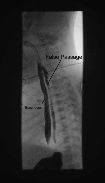
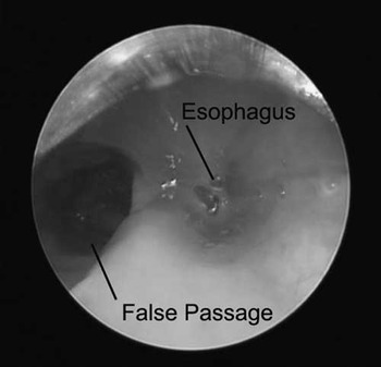
An oesophagram was performed, which revealed a contained false passage slightly to the left and posterior to the native oesophagus (Fig 1). This false passage extended from the level of the neck, left and posterior to the native oesophagus, caudally to approximately the level of the gastro-oesophageal junction. No free mediastinal spill of contrast beyond the false passage was evident. Nasogastric feeds were stopped, and empiric antibiotic treatment was initiated with piperacillin–tazobactam 70 mg/kg q 8 hours, clindamycin 10 mg/kg q 6 hours, and 1 dose of vancomycin 15 mg/kg. A microlaryngoscopy, bronchoscopy, and oesophagoscopy demonstrated a large left-sided false passage through the oesophageal wall just distal to the cricopharyngeus muscle (Fig 2). A drain (Replogle suction catheter; Covidien AG, Mansfield, MA USA) was placed via the right nares under direct visualisation ∼8 cm into the false passage. The nasogastric feeding tube was then advanced to a nasojejunal position and enteral feeds resumed.
Oesophagram postoperative day 4 showing oesophageal false passage extending to the level of the diaphragm.

Oesophagoscopy postoperative day 5 showing perforation and false passage to the left of the oesophagus, with the feeding tube in place.

The drain was subsequently kept to low wall suction and after 2 days it was withdrawn 1 cm daily with removal after 10 days. During this time, the infant continued on prophylactic antibiotic therapy with piperacillin–tazobactam and clindamycin to minimise the risk of mediastinitis. An oesophagram, 1 day after removal of the drain, showed no extravasation of contrast into the former false passage. Swallowing was noted to be grossly normal. The oesophagus was smooth with normal calibre and motility.
On postoperative day 19, follow-up oesophagoscopy demonstrated well-healed mucosa with only a small area of the granulation tissue at the site of the previous oesophageal defect. There was no evidence of false passage extending from the defect. Oral feeds were initiated. At the time of her most recent follow-up she was feeding without difficulty.
Discussion
Iatrogenic neonatal oesophageal perforation – typically from nasogastric tube placement – is not uncommon and has been reported to “masquerade” as oesophageal atresia.Reference Knight, Webb and Coppola 4 Initial transoesophageal echo probe insertion in this patient was not difficult and routine for inserting a 10.7-mm probe into a neonatal oesophagus. However, failure of passage into the stomach was clearly unusual and prompted concern.
The first report of oesophageal perforation by a transoesophageal echocardiography (echo) probe in a neonate was in a full-term 3.7-kg infant with 22q11 deletion on prostaglandin infusion undergoing interrupted aortic arch repair.Reference Muhiudeen-Russell, Miller-Hance and Silverman 5 Like our case, the probe could not be passed easily across the gastro-oesophageal junction. A limited transoesophageal echo examination was conducted, and no transgastric images could be obtained. The quality of the echocardiographic images was “excellent” and did not cause any suspicion that there was a problem. However, the transoesophageal echo probe was found during surgery to be lying freely in the posterior mediastinum. On oesophagoscopy, the transoesophageal echo probe was noted to have perforated the oesophagus at the level of the cricopharyngeus muscle. The perforation was successfully managed conservatively with antibiotics and tube feedings. Another report involved a 3.3-kg neonate with 22q11 deletion for truncus arteriosus repair. Transgastric views could not be obtained and a contained right-sided posterior mediastinal false passage was discovered several days later. Conservative management was followed by complete recovery.Reference Sasaki, Culham and Gandhi 6
Risk factors for failure of transoesophageal echo insertion in children have been defined.Reference Hilberath, Oakes, Shernan, Bulwer, D’Ambra and Eltzschig 2 , Reference Wellen, Glatz, Gaynor, Montenegro and Cohen 7 This patient had two possible contributing factors. First, the patient size was small for the selected transoesophageal echo probe (recommended minimum 3.5 kg). 3 Second, she had Trisomy 21 with marked excess nuchal folds and a prior flexible endoscopy showing “floppy collapsible” pharyngeal mucosa. She did not have other suggested risk factors: 22q11 deletion, craniofacial anomaly, prior surgical intervention of the oesophagus, the presence of (known) abnormal tracheoesophageal anatomy, micrognathia, prematurity, or small size for gestational age.Reference Wellen, Glatz, Gaynor, Montenegro and Cohen 7
The cricopharyngeal region may allow entry into the mediastinum because the posterior oesophageal mucosa above (Killian’s triangle) and below (Lannier’s triangle) the cricopharyngeus muscle is only covered by fascia or “muscular aponeurosis”.Reference Muhiudeen-Russell, Miller-Hance and Silverman 5 In children, if recognised early and found to be non-purulent, conservative management of oesophageal perforation may be attempted.Reference Gander, Berdon and Cowles 8
The complication of perforation by transoesophageal echo probe in children, although truly rare, may go unreported. The authors are anecdotally aware of two other oesophageal perforations by different providers during transoesophageal echo probe insertion for congenital heart surgery: one in a neonate, which ultimately terminated in the pleural space – upper oesophageal entry repaired surgically by cervical incision – and one in an older paediatric congenital cardiac patient – probe visible in the mediastinum during cardiac surgery.
Greene reported that 64% of children who underwent transoesophageal echo examination showed evidence of acute mucosal injury by upper endoscopy, but had no long-term feeding or swallowing difficulties.Reference Greene, Alexander and Knauf 9 Patients with CHD often have associated anomalies, which increase oesophageal injury risk. Congenital lateral pharyngeal diverticuli occur rarely as embryological remnants of the branchial clefts with the third and fourth clefts nearest to the pyriform fossae. These could simulate oesophageal atresia and provide a favourable site for pharyngeal perforation.
A high index of suspicion is necessary to identify iatrogenic oesophageal injury during transoesophageal echo probe placement. Our case provides further evidence that with prompt identification and early initiation of parenteral feeds and broad-spectrum antibiotics, even large oesophageal perforations may be managed conservatively.
Acknowledgments
Sara Powell, D.O. pediatric anesthesia fellow assisted in the preparation of this report.
Financial Support
This research received no specific grant from any funding agency, commercial or not-for-profit sectors.
Conflicts of Interest
None.
Ethical Standards
The author states that the report describes the care of one or more patients. A family member, next of kin, or legal representative of the patient consented to publication.