Introduction
Structural neuroimaging studies have confirmed the presence of neuroanatomical abnormalities in schizophrenia (Wright et al. Reference Wright, Rabe-Hesketh, Woodruff, David, Murray and Bullmore2000). The most consistent findings are reductions in medial temporal and prefrontal regions, superior temporal gyri, parietal areas and subcortical regions, together with ventricular enlargement (Shenton et al. Reference Shenton, Dickey, Frumin and McCarley2001). Previous studies have also reported that alterations in brain morphology are correlated with positive psychotic symptoms in schizophrenia (Young et al. Reference Young, Blackwood, Roxborough, McQueen, Martin and Kean1991; Flaum et al. Reference Flaum, O'Leary, Swayze, Miller, Arndt and Andreasen1995; Suzuki et al. Reference Suzuki, Zhou, Hagino, Niu, Takahashi, Kawasaki, Matsui, Seto, Ono and Kurachi2005a), particularly in the case of auditory hallucinations (see Allen et al. Reference Allen, Laroi, McGuire and Aleman2008 for review).
Subjects with an at-risk mental state (ARMS; Yung et al. Reference Yung, Phillips, McGorry, McFarlane, Francey, Harrigan, Patton and Jackson1998) show subthreshold psychotic symptoms, although only a proportion of them will ultimately progress to psychosis (McGorry & Singh, Reference McGorry, Singh, Raphael and Burrows1998; Yung et al. Reference Yung, Phillips, Yuen, Francey, McGarlane, Hallgren and McGorry2003). Magnetic resonance imaging (MRI) scanning of the brain in ARMS subjects has shown gray-matter volume (GMV) deficits compared to healthy controls, which overall do not seem to differ significantly from known deficits in schizophrenia (Pantelis et al. Reference Pantelis, Velakoulis, McGorry, Wood, Suckling, Phillips, Yung, Bullmore, Brewer, Soulsby, Desmond and McGuire2003; Velakoulis et al. Reference Velakoulis, Wood, Wong, McGorry, Yung, Phillips, Smith, Brewer, Proffitt, Desmond and Pantelis2006; Borgwardt et al. Reference Borgwardt, Riecher-Rössler, Dazzan, Chitnis, Aston, Drewe, Gschwandtner, Haller, Pflüger, Rechsteiner, D'Souza, Stieglitz, Radü and McGuire2007b). Abnormalities have been found mainly in the medial temporal lobe (Lawrie et al. Reference Lawrie, Whalley, Kestelman, Abukmeil, Byrne, Hodges, Rimmington, Best, Owens and Johnstone1999; Pantelis et al. Reference Pantelis, Velakoulis, McGorry, Wood, Suckling, Phillips, Yung, Bullmore, Brewer, Soulsby, Desmond and McGuire2003; Velakoulis et al. Reference Velakoulis, Wood, Wong, McGorry, Yung, Phillips, Smith, Brewer, Proffitt, Desmond and Pantelis2006; Meisenzhal et al. Reference Meisenzhal, Koutsouleris, Gaser, Bottlender, Schmitt, McGuire, Decker, Burgermeister, Born, Reiser and Möller2008), superior temporal gyrus (Job et al. Reference Job, Whalley, Johnstone and Lawrie2005; Borgwardt et al. Reference Borgwardt, Riecher-Rössler, Dazzan, Chitnis, Aston, Drewe, Gschwandtner, Haller, Pflüger, Rechsteiner, D'Souza, Stieglitz, Radü and McGuire2007b; Meisenzhal et al. Reference Meisenzhal, Koutsouleris, Gaser, Bottlender, Schmitt, McGuire, Decker, Burgermeister, Born, Reiser and Möller2008), prefrontal cortex (Pantelis et al. Reference Pantelis, Velakoulis, McGorry, Wood, Suckling, Phillips, Yung, Bullmore, Brewer, Soulsby, Desmond and McGuire2003; Meisenzhal et al. Reference Meisenzhal, Koutsouleris, Gaser, Bottlender, Schmitt, McGuire, Decker, Burgermeister, Born, Reiser and Möller2008), cerebellum (Pantelis et al. Reference Pantelis, Velakoulis, McGorry, Wood, Suckling, Phillips, Yung, Bullmore, Brewer, Soulsby, Desmond and McGuire2003; Job et al. Reference Job, Whalley, Johnstone and Lawrie2005), anterior cingulate gyrus (Pantelis et al. Reference Pantelis, Velakoulis, McGorry, Wood, Suckling, Phillips, Yung, Bullmore, Brewer, Soulsby, Desmond and McGuire2003; Borgwardt et al. Reference Borgwardt, McGuire, Fusar-Poli, Radue and Riecher-Rössler2008b; Fornito et al. Reference Fornito, Yung, Wood, Phillips, Nelson, Cotton, Velakoulis, McGorry, Pantelis and Yücel2008), posterior midline structures (Pantelis et al. Reference Pantelis, Velakoulis, McGorry, Wood, Suckling, Phillips, Yung, Bullmore, Brewer, Soulsby, Desmond and McGuire2003; Borgwardt et al. Reference Borgwardt, McGuire, Aston, Berger, Dazzan, Gschwandtner, Pflüger, D'Souza, Radue and Riecher-Rössler2007a, Reference Borgwardt, Riecher-Rössler, Dazzan, Chitnis, Aston, Drewe, Gschwandtner, Haller, Pflüger, Rechsteiner, D'Souza, Stieglitz, Radü and McGuireb) and the insula lobe (Pantelis et al. Reference Pantelis, Velakoulis, McGorry, Wood, Suckling, Phillips, Yung, Bullmore, Brewer, Soulsby, Desmond and McGuire2003; Borgwardt et al. Reference Borgwardt, Riecher-Rössler, Dazzan, Chitnis, Aston, Drewe, Gschwandtner, Haller, Pflüger, Rechsteiner, D'Souza, Stieglitz, Radü and McGuire2007b, Reference Borgwardt, Fusar-Poli, Radue and Riecher-Rössler2008a). Notably, not only decreases but also increases in GMV have been observed (Pantelis et al. Reference Pantelis, Velakoulis, McGorry, Wood, Suckling, Phillips, Yung, Bullmore, Brewer, Soulsby, Desmond and McGuire2003; Shin et al. Reference Shin, Lee, Kang, Kim, Bae and Jung2005; Borgwardt et al. Reference Borgwardt, McGuire, Aston, Berger, Dazzan, Gschwandtner, Pflüger, D'Souza, Radue and Riecher-Rössler2007a). Taken together, these studies suggest that some anatomical abnormalities are present before illness onset, presumably independent of outcome.
Nevertheless, the neuroanatomical correlates of the distinctive positive psychotic symptoms often present in these populations remain poorly understood. Psychotic experiences are not encountered exclusively as part of a fully developed psychotic disorder or in high-risk subjects, as they may also be found in schizotypal personality disorder (SPD; APA, 1994) and in healthy individuals who can be psychometrically identified from the general population (Chapman et al. Reference Chapman, Chapman, Kwapil, Eckblad and Zinser1994). The few available neuroanatomical studies in SPD thus far align well with studies on schizophrenia and ARMS patients reporting general structural brain abnormalities compared to controls. These include the temporal lobe (Dickey et al. Reference Dickey, McCarley, Voglmaier, Niznikiewicz, Seidman, Hirayasu, Fischer, Teh, Van Rhoads, Jakab, Kikinis, Jolesz and Shenton1999; Downhill et al. Reference Downhill, Buchsbaum, Hazlett, Barth, Lees Roitman, Nunn, Lekarev, Wei, Shihabuddin, Mitropoulou, Silverman and Siever2001; Kawasaki et al. Reference Kawasaki, Suzuki, Nohara, Hagino, Takahashi, Matsui, Yamashita, Chitnis, McGuire, Seto and Kurachi2004; Suzuki et al. Reference Suzuki, Zhou, Takahashi, Hagino, Kawasaki, Niu, Matsui, Seto and Kurachi2005b), inferior frontal gyrus and insula (Kawasaki et al. Reference Kawasaki, Suzuki, Nohara, Hagino, Takahashi, Matsui, Yamashita, Chitnis, McGuire, Seto and Kurachi2004) and the thalamic pulvinar nucleus (Byne et al. Reference Byne, Buchsbaum, Kemether, Hazlett, Shinwari, Mitropoulou and Siever2001). In addition, the prefrontal cortex was implicated in one investigation (Raine et al. Reference Raine, Sheard, Reynolds and Lencz1992), although later studies failed to replicate this finding (Raine et al. Reference Raine, Lencz, Yaralian, Bihrle, LaCasse, Ventura and Colletti2002; Siever et al. Reference Siever, Koenigsberg, Harvey, Mitropoulou, Laruelle, Abi-Dargham, Goodman and Buchsbaum2002; Kawasaki et al. Reference Kawasaki, Suzuki, Nohara, Hagino, Takahashi, Matsui, Yamashita, Chitnis, McGuire, Seto and Kurachi2004; Suzuki et al. Reference Suzuki, Zhou, Takahashi, Hagino, Kawasaki, Niu, Matsui, Seto and Kurachi2005b; Zhou et al. Reference Zhou, Suzuki, Takahashi, Hagino, Kawasaki, Matsui, Seto and Kurachi2007), and further evidence suggests that this region might instead be involved in transition (Kawasaki et al. Reference Kawasaki, Suzuki, Nohara, Hagino, Takahashi, Matsui, Yamashita, Chitnis, McGuire, Seto and Kurachi2004; Suzuki et al. Reference Suzuki, Zhou, Takahashi, Hagino, Kawasaki, Niu, Matsui, Seto and Kurachi2005b).
Epidemiological research has found a prevalence of 17.5% of any type of positive psychotic symptoms in the general population (Van Os et al. Reference Van Os, Hanssen, Bijl and Ravelli2000), supporting the notion that psychometric schizotypy (PS) represents a dimensional trait, ranging from ‘normality’ to clinical cases of psychosis (Strauss, Reference Strauss1969). This is in agreement with continuity models of psychosis proneness, which understand such schizotypal features as part of the normal diversity of personality expression found in healthy individuals, although these features entail a cognitive vulnerability that predisposes towards psychosis (Chapman & Chapman, Reference Chapman and Chapman1980). Hence, MRI findings in these subjects could inform the pathophysiology of psychotic-like experiences without the confounding effects of illness chronicity or medication. To date, however, no studies have investigated the brain volumetric characteristics of subjects with positive PS.
The aim of the present study was to compare GMV in people with high and low positive PS using voxel-based morphometry (VBM). VBM has been used successfully in prior research to ascertain brain morphology in patients with schizophrenia (Wright et al. Reference Wright, Ellison, Sharma, Friston, Murray and McGuire1999) and also in ARMS (Borgwardt et al. Reference Borgwardt, McGuire, Aston, Berger, Dazzan, Gschwandtner, Pflüger, D'Souza, Radue and Riecher-Rössler2007a, Reference Borgwardt, Riecher-Rössler, Dazzan, Chitnis, Aston, Drewe, Gschwandtner, Haller, Pflüger, Rechsteiner, D'Souza, Stieglitz, Radü and McGuireb; Meisenzhal et al. Reference Meisenzhal, Koutsouleris, Gaser, Bottlender, Schmitt, McGuire, Decker, Burgermeister, Born, Reiser and Möller2008) and SPD subjects (Kawasaki et al. Reference Kawasaki, Suzuki, Nohara, Hagino, Takahashi, Matsui, Yamashita, Chitnis, McGuire, Seto and Kurachi2004). Based on the findings in these previous studies, we tested the hypothesis that individuals with high and low positive PS scores would show volumetric differences in the inferior frontal, insular, cingulate, medial and lateral temporal, and parietal cortex. We further hypothesized that gray-matter deficits in subjects with high levels of positive schizotypy would be qualitatively similar to those in ARMS or SPD subjects, yet quantitatively less pronounced.
Method
Subjects
A total of 600 subjects from a university sample completed the positive subscale of the Community Assessment of Psychic Experiences (CAPE; Stefanis et al. Reference Stefanis, Hanssen, Smirnis, Avramopoulos, Evdokimidis, Stefanis, Verdoux and Van Os2002). This questionnaire was completed voluntarily by first- and second-year undergraduate students to match them on social class and educational level.
Potential participants were identified when scoring above the 75th percentile of the distribution of individual positive CAPE scores for the high PS group, and below the 25th percentile for the comparison group (low PS). These subjects were invited to participate and were screened for exclusion criteria using a self-report checklist for healthy subjects, comprising the following points: (1) no personal history of neurological or psychiatric illness; (2) no family history of psychotic or neurological illness in first-degree relatives; (3) no use of illicit substances; and (4) no changes in overall level of functioning, including academic performance over the past 6 months. After receiving a complete description of study procedure, a total of 38 subjects were recruited (18 high PS, 20 low PS) and written informed consent was obtained from all of them. All subjects were paid for their participation. The study was approved by the Medical Ethical Committee of the University of Groningen.
Instrument
The CAPE was used to measure positive schizotypal signs. This instrument was chosen on the basis of the following characteristics: (1) efficient assessment of subclinical schizotypal experiences in the general population (Hanssen et al. Reference Hanssen, Bak, Bijl, Vollebergh and Van Os2005), (2) good concurrent validity with interview-based measures (Konings et al. Reference Konings, Bak, Hanssen, van Os and Krabbendam2006), and (3) it was developed and standardized on a Dutch population. A detailed description of its psychometric properties and administration procedure can be found elsewhere (Stefanis et al. Reference Stefanis, Hanssen, Smirnis, Avramopoulos, Evdokimidis, Stefanis, Verdoux and Van Os2002). In brief, this is a 42-item self-report questionnaire that measures the frequency of attenuated psychotic symptoms, on a four-point scale of ‘never’, ‘sometimes’, ‘often’ and ‘nearly always’. Originally, the CAPE was based on a three-factor structure of positive, negative and depressive factors. For the purpose of the present study, we only used the scores on the positive factor because several studies have reported a relationship between the severity of positive psychotic symptoms and GMV in schizophrenia. In addition, all subjects completed the Beck Depression Inventory (BDI; Beck et al. Reference Beck, Steer, Ball and Ranieri1996), which is a widely used, well-validated measure for depression, administered in our study to control for other psychiatric symptomatology that could act as confound for schizotypal signs (i.e. negative symptoms).
MRI data acquisition
High-resolution MR images were acquired on a 3-T Philips Intera MR scanner (Philips Medical Systems, The Netherlands). Subjects were scanned with a T1-weighted 3D fast-field echo (FFE) sequence [repetition time (TR)=25 ms, echo time (TE)=4.6 ms, field of view (FOV)=260 mm, matrix=256×256, 160 contiguous axial slices of 1-mm thickness, voxel size=1×1×1 mm.
MRI data preprocessing
Structural images were pre-processed using optimized VBM implemented on the SPM5 software package (Wellcome Department of Cognitive Neurology, UK) running under Matlab 7.4 (The MathWorks, USA). VBM is a whole-brain, unbiased, semi-automated technique for characterizing regional cerebral differences in structural MR images (Ashburner & Friston, Reference Ashburner and Friston2000; Good et al. Reference Good, Johnsrude, Ashburner, Henson, Friston and Frackowiak2001; Mechelli et al. Reference Mechelli, Price, Friston and Ashburner2005). Structural images were segmented to extract gray matter and then normalized to an asymmetric T1-weighted template in Montreal Neurological Institute (MNI) stereotactic space, in a recursive fashion. Image segmentation incorporated an intensity non-uniformity correction to account for smooth intensity variations caused by gradient distortions and different positions of cranial structures within the MRI coil (Ashburner & Friston, Reference Ashburner and Friston2000). A further step was added to ensure that the total amount of gray matter in each voxel was conserved before and after spatial normalization (Good et al. Reference Good, Johnsrude, Ashburner, Henson, Friston and Frackowiak2001). This ‘modulation’ step involves multiplying the spatially normalized gray matter by its relative volume before and after spatial normalization. The resulting gray-matter images were finally smoothed with a 12-mm isotropic Gaussian kernel. Smoothing is required to compensate for the inexact nature of spatial normalization and to maximize the chance that regional effects are expressed at a spatial scale where homologies in structural anatomy exist over subjects. After smoothing, each voxel represents the local average amount of gray matter in the region, the size of which is defined by the smoothing kernel.
Statistical analysis
An analysis of covariance (ANCOVA) was designed by applying the general linear model (GLM) to compare the modulated gray-matter images of subjects with high and low positive PS. Global GMV, BDI scores and age were modeled as covariates of no interest to identify regionally specific differences that were not confounded by these variables. Global GMV was estimated using the total amount of gray matter in the gray-matter images obtained from segmentation, including the cerebellar gray matter.
Inferences were made using a statistical threshold of p<0.05 after false-discovery rate (FDR) correction for multiple comparisons across the whole brain. In addition to the whole-brain analysis, we performed six region-of-interest (ROI) analyses. The ROIs were defined on the basis of the reported stereotactic coordinates of ROIs from the study of SPD by Kawasaki et al. (Reference Kawasaki, Suzuki, Nohara, Hagino, Takahashi, Matsui, Yamashita, Chitnis, McGuire, Seto and Kurachi2004) and from VBM reports in high-risk subjects (Pantelis et al. Reference Pantelis, Velakoulis, McGorry, Wood, Suckling, Phillips, Yung, Bullmore, Brewer, Soulsby, Desmond and McGuire2003; Borgwardt et al. Reference Borgwardt, McGuire, Aston, Berger, Dazzan, Gschwandtner, Pflüger, D'Souza, Radue and Riecher-Rössler2007a). These included: (1) medial temporal structures (hippocampus, parahippocampus and surrounding area: x=−26, y=−12, z=−14); (2) left inferior frontal gyrus (x=36, y=18, z=−4); (3) insula (left: x=−40, y=−26, z=16; right: x=42, y=12, z=3); (4) superior temporal gyrus (x=32, y=6, z=−32); (5) cingulate gyrus (anterior: x=−6, y=0, z=43; posterior: x=2, y=−50, z=24); and (6) precuneus (x=2, y=−52, z=52). ROIs were defined using a sphere of radius 10 mm around the focus, using the Small Volume Correction (SVC) tool in SPM5. Significance was set at p<0.05, FDR corrected.
For regions showing significant between-group differences, we extracted the individual GMVs for each participant and subjected them to correlation analysis (Pearson) with the scores on the positive factor of the CAPE questionnaire, in order to further explore the nature of their associations. To this end, we used the SPSS 16.0 statistical package for Mac (SPSS Inc., USA), with a two-tailed α level of 0.05.
Results
Demographic and clinical results
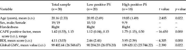
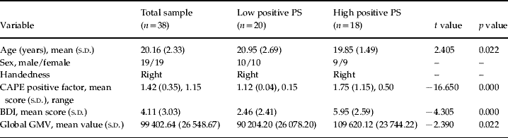
Significant differences in age, BDI scores and global GMV were observed between the two study groups. Hence, these three variables were entered in the statistical design as covariates of no interest to control for potential effects on between-group GMV differences. There were no group differences in gender or handedness (see Table 1).
Demographic and clinical characteristics, and global gray-matter volumes (GMVs) of study sample
PS, Psychometric schizotypy; s.d., standard deviation; CAPE, Community Assessment of Psychic Experiences; BDI, Beck Depression Inventory.
There was a significant negative correlation between CAPE positive factor scores and age (r=−0.356, p=0.028). Although none of the subjects in the study scored above the cut-off on the BDI, there was a significant correlation between this test and the CAPE positive factor (r=0.615, p<0.001). Thus, higher scores on positive PS were associated with younger age, and higher ratings of depression.
VBM results: low PS versus high PS
Global GMV correlated significantly with CAPE positive factor scores (r=0.325, p=0.047). A direct comparison between the high and the low PS groups confirmed larger global GMV in subjects with high positive PS (t score=−2.390, p=0.022).
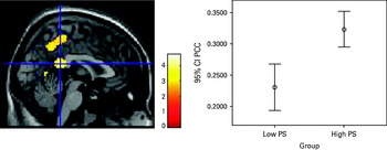
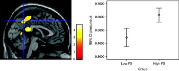
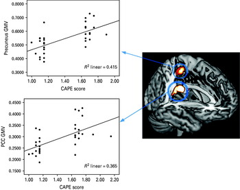
With regard to regional GMV, we detected two regions of greater volume in the high relative to the low positive PS group: the medial posterior cingulate cortex (PCC) and the precuneus (see Table 2, Figs 1 and 2). Conversely, there were no areas where volume was significantly smaller in subjects with high positive PS. Finally, the GMV in each of these two regions was extracted and subjected to correlation analysis with the CAPE positive factor scores. Significant correlations were found between the CAPE positive factor and both the PCC (r=0.503, p=0.001) and precuneus (r=0.484, p=0.001) volumes. Cook's D test was applied to rule out the influence of potential outliers on the correlation. This correction strengthened the significance of the correlations in both cases (PCC: r=0.604, p<0.001; precuneus: r=0.644, p<0.001) (Fig. 3).
Increased gray-matter volume in the medial posterior cingulate cortex (PCC) in subjects with high scores on positive psychometric schizotypy (PS) relative to subjects with low scores, at a p-uncorrected level of 0.005. Between-group differences were considered significant at p<0.05, false-discovery rate corrected.

Increased gray-matter volume in the medial precuneus in subjects with high scores on positive psychometric schizotypy (PS) relative to subjects with low scores, at a p-uncorrected level of 0.005. Between-group differences were considered significant at p<0.05, false-discovery rate corrected.

Areas of increased gray-matter volume (GMV) in subjects with high scores on positive psychometric schizotypy (PS) at a p-uncorrected level of 0.005. The scatterplots show the correspondent positive relationships between individual scores in PS [Community Assessment of Psychic Experiences (CAPE) positive factor] and GMV in the medial posterior cingulate cortex (PCC) and medial precuneus.

Regions of gray-matter volume differences between subjects with high versus subjects with low positive psychometric schizotypy (p<0.05, FDR corrected)
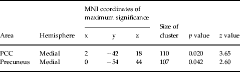
PCC, Posterior cingulate cortex; FDR, false-discovery rate; MNI, Montreal Neurological Institute.
Conclusions
The present study investigated GMV in positive PS. Using a series of ROI analyses, we examined the brain regions that have been previously implicated in volumetric studies of schizophrenia spectrum disorders. The approach used here showed significant global GMV differences between subjects with high compared to low positive PS, with high schizotypal subjects showing larger volume. We also found significant differences in focal GMV in the PCC and the medial precuneus. These global and regional MRI findings cannot be attributed to differences in BDI scores or age as these variables were modeled as covariates of no interest in the statistical analysis.
To our knowledge, this is the first study to investigate brain morphology with VBM in subjects with high positive PS. We tested the hypothesis that alterations in GMV would be located within the same regions that are sites of abnormal GMV in ARMS, SPD and schizophrenia. Our results are consistent with this hypothesis, as the PCC had previously been implicated in structural MRI studies of chronic schizophrenia (Hulshoff Pol et al. Reference Hulshoff Pol, Schnack, Mandl, van Haren, Koning, Collins, Evans and Kahn2001; Mitelman et al. Reference Mitelman, Shihabuddin, Brickman, Hazlett and Buchsbaum2005), first-episode schizophrenia (Kubiki et al. Reference Kubicki, Shenton, Salisbury, Hirayasu, Kasai, Kikinis, Jolesz and McCarley2002; Job et al. Reference Job, Whalley, McConell, Glabus, Johnstone and Lawrie2003; Koo et al. Reference Koo, Levitt, Salisbury, Nakamura, Shenton and McCarley2008), early-onset schizophrenia (Sowell et al. Reference Sowell, Levitt, Thompson, Holmes, Blanton, Kornsand, Caplan, McCracken, Asarnow and Toga2000), and high-risk subjects (Pantelis et al. Reference Pantelis, Velakoulis, McGorry, Wood, Suckling, Phillips, Yung, Bullmore, Brewer, Soulsby, Desmond and McGuire2003; Borgwardt et al. Reference Borgwardt, McGuire, Aston, Berger, Dazzan, Gschwandtner, Pflüger, D'Souza, Radue and Riecher-Rössler2007a, Reference Borgwardt, Riecher-Rössler, Dazzan, Chitnis, Aston, Drewe, Gschwandtner, Haller, Pflüger, Rechsteiner, D'Souza, Stieglitz, Radü and McGuireb). Moreover, most of these studies also reported abnormalities in the precuneus (Hulshoff Pol et al. Reference Hulshoff Pol, Schnack, Mandl, van Haren, Koning, Collins, Evans and Kahn2001; Shapleske et al. Reference Shapleske, Rossell, Chitnis, Suckling, Simmons, Bullmore, Woodruff and David2002; Pantelis et al. Reference Pantelis, Velakoulis, McGorry, Wood, Suckling, Phillips, Yung, Bullmore, Brewer, Soulsby, Desmond and McGuire2003; Borgwardt et al. Reference Borgwardt, McGuire, Aston, Berger, Dazzan, Gschwandtner, Pflüger, D'Souza, Radue and Riecher-Rössler2007a, Reference Borgwardt, Riecher-Rössler, Dazzan, Chitnis, Aston, Drewe, Gschwandtner, Haller, Pflüger, Rechsteiner, D'Souza, Stieglitz, Radü and McGuireb; Zhou et al. Reference Zhou, Suzuki, Takahashi, Hagino, Kawasaki, Matsui, Seto and Kurachi2007). Although these previous findings were of reduced GMV in these regions in schizophrenia and the ARMS, in the present study high positive PS was associated with relatively greater GMV in the PCC and precuneus.
Increases, rather than decreases, in GMV have been found in other schizophrenia spectrum disorders. Increased GMV has been described in first-episode schizophrenia patients with hallucinations compared to non-hallucinators in frontal and temporal regions (Shin et al. Reference Shin, Lee, Kang, Kim, Bae and Jung2005). Furthermore, two studies have reported larger prefrontal volume in subjects with SPD compared to controls (Suzuki et al. Reference Suzuki, Zhou, Takahashi, Hagino, Kawasaki, Niu, Matsui, Seto and Kurachi2005b; Hazlett et al. Reference Hazlett, Buchsbaum, Haznedar, Newmark, Goldstein, Zelmanova, Glanton, Torosjan, New, Lo, Mitropoulou and Siever2008), and relatively increased regional volumes have been found in ARMS subjects who later developed psychosis. Individuals who later transited to psychosis showed larger volumes in the left hippocampus (Phillips et al. Reference Phillips, Velakoulis, Pantelis, Wood, Yuen, Yung, Desmond, Brewer and McGorry2002), in the parahippocampal, parietal and posterior temporal cortex, and also in the thalamus (Borgwardt et al. Reference Borgwardt, Riecher-Rössler, Dazzan, Chitnis, Aston, Drewe, Gschwandtner, Haller, Pflüger, Rechsteiner, D'Souza, Stieglitz, Radü and McGuire2007b), the pituitary gland (Garner et al. Reference Garner, Pariante, Wood, Velakoulis, Phillips, Soulsby, Brewer, Smith, Dazzan, Berger, Yung, van den Buuse, Murray, McGorry and Pantelis2005) and the cuneus (Pantelis et al. Reference Pantelis, Velakoulis, McGorry, Wood, Suckling, Phillips, Yung, Bullmore, Brewer, Soulsby, Desmond and McGuire2003), compared to those who did not transit to psychosis. The reason for relatively greater volumes in these regions is not yet understood, but it has been suggested they may reflect functional compensation for early disturbances in other brain regions (Kawasaki et al. Reference Kawasaki, Suzuki, Nohara, Hagino, Takahashi, Matsui, Yamashita, Chitnis, McGuire, Seto and Kurachi2004), or transient increases due to an active pathological process (Meisenzhal et al. Reference Meisenzhal, Koutsouleris, Gaser, Bottlender, Schmitt, McGuire, Decker, Burgermeister, Born, Reiser and Möller2008).
The precuneus, which is part of the parietal lobe, has been implicated in discriminating agency and, in patients with schizophrenia, in delusions of control (Sirigu et al. Reference Sirigu, Daprati, Pradat-Diehl, Franck and Jeannerod1999; Blakemore et al. Reference Blakemore, Wolpert and Frith2002). The cingulate cortex is thought to mediate cognitive and emotional impairments in patients with schizophrenia, although this seems to be more true of its anterior than its posterior portion (Baiano et al. Reference Baiano, David, Versace, Churchill, Balestrieri and Brambilla2007). Both the precuneus and PCC are key components of the ‘default mode’ network of the human brain (Cavanna & Trimble, Reference Cavanna and Trimble2006; Fransson & Marrelec, Reference Fransson and Marrelec2008). Patients with schizophrenia show aberrant functional connectivity within this network, which may contribute to the positive and negative symptoms of the disorder (Garrity et al. Reference Garrity, Pearlson, McKiernan, Lloyd, Kiehl and Calhoun2007).
The significant negative correlation between age and CAPE is consistent with research on self-reported experiences in non-clinical populations showing a reduction in the endorsement of such experiences as age increases (Van Os et al. Reference Van Os, Hanssen, Bijl and Ravelli2000; Verdoux & Van Os, Reference Verdoux and van Os2002). According to these studies, this could reflect an increased propensity of subjects at a younger age from the general population to endorse unusual ideas or strange beliefs (Verdoux & Van Os, Reference Verdoux and van Os2002). In addition, CAPE and BDI scores were positively correlated, in keeping with previous findings of an association between symptoms of depression and the positive symptom dimension of schizotypy (Lewandowski et al. Reference Lewandowski, Barrantes-Vidal, Nelson-Gray, Clancy, Kepley and Kwapil2006). Nonetheless, we observed an effect of the positive psychotic-like experiences on GMV after controlling for the effect of age and BDI in the statistical analyses (between-group differences remained significant).
Finally, it has been proposed that schizophrenia could result from a disruption of brain maturation during adolescence (Rapoport et al. Reference Rapoport, Giedd, Blumenthal, Hamburger, Jeffries, Fernandez, Nicolson, Bedwell, Lenane, Zijdenbos, Paus and Evans1999; Thompson et al. Reference Thompson, Vidal, Giedd, Gochman, Blumenthal, Nicolson, Toga and Rapoport2001). Given that the average age of our participants was around 20 years, increased GMV in high PS subjects might reflect a delay in synaptic pruning (Gotay et al. Reference Gotay, Giedd, Lusk, Hayashi, Greenstein, Vaituzis, Nugent, Herman, Clasen, Toga, Rapoport and Thompson2004). Moreover, increased global GMV has been described in other disorders of neurodevelopmental origin, such as autism (Palmen et al. Reference Palmen, Hulshoff Pol, Kemner, Schnack, Janssen, Kahn and van Engeland2004).
As in previous neuroimaging studies of other high-risk groups, the sample size in the present study was relatively small. Groups were formed with high and low scorers to allow consistency with previous literature on PS, which has typically compared behavioral performance between groups selected upon the same criterion (e.g. Langdon & Coltheart, Reference Langdon and Coltheart1999, Reference Langdon and Coltheart2001, Reference Langdon and Coltheart2004; Platek & Gallup, Reference Platek and Gallup2002; Jahshan & Sergi, Reference Jahshan and Sergi2007; Fernyhough et al. 2008). The distribution of schizotypy in non-clinical samples does not show a continuous normal distribution but a continuous half-normal distribution, with the majority of the population having very low values, although a significant proportion also has progressively higher values (Van Os et al. Reference Van Os, Linscott, Myin-Germeys, Delespaul and Krabbendam2009). In addition, compared to other measures of schizotypal signs (e.g. Paranoia Scale, Launay–Slade Hallucination Scale), the CAPE comprises more ‘pathological’ items (e.g. does not tap on phenomena such as daydreaming), so that very low scores are ‘normal’. Thus, using average scorers would not be expected to significantly alter the present findings. However, future studies on PS including such control group should help expand our findings. Another potential limitation of the present study is that the subjects were recruited from a university sample, and caution should therefore be used when extrapolating the present findings to schizotypy in the general population. Nevertheless, this study demonstrated, for the first time, altered GMV in the PCC and precuneus in a group of subjects with high scores on positive PS, consistent with volumetric findings in these regions in schizophrenia spectrum disorders. These findings can be interpreted as correlates of the increased risk of schizophrenia in these subjects.
Acknowledgments
This work was supported by the European Science Foundation EURYI grant (NWO no. 044035001) to Professor A. Aleman. We thank Dr U. Ettinger for the critical comments on the findings presented in this study.
Declaration of Interest
None.