Introduction
Lichens cover about 6% of the Earth's land surface and play a major role in the biogeochemical cycling of elements (Haas & Purvis Reference Haas, Purvis and Gadd2006; Nash Reference Nash2008; Purvis Reference Purvis, Batty and Hallberg2010). Reports of high metal contents in lichen hyperaccumulators (in excess of 1000 ppm) are normally due to the trapping of particulate matter and formation of biominerals (Purvis & Pawlik-Skowrońska Reference Purvis, Pawlik-Skowroñska, Avery, Stratford and van West2008). Possibly the highest copper concentrations (c. 16% dry weight) ever recorded were from a sample of Acarospora rugulosa Körb. found growing on the mineral brochantite [Cu4SO4(OH)6] in Sweden, corresponding to about 40% copper oxalate in an extracellular form (Purvis Reference Purvis1984; Chisholm et al. Reference Chisholm, Jones and Purvis1987). Lange & Ziegler (Reference Lange and Ziegler1963) reported unusually high concentrations of both copper and iron (between 10 and 30 thousand ppm ash dry weight) in rust-coloured Acarospora sinopica and Rhizocarpon oederi growing on slag dumps in the Harz Mountains.
Some lichens are brightly coloured, normally due to the presence of organic substances (Huneck & Yoshimura Reference Huneck and Yoshimura1996). Various biological roles for cortical lichen substances are implicated, including responses to light (Gauslaa & McEvoy Reference Gauslaa and McEvoy2005). A striking effect of mineralization is the bright green colour of certain lichen species on cupriferous (and adjacent) substrata. Sixteen lichen species belonging to 9 genera (Acarospora, Aspicilia, Bellemerea, Hypotrachyna, Lecanora, Lecidea, Miriquidica, Psilolechia and Tephromela) have been reported to display a green colour or contain blue-green inclusions in cupriferous habitats (Purvis Reference Purvis1996). Vivid blue inclusions of copper oxalate hydrate, moolooite, have been found in specimens sampled from mining regions in Scandinavia and the UK: Acarospora rugulosa, Lecidea lapicida (=L. lactea), L. inops (Purvis Reference Purvis1984) and Lecanora polytropa (Purvis et al. Reference Purvis, Pawlik-Skowroñska, Cressey, Jones, Kearsley and Spratt2008). Copper lichen-acid complexes are also known to be responsible for the unusual greenish colours observed in lichens occurring in cupriferous habitats across taxonomically unrelated groups, [Acarospora smaragdula and Lecidea lapicida (Purvis et al. Reference Purvis, Elix, Broomhead and Jones1987) and Lecidella bullata (Purvis et al. Reference Purvis, Elix and Gaul1990)]. Element localization and microscopy confirmed copper is present and responsible for the vivid turquoise-blue colour of Lecanora polytropa thalli sampled from a Swedish mine (Purvis et al. Reference Purvis, Pawlik-Skowroñska, Cressey, Jones, Kearsley and Spratt2008), although the identity of the compound(s) or process(es) involved was unknown.
Dark green samples of the lichen Lecanora sierrae B. D. Ryan & T. H. Nash (formerly L. cascadensis H. Magn.) were observed near copper mines in 1976 during a geochemical examination of the Lights Creek District situated in Northern California (Czehura Reference Czehura1977). Colour differences in thalli ranging from light green (containing 1% Cu in ash), to green-rimmed (containing 2% Cu in ash) to dark green (containing 4% Cu in ash) were found at Warren Canyon to correspond with copper mineralization in the soil. Field observations suggested the lichen's colour was caused by the accumulation of insoluble copper salts in the cortex. Since Czehura's study, a critical revision of the Lecanora garovaglii group was carried out and two new species were described: yellow to blue-green Lecanora sierrae and orange or reddish brown L. pseudomellea B. D. Ryan and a range of chemotypes (Ryan & Nash Reference Ryan and Nash1993). Different techniques, including infrared absorption spectroscopy, were applied to investigate lichen material sampled by Czehura from Warren Canyon (and other lichens) and of a synthetic copper-psoromic acid complex to test the hypothesis that complexing of copper by psoromic acid occurs within specific areas of the lichens examined and that this leads to their unusual coloration. The psoromic acid-containing Lecanora sierrae, which also contains usnic acid, was anomalous in not apparently forming a copper-psoromic acid complex (Purvis et al. Reference Purvis, Elix and Gaul1990).
The highest copper concentration in dark coloured material (4% Cu in ash) is about 36 times that of associated species collected at the same site (Czehura Reference Czehura1977). Assuming ash weight to be about 10% of dry weight, L. sierrae would appear to contain between 1 to 2 orders of magnitude lower copper contents than Scandinavian crustose lichens with anomalous coloration in cupriferous habitats (Purvis Reference Purvis1984).
Lecanora sierrae is known to contain several secondary metabolites, one of which, usnic acid, is a lemon yellow colour (Nash et al. Reference Nash, Ryan, Diederich, Gries and Bungartz2004). This colour is sometimes evident in older thalli, but more typically is masked by the blue-green colour. It is therefore unknown if the blue-green colour is due to a pigmented secondary compound or copper mineralization. This study addresses the hypothesis that the colour of a sample of dark green Lecanora sierrae collected from the Lights Creek District in 2008 reflects copper localization rather than the presence of lichen pigments. A dark green sample from Warren Creek was selected for initial study because Czehura (Reference Czehura1977) had previously found specimens there which correlated with high Cu contents in soil. We tested this hypothesis using optical microscopy and electron microprobe analysis, and supplemented it with preliminary elemental analyses of lichen tissues including L. sierrae and other species from the same habitat for comparison.
Methods
Study area and material
The Lights Creek District is situated in northern California, approximately 190 km north-east of Sacramento, in the northern portion of the Plumas Copper Belt. Pre-intrusive rocks within the Lights Creek District consist mostly of upper Palaeozoic metasedimentary rocks and Mesozoic metavolcanics. Copper mineralization occurs in the southern portion of the Lights Creek stock associated with metavolcanics and mineralized sheer zones exist in the surrounding plutons. Two major mines have been developed in the Lights Creek District, the Engels and the larger Superior mine, a porphyry copper deposit. Both were active between 1916–1929, and in 1930 operations were suspended (Czehura Reference Czehura1977). Today, abandoned spoil heaps form a prominent feature in the mining region. Warren Canyon, where Czehura mapped “3 colour variants of L. cascadensis” is a steep ravine with scree slopes just south-east of the two mines. The climate of the Lights Creek district is classed as ‘moderate’ with summer temperatures often reaching 27–30°C, while winter extremes are well below freezing. The vegetation belongs to the Middle Mountain Forest zone of the central Sierra Nevada, the tree layer dominated by ponderosa pine (Pinus ponderosa). In August 2007, the Moonlight Fire burned over 65 000 acres but saxicolous lichens on scree slopes in Warren Canyon largely escaped damage. Dark green specimens of Lecanora sierrae (Czehura Reference Czehura1977; Ryan & Nash Reference Ryan and Nash1993) are frequently accompanied by other lichen species, including the dark brown L. pseudomellea and Lecidea atrobrunnea, and yellow-green Rhizocarpon species, Cheilanthes sp. (a fern) and an Eriogonum sp., a flowering plant belonging to the Polygonaceae. Samples of the dark green L. sierrae were collected together with their substratum: a basalt with abundant plagioclase phenocrysts and wrapped in tissue to avoid damage to the lichen surface (Fig. 1). The specimen used for microscopy is a fragment of a larger specimen with iron staining corresponding to brittle fractures along planar surfaces. The feldspars are quite fresh and unaltered. Slight alteration of the rock is apparent adjacent to the lichen and lower fracture surface.
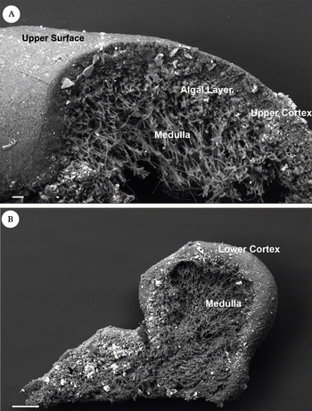
Lecanora sierrae, SEM (back-scattered) images, bright areas indicate areas with a higher average atomic number. A, general section through thallus showing dense cortex overlying algal and medullary layers; B, lower surface with distinct marginal lower cortex. Brighter areas correspond to mineral grains. Scales: A = 20 µm; B = 100 µm.

Microscopy and electron microprobe analysis
Fractured sections were prepared for scanning electron microscopy (SEM) to prevent section smearing. Sections were mounted on an adhesive carbon tab and examined in a LEO 1455 variable pressure (VP) SEM utilizing a 4 quadrant solid-state back-scattered electron (BSE) detector, operated at 15 kV. A sample was mounted in a resin block, ground down to reveal a suitable transverse section and polished to produce a flat surface suitable for X-ray elemental mapping (Williamson et al. Reference Williamson, McLean and Purvis1998). The resin-embedded block was examined using a Zeiss Axioplan reflected light microscope under cross polarizers with digital imaging, and images were processed using auto montage software to create pseudo-3D images.
Blocks were carbon coated and X-ray emission maps generated for the elements Al, Ca, Cu, Fe, K, Mg, N, Na, P, Si and Ti to show element distribution using a CAMECA SX100 electron microprobe operating at 20 kV and 10 nA with a count time of 600 ms for each pixel (512 × 512) using a 2 µm step size giving a total area of 1024 × 1024 µm. The element maps for Ca, Na, K, Fe and Cu were obtained using the WD spectrometers and the element maps for N, Mg, Al, Si, P, S, Ti and Fe were obtained using a Bruker XFLASH 4010 SDD ED detector.
Images taken under reflected light were merged using Adobe Photoshop CS3 to create a montage and X-ray emission maps for each element were superimposed as layers. Using the opacity function, the relationship between element distribution and lichen tissues was assessed.
Elemental analyses
Seventeen random bulk samples (10–20 g d. wt.) of Letharia columbiana (Nutt.) J. W. Thomson, Letharia vulpina (L.) Hue, and Umbilicaria phaea Tuck. were collected at Warren Canyon and Superior mine for elemental analysis. One small sample of the blue-green L. sierrae was collected where it occurred at the Superior mine, while all other species were collected at both sites at various localities. It was difficult to get enough of L. sierrae to sample as it is very adnate. Only the dark green form was sampled based on Czehura's (Reference Czehura1977) findings. We included species other than L. sierrae in our sampling to determine if elevated levels of any elements were restricted to L. sierrae in any of the localities. Samples were ground to a powder and analyzed by ICP-MS for 20 elements including both macro- and micro- elements. More information on these methods is found in Bennett (Reference Bennett2008). Enrichment factors were determined for each element, based on the ratio of element concentration to Al concentration ([x/Al]lichen/[x/Al]soil] where x = the element of interest; Taylor & McLennan Reference Taylor and McLennan1985; Garty Reference Garty and Markert1993; Bargagli Reference Bargagli1998). It is generally accepted that an enrichment factor near unity indicates the element's source is from the environment of the lichen and not anthropogenic inputs. However, if the factor is greater than 100, it suggests there are anthropogenic or external inputs in the lichen environment, although there are problems with this interpretation (Reimann & de Caritat Reference Reimann and de Caritat2000; Nash Reference Nash2008). We used these measures to examine possible enrichment from the mining environment. The goal was to determine if elemental concentrations were high enough to warrant further, systematic sampling. Simple t tests comparing mean element concentrations between the two Letharia species revealed no significant differences so the data were combined. We tested the significance of the enrichment factors by one-way analysis of variance using the five species × locality treatments, followed by Tukey-Kramer Honestly Significant Difference (HSD) post-hoc mean separation tests (SAS Institute 2005) to determine where significant differences were among elements. In addition, we tested if the means were significantly greater than one using one-tailed t tests. All statistical tests were performed using JMP 8 (http://www.jmp.com/).
Results
Microscopy and electron microprobe analysis
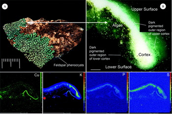
The outer surface of our dark green specimen of Lecanora sierrae was smooth at low magnification, the cortex consisting of densely packed hyphal cells surrounded by a gelatinous matrix extending from the upper surface to the lower within c. 500 µm from the lobe tip (Fig. 1). Irregular mineral grains were present, especially scattered on the upper and lower lichen surfaces (Fig. 1). In contrast, the medulla consisted of loose hyphae with intracellular spaces. The cortex in the embedded section was birefringent, the outermost cortical cells capped by a bright blue-green pigment, the colour appearing darker on the lichen's lower cortical surface adjacent to the mineral surface, a region extending c. 500 µm from the lobe apex (Fig. 1).
In the untreated lichen, Cu was readily detected in the darkest blue-green coloured lower cortex near the lobe margin (Fig. 2) using the ED spectrometer [LEO 1455 variable pressure (VP) SEM)]. All elements analyzed apart from N were detected in lichen tissues using the more sensitive EPMA WD spectrometer. X-ray element maps (CAMECA SX100) for Cu, S, and P showed strong localization within lichen tissues. Copper was also detected in all other lichen tissues as discrete phases (Fig. 2). Sulphur and P were correlated throughout, and the highest contents were present in the algal layer. The highest K concentration was recorded in a feldspar phenocryst at the medulla-rock interface (Fig. 2). The elements Al, Ca, Fe, Mg and Na were mostly present as discrete particulate phases, either occurring on, within or beneath the lichen surface. Cu, K, P and S were correlated at the outermost region of the pigmented lower surface lobe tip.
Lecanora sierrae, Warren Creek, Lights Creek District, 21 July 2008 (BM). A, close-up of a dark green variant found growing on a weathered, iron-stained basalt with plagioclase phenocrysts, lobe embedded in resin (arrowed); B, close-up of a section of a lobe of same specimen embedded in resin (arrowed in Fig. 2A) photographed under transmitted (polarized) light using and processed with auto montage software to create pseudo- 3D images; Cu, K, P, & S, X-ray emission maps showing element distribution where elements concentrated in specific lichen tissues (generated using an automated wavelength dispersive CAMECA SX100 microprobe). Scales: A = 10 mm; B = 50 µm; Cu, K, P & S = 500 µm.

Elemental analyses
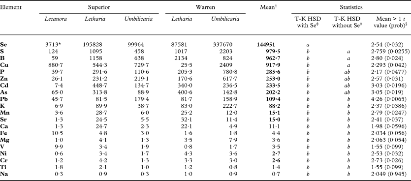
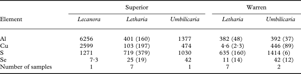
The analysis of variance of the enrichment factors revealed that there were significant differences among the 20 elements (F = 6·45, P = <0·0001, df = 19·80). The Tukey HSD mean separation test on all 20 elements indicated that Se (selenium) differed significantly from all other elements, and none of the remaining differed among them (Table 1). This was due to the very large size of the Se enrichment factor. A second Tukey HSD test on 19 elements without Se indicated that S, B and Cu were significantly greater than 12 other elements, and equal to 4 other elements (Table 1). Nine elements had mean enrichment factors greater than 100: Se, S, B, Cu, P, Zn, Cd, As and Pb (Table 1). Fourteen elements had mean enrichment factors significantly greater than one: Se, S, B, Cu, P, Zn, Cd, As, Pb, K, Mn, Sr, Ni and Cr (Table 1). The largest individual enrichment factors across species and mines were for Se and Cu. Actual element concentrations for Al, Cu, S and Se (Table 2), were unusually high for lichens (Garty Reference Garty and Markert1993; Bargagli Reference Bargagli1998).
Enrichment factors for 20 elements in descending order by mean in lichens at the Superior Mine and Warren Canyon in northern California. Elements with mean enrichment factors significantly greater than one are in bold font. Genera refer to specific specimens (see methods)

* factors are the ratios of the given element to Al divided by the same ratio for the earth's crust, and have no units, the factors are calculated as the ratios of means for each cell.
† grand means across the five species × locality cells.
‡ Tukey-Kramer (T-K) HSD mean separation tests with and without Se, means not connected by same letter are significantly different at the 0·05 probability level.
§ t test value and probability (one-tailed) for the mean being significantly greater than one.
Mean concentrations (ppm) of four elements in lichens at Superior Mine and Warren Canyon in northern California. Standard deviations are in parentheses. Genera refer to specific specimens (see methods)

Discussion
Results from both optical microscopy and from element analyses generally supported our hypothesis that the vivid pigmentation in dark green forms of L. sierrae is due to copper localization. Spot analyses of the untreated lichen using the JEOL 5900 LV (ED spectrometer), element mapping of the embedded section using the CAMECA SX100 (WD spectrometer) of embedded sections and image analysis confirm Cu is correlated with the outer blue-green cortical layer. The hyphal tips of outer cells within the bright birefingent cortex are clearly capped by vivid blue pigmentation. This is most apparent on the upper surface, the effect emphasized by the use of auto-montage software. Although it appears to be extensive, the blue pigmentation is an illusion created by light reflectance and transmission through the clear resin. The embedded section is not perpendicular throughout the lobe length but at a slight angle towards the lobe tip. In resin-embedded sections X-ray generation is limited to c. 10 µm below the surface (Williamson et al. Reference Williamson, McLean and Purvis1998) and the area available for analysis consequently was not as extensive as would appear from the reflected light image. In contrast, the dark colour of the hyphal cells on the lower cortical surface (< 500 µm) is less clear in view of the section orientation and surrounding dark tissues.
Of the elements with enrichment factors greater than one, eight are of interest, those with values of 200 or greater (the enrichment factor for Pb, 109, was barely over the 100 threshold and will not be discussed). Four elements had enrichment factors between 200–300: As, Cd, Zn and P. The first three are elements probably associated with crustal minerals in the copper district, while P is enriched because it is essential to lichens which scavenge it from the environment where it is poorly retained (Nash Reference Nash2008). There are four elements with enrichment factors greater than 900: B, Cu, S and Se. The last three will be discussed in detail below. We do not know, however, why B is elevated. There are two possible reasons: first, B is mined in southern California extensively (Angulo Reference Angulo2010) and fugitive dust from those mines may be transported to the northern Sierra Mountains. Second, B is used in fire retardants (Angulo Reference Angulo2010), and these may have been applied in our study area to suppress the Moonlight fire in 2007.
The elemental analyses suggested that the colour of L. sierrae may be caused not only by copper in the lichens, but also by mineralizations in the form of selenates, selenites or sulphates. Copper sulphate, selenate and selenite are all a bright blue-green colour (Weast Reference Weast1968) and are strong candidates for this given the elemental analysis. It is worth noting that Se is produced as a by-product of copper refining from copper ores because of its strong association with that element (Butterman & Brown Reference Butterman and Brown2004), so it is logical to expect a copper selenate/selenite to occur in the lichens. There are numerous reports of the element selenium in lichens, but none of selenates or selenites. Further testing of mineral extracts from specimens by inorganic chemical analysis is needed to verify which compounds are present.
We found similar element enrichments in both epiphytic (Letharia) and saxicolous (Umbilicaria) species collected from the same habitats as the dark green form of Lecanora sierrae. The presence of copper in epiphytic species at both sites suggests it may be occurring in fugitive dust in the area as well as the rocks. It is also worth noting that the highest Cu enrichment factors were in the two saxicolous species, while the highest Se and S enrichment factors were in both a saxicolous and epiphytic species, suggesting that possibly some of the S and Se originated in fugitive dust.
We were unable to identify the copper compound(s) responsible for the observed pigmentation and particulates in the examined material of Lecanora sierrae. In addition to copper minerals and copper lichen acid complexes, copper could also be complexed organically in a laccase or tyrosinase as in the Peltigerineae (Laufer et al. Reference Laufer, Beckett, Minbayeva, Lüthje and Böttger2009). The considerable diversity that exists in extracellular lichen laccases suggests that they may have more than one role in lichen biology. Blue-green pigments are frequent in Lecanora, especially in species exposed to high illumination in alpine habitats (Obermayer & Poelt Reference Obermayer and Poelt1992). The extent to which blue pigmentation in other taxa belonging to the L. garovaglii group present at Warren Creek and Superior Mine reflects copper localization remains to be established. This study confirms copper localization correlates with pigmentation in L. sierrae, but not other lichens.
We are grateful for support from a UK Royal Society Conference Grant for OWP to attend IAL6 and particularly to Linda Geiser for her support, to Norman Lamb for his interest, advice, and for granting access and permissions, to Abigail Stephens, John Dilles, Robert Wetzel and Steve Czehura for field assistance, Tony Wighton for sample preparation, Chris Stanley and Gordon Cressey for assistance with reflected light microscopy, Alex Ball and Lauren Howard with FESEM, Harry Taylor for macrophotography (Fig. 1A), Juliet Brodie for helpful discussions, and James McNeal for help on mineral colours. The mention of commercial equipment or products does not imply endorsement by the U.S. Geological Survey.