I. INTRODUCTION
Recent advances in synthetic polymers in the biomedical field have increased the need for designing materials with high biocompatibility, easy processing, and antibacterial properties, avoiding additional processing steps related to the modification of the polymer surface. One biomedical application of synthetic polymers is bioadhesives. In the last five years, surgical tissue adhesives have undergone significant advances. Reference Ates, Koytepe, Karaaslan, Balcioglu and Gulgen1,Reference Bouten, Zonjee, Bender, Yauw, Van Goor, Van Hest and Hoogenboom2 Bioadhesive is understood as the adhesion of a polymer (natural or synthetic) with biological tissues such as skin or mucous membranes. Adhesion is the attachment between the surfaces of two substances that can be similar or dissimilar in structure. The process of adhesion can occur from energy obtained from chemical or physical linkages of chemical structures. Reference Patel3 Bioadhesives are used in drug delivery, implantation of biomedical devices, tissue engineering, dental and bone applications, and as alternative internal and external wound closures in which healing and infection prevention are encouraged. Reference Guo, Kim, Shan, Kim, Hu, Wang, Hamad, Qian, Rizk and Yang4–Reference Jeon, Samorezov and Alsberg7 The use of bioadhesives has been enhanced by the search for non-invasive biomedical devices for surgical practice, which help control blood loss and promote tissue healing. Reference Wheat and Wolf8 Biomedical adhesives must have strong binding strength, ease of application, tissue biocompatibility, biodegradable by products, minimal tissue reactivity, and reasonable cost. Reference Ates, Koytepe, Karaaslan, Balcioglu and Gulgen1
The most common bioadhesives are fibrin, cyanoacrylates, and an albumin-glutaraldehyde blend, which are characterized by rapid curing times, biodegradability, and biocompatibility. Reference Marques, Santos, Ferreira, Correia, Correia, Gil and Baptista6,Reference Jeon, Samorezov and Alsberg7,Reference Mehdizadeh, Weng, Gyawali, Tang and Yang9 These bioadhesive materials exhibit disadvantages such as poor adhesion to tissues, rapid degradation, inflammation problems, and allergic reactions. Reference Marques, Santos, Ferreira, Correia, Correia, Gil and Baptista6,Reference Jeon, Samorezov and Alsberg7,Reference Mehdizadeh, Weng, Gyawali, Tang and Yang9 Traditional bioadhesives do not have antibacterial properties, so it is necessary to add compounds that fulfill this function. Reference Guo, Wang, Hu, Xie, Gerhard, Nisic, Shan, Qian, Zheng and Yang10 There are reasons to study polymeric materials to mitigate these drawbacks. In addition, polymer-based adhesives have physical, mechanical, and chemical properties that promote adhesion. Reference Patel3
One of these polymers corresponds to polyurethanes (PUs). PUs are a class of synthetic polymers that are used as biomaterials. PUs have advantages such as flexibility, elasticity, durability, ease of processing, and biocompatibility. Reference Seeni Meera, Murali Sankar, Paul, Jaisankar and Mandal11 These properties are generated by the block structure that contains hard and flexible segments joined together by urethane groups. Reference Szycher12,Reference Alves, Ferreira, Gil, Cavaco and Melo13 The mechanical, thermal, chemical, and biological properties of PUs can be modified in synthesis, Reference Alves, Ferreira, Gil, Cavaco and Melo13 through the type of polyol or polymer addition, such as chitosan (Ch) which modifies the properties such as the biocompatibility. Reference Usman, Zia, Zuber, Tabasum, Rehman and Zia14 Ch is a polysaccharide that is obtained from renewable sources, as it is part of the structure of some crustaceans. Ch is characterized as being biocompatible, biodegradable, bioadhesive, and non-toxic and has antimicrobial properties. Reference Usman, Zia, Zuber, Tabasum, Rehman and Zia14–Reference Liu, Zhou and Sun17 These properties facilitate the use of this material in the design of biomaterials. Some natural polysaccharides, such as Ch, are characterized by forming covalent bonds with biological tissues via bio-adhesion due to hydroxyl, carboxyl, or amino groups in the structure. Reference Kaur, Mahajan and Bassi15
The selection of the polyol in synthesis allows the modification of the properties of PUs. Polyols derived from vegetable oil are currently used in PU synthesis. Vegetable oils have different chemical structures and reactive sites that allow for the generation of different modified polyols. In addition, polyols derived from vegetable oils can replace the use of polyols derived from fossil sources. Reference Maisonneuve, Chollet, Grau and Cramail18,Reference Narute and Palanisamy19 One oil used in the synthesis of PUs is castor oil (CO). CO is characterized by hydroxyl groups in the chain that react with isocyanates generating different morphological and mechanical properties. Reference Uscátegui, Arévalo, Díaz, Cobo and Valero20,Reference Shaik, Narayan and Raju21 PUs derived from CO may exhibit antibacterial activity and applications of these polymers for wound closure have been reported. Reference Seeni Meera, Murali Sankar, Paul, Jaisankar and Mandal11
According to the previous information, there are works related to bioadhesive designs. But to the best of our knowledge, there are no works about PU-based bioadhesives from CO polyols and chitosan describing the effect of composition on biological properties such as cell viability, cell adhesion, and antimicrobial activity. PUs with these properties avoid subsequent processes of adaptation as biomedical devices. Therefore, the objective of this work was to synthesize PU-based bioadhesives for external applications using polyols derived from CO (Ricinus communis) and hexamethylene diisocyanate (HDI) with Ch addition. We evaluated the effect of the chemical modification of polyols derived from CO and Ch addition on the mechanical, morphological, and biological properties of polymeric matrices that may be useful in biomedical applications such as bioadhesives.
II. EXPERIMENTAL
A. Materials
CO was obtained from Químicos Campota y Cía, Ltda. (Bogotá D.C., Colombia). Low-molecular-weight chitosan (Ch) with 75–85% deacetylated and hexamethylene diisocyanate (HDI) was obtained from Sigma-Aldrich Chemical Co. (St. Louis, Missouri). Phosphate buffered saline (PBS) was prepared by dissolving KH2PO4 and NaHPO4 0.01 M in 0.8% NaCl at pH 7.4 and pentaerythritol was obtained from Merck KGaA (Darmstadt, Germany). MTT (3-[4,5-dimethylthiazol-2-yl]-2,5-diphenyl-2H-tetrazolium bromide), trypsin 2.5% (10×), penicillin-streptomycin (10,000 μg of penicillin and 10,000 μg streptomycin per milliliter) for cell culture, and RPMI 1640 (Thermo Fisher Scientific, Springfield, New Jersey) were obtained from Gibco/Invitrogen (Paisley, United Kingdom). Fetal bovine serum (FBS) was obtained from Eurobio, Les Ulis, France. Trypticase soy broth (TSB) and trypticase soy agar (TSA) were obtained from Scharlau Co. (Barcelona, Spain). Embryonic mouse fibroblasts L-929 (ATCC® CCL-1) and Escherichia coli (ATCC® 2469) were obtained from the strain library of the Universidad de La Sabana.
B. Synthesis of PUs
1. Transesterification of CO using pentaerythritol
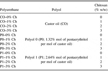
In PU synthesis, 3 polyols derived from CO (CO, P0, and P1) were used. CO is unmodified castor oil, and P0 and P1 were obtained by the chemical modification of CO by transesterification with pentaerythritol (1.32% and 2.64 mol% of pentaerythritol per mole of CO, respectively). Reference Valero and Gonzalez22 The transesterification reaction was performed in a 3-neck reactor with stirring (300 rpm) and temperature control. In the first stage, CO was dehydrated at 120 °C for 10 min. Then, pentaerythritol was added, and 0.05% lead oxide was used as catalysts at 210 °C for 2 h. Reference Valero and Ortegón23 The polyol was allowed to decant for 24 h and a filtration process was performed to remove catalysts. The hydroxyl number of polyols was determined using the standard ASTM D1957-86 method.
2. Synthesis of PUs of polyols derived from CO
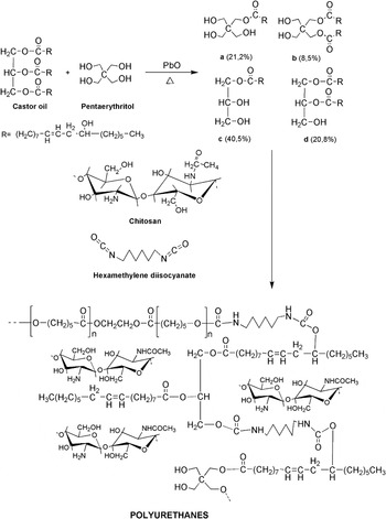
PUs were synthesized using the prepolymer method with HDI maintaining a constant NCO/OH ratio (1:1) (see Table I). Reference Valero and Díaz24 The synthesis was performed in 250-mL glass reactors. HDI was mixed with polyol (CO, P0, or P1) at 60 °C and 300 rpm for 10 min. Then, Ch (0, 1, 2, and 3% w/w with respect to the mass of the oil polyol) was added as a filler. The prepolymer was subjected to vacuum and poured into a steel mold (15.0 × 9.0 × 0.3 cm). The curing process took place at 110 °C for 12 h in a drying and heating chamber (Tuttlingen, Germany). The reaction scheme for the synthesis of PUs is provided in Fig. 1.
Polyurethane compositions.

Reaction scheme for the synthesis of PUs with HDI and Ch.

C. Characterization of PUs
1. Mechanical properties
The tensile strength and elongation at break of the PUs were determined following the ASTM D638-10 method. A universal traction machine EZ-LX (SHIMADZU, Tokyo, Japan) was used with a 5 kN load cell and a jaw displacement speed of 25 mm/min. Three samples of each formulation with dimensions of 40 × 6 × 3 mm (length × width × thickness) were tested. The dynamic hardness test was performed using a shore A durometer (SHR-MARK-III) according to the ASTM D676-59 method. Ten determinations were evaluated for each material. Reference Valero and Díaz24
2. Adhesive properties
The adhesive properties were assessed using the scratch resistance according to BS 3900. The impact strength and tensile lap/shear were determined according to ASTM D2794-93 (2010) and ASTM D906-98 (2011), respectively. The adhesion tests were performed on a wood substrate in accordance with ASTM D2651-01 (2016) to determine the type of resulting failure. Reference Arevalo, Uscategui, Diaz, Cobo and Valero25
3. Thermogravimetric analysis (TGA)
The thermal behavior of the PUs was evaluated using a thermogravimetric analyzer, model TGA/DCS1 (Mettler Toledo, Columbus, Ohio). The heating rate was 25 °C/min from 25 to 600 °C under a nitrogen atmosphere according to ASTM D6370. The weight of the samples was 15 ± 2 mg. Reference Cakić, Ristić, Cincović, Nikolić, Nikolić and Cvetinov26
4. Differential scanning calorimetry (DSC) analysis
The transition temperature of the samples was analyzed using a DSC analyzer PYRIS 1 DSC (Perkin-Elmer, Waltham, Massachusetts). The temperature range was −70 to 150 °C under nitrogen at a flow rate of 20 mL/min. Reference Conejero-García, Gimeno, Sáez, Vilariño-Feltrer, Ortuño-Lizarán and Vallés-Lluch27
5. Water absorption and contact angle
The water absorption percentage and contact angle of the polymeric materials evaluate the hydrophilic behaviors of the PUs. The water absorption percentage was determined by immersing PU in PBS at 37 °C until reaching a constant weight. Reference Zia, Zuber, Saif, Jawaid, Mahmood, Shahid, Anjum and Ahmad28 All assays were performed in triplicate. Equation (1) calculated the percentage of absorption, where m 0 is the initial mass of the PU and m w corresponds to the mass of the sample after obtaining a constant weight. The contact angle was determined using the sessile drop method in a Drop Shape Analysis (DSA) system (GH11, Krüss, Germany) with distilled water at 20 °C according to ASTM-D7334-08 (2013). The average contact angle corresponds to ten measurements for each polymeric material, Reference Skrobot, Zair, Ostrowski and Fray29
 $$\% \;{\rm{water}}\;{\rm{absorbed}} = {{\left( {{m_0} - {m_{\rm{w}}}} \right)} \over {{m_{\rm{w}}}}} \times 100\quad .$$
6. Scanning electron microscopy (SEM)
The morphology of the polymeric materials was determined by SEM using a JEOL JSM6300 (JEOL Ltd., Akishima Japan) microscope operated at a voltage acceleration of 10 kV, a magnification of 500×, and a working distance of 15 mm. All samples were gold-plated prior to observation. Reference Riaz, Ahmad, Saleemi, Adrees, Jamshed, Moqeet and Jamil30
D. In vitro cytotoxicity of PUs
1. Cell culture
L-929 mouse embryonic fibroblasts were cultured in RPMI 1640 medium supplemented with FBS (10%) and penicillin-streptomycin (1%) under 5% CO2 at 37 °C until 100% confluence. Reference Pignatello, Impallomeni, Pistarà, Cupri, Graziano, Cardile and Ballistreri31 Trypsin EDTA was added to the cells for 3 min at 37 °C prior to centrifugation at 2000 rpm for 10 min and culturing in a fresh RPMI 1640 medium. Reference Arnal-Pastor, Comin-Cebrian, Martinez-Ramos, Monleon Pradas and Valles-Lluch32
2. In vitro cytotoxicity of PUs
The cellular viability of the PUs was evaluated using the MTT method defined by ISO/CD 10993-5. Reference Bakhshi, Yeganeh, Mehdipour-Ataei, Shokrgozar, Yari and Saeedi-Eslami33 Cylindrical samples with a 3 mm diameter and 2 mm thickness were sterilized with Ultraviolet (UV) light (260 nm) for 30 min on each side. Reference Hou, Zhang, Qu, Xu and Han34 Samples of PUs were plated on pre-cultured cells (4.0 × 10⁴ cells per well) in 96-well plates with the RPMI 1640 medium at 37 °C with 5% CO2 for 24 h. The supernatant was removed, and 100 μL of MTT solution (12 mM in PBS) was added for 4 h at 37 °C. The supernatant was discarded, and 100 μL of dimethylsulfoxide (DMSO) was added for 15 min at 37 °C. Optical density measurement was conducted on an iMark™ Microplate Reader (Bio-Rad, Hercules, California) at a wave length of 595 nm. All assays were independently performed in triplicate. Cells seeded without polymers and with DMSO were used as control. A commercial polymer (polypropylene) was used as a reference. The cell viability percentage was calculated using Eq. (2) Reference Gentile, Bellucci, Sola, Mattu, Cannillo and Ciardelli35 :
 $${\rm{Cell}}\;{\rm{viability}}\;\left( \% \right) = {{\left( {{\rm{Ab}}{{\rm{s}}_{{\rm{sample}}}}} \right)} \over {{\rm{Ab}}{{\rm{s}}_{{\rm{control}}}}}} \times 100\quad ,$$
where Abssample corresponds to the absorbance of cells after contact with PU and Abscontrol is the absorbance of cells without polymeric material.
E. Cell adhesion testing of PUs
Cell adhesion assay of the PUs was evaluated using mouse embryonic fibroblast cell lines of L-929 in the RPMI 1640 medium as described in the previous item. Cell adhesion was performed using a methodology described by Pitchaimani et al. Reference Pitchaimani, Duong, Nguyen and Koirala36 with modifications. Cells were seeded at a density of 4.0 × 10⁴ cells per well in 96-well plates, with the RPMI 1640 medium on polymers fixed to the bottom of the plate. Samples were incubated at 37 °C with 5% CO2 for different times (6, 24, and 48 h). After the incubation time, non-adhered cells were removed. Cells were washed three times with PBS. 100 μL of MTT solution (12 mM in PBS) was added for 4 h at 37 °C. The supernatant was discarded, and 100 μL of DMSO was added for 15 min at 37 °C. The resulting solution was transferred into the new 96-well plates. Optical density measurement was conducted on an iMark™ Microplate Reader at a wave length of 595 nm. All assays were independently performed in triplicate. Cells seeded without polymers were used as control. Cell adhesion was determined with the cell viability percentage [Eq. (2)].
F. Antibacterial testing of PUs
Antibacterial activity assays of the polymeric materials were performed according to the methodology described by Kara et al. using E. coli (ATCC 2469) bacteria. Reference Kara, Aksoy, Yuksekdag, Hasirci and Aksoy37 The bacterial suspension was grown in TSB at 37 °C for 16 h in an incubator (Memmert GmbH + Co.KG, Schwabach, Germany). Aliquots of the bacterial suspension were inoculated in fresh TSB at 37 °C for 6 h. The bacteria cells were separated by centrifugation of the bacterial suspension at 3000 rpm for 10 min at 4 °C in a Sorvall™ Centrifuge (Thermo Scientific, Waltham, Massachusetts). The cells were washed twice with PBS after removing the supernatant. The bacteria were dissolved in PBS at 1.17 × 10⁷ colony forming units (CFU) mL−1 of E. coli.
Sterile PUs were placed in microcentrifuge tubes with 100 μL of bacterial suspension and incubated at 37 °C for 24 h. Then 900 μL of sterile PBS was added. The CFU were determined using serial dilutions seeded in TSA at 37 °C for 18 h in a Memmert incubator. A bacterial suspension without polymers was used as a control.
G. Statistical analysis
The results were analyzed using the analysis of variance (ANOVA) and significant differences were determined to be P < 0.05. The comparison of means was performed using Student’s t-test with SPSS Statistics 23 software (IBM, Armonk, New York). The experiments were conducted using three independent replicates. The results are reported as the mean ± standard deviation (SD).
III. RESULTS AND DISCUSSION
A. Characterization of PUs
1. Mechanical and adhesive properties
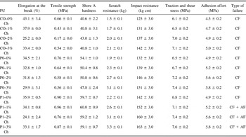
Polymeric matrices synthesized with polyols derived from CO (Ricinus communis) and HDI with Ch addition were characterized to determine the mechanical and adhesive properties. The results are presented in Table II.
Mechanical and adhesive properties of the PUs.

Notes: The results of mechanical and adhesive properties are given as the mean ± SD (n = 3). The shore A hardness result is given as the mean ± SD (n = 10).
CF: cohesive failure of the adhesive; AF: adhesive failure of the adhesive.
The hydroxyl number of each polyol was obtained after chemical modification (160, 191, and 236 mg KOH g−1 sample for CO, P0, and P1, respectively). The analysis of the elongation at break presented statistical differences (P < 0.05) between the polyol CO and the other polyols (P0 and P1). The percentages of elongation at break decrease with the chemical modification of the polyols. In polymers from the polyol CO, it was observed that increasing in the concentration of Ch tends to decrease the percentage of elongation. There were no differences related to the effect of Ch addition into matrices of P0 polyol. In P1 polyol-based PUs, the concentration of 2% Ch presented a statistically significant difference with a smaller percentage of elongation.
The tensile strength properties that were studied using the statistical analysis showed that there were significant differences (P < 0.05) between the type of polyols used in synthesis. The chemical modification performed on the polyols increases the maximum stress due to an increase in the physical cross-links of the polymers. The effects of Ch addition on the tensile force were determined. Statistically, they do not present differences so it can be inferred that Ch addition does not influence the maximum tension and thus, the factor that influences the tensile force depends on the type of polyol used in synthesis.
The tensile strength results from a previous study with polymer matrices synthesized using the same polyols (CO, P0, and P1) with isophorone diisocyanate Reference Uscátegui, Arévalo, Díaz, Cobo and Valero20 show lower values when HDI is used. This may be due to the chemical structure of HDI because it is a linear aliphatic diisocyanate, whereas isophorone diisocyanate has a cyclic structure that allows higher tensile strength than materials from linear diisocyanates.
Anirudhan et al. Reference Anirudhan, Nair and Nair16 designed a transdermal bioadhesive based on chitosan and hyaluronic acid for controlled release of lidocaine. The authors determined that the tensile force was approximately 0.5 MPa.
As part of the mechanical property evaluation, the dynamic hardness test of materials was conducted and the results are presented in Table II. According to the statistical analysis, there were significant differences (P < 0.05) between the polyols used. It can be seen that increasing the functionality of the polyol increases the material response, so PUs derived from P1 presented higher hardness than P0 and CO. The polyols derived from transesterification with pentaerythritol produce materials with a higher cross-link density which is related to better mechanical performance. These values are similar to those described by Valero et al., Reference Valero, Pulido, Ramírez and Cheng38 who studied the mechanical performance of PUs with polyols derived from CO obtained by transesterification with pentaerythritol and diphenyl methane diisocyanate (MDI) at NCO/OH = 0.7. The difference between the ratios and the hardness response is caused by the lower reactivity of HDI with respect to MDI; MDI is an aromatic diisocyanate used in industry that is characterized by high reactivity. Reference Szycher12 The decrease in the hard segments of the PUs obtained by the reaction of the –OH groups of the polyol and NCO of the HDI causes a reduction in the mechanical performance.
For the adhesion properties, the scratch resistance of the PUs was determined and the results are shown in Table II. The statistical analysis showed significant differences for each polyol used. An increase in resistance was observed as the physical PU cross-linkage increased. This was achieved by the chemical modification of the polyols used in synthesis. These results agree with those reported by Cakić et al., Reference Cakić, Ristić, Cincović, Stojiljković, János, Cvetinov and Stamenković39 who synthesized PU-based adhesives from polyols derived from CO with residues of polyethylene terephthalate. The authors attributed the increase in hardness to the cross-link density achieved by modifying the polyols by glycosylation of CO, which generated an increase in the stiffness of the main chain. Reference Cakić, Ristić, Cincović, Stojiljković, János, Cvetinov and Stamenković39
Kathalewar et al. Reference Kathalewar, Sabnis and D’Mello40 synthesized isocyanate-free PUs to obtain eco-friendly materials for applications in coatings. The authors evaluated the scratch resistance, and the results ranged from 0.9 to 2.8 kg. The authors affirm that the results are due to the crosslinked structure of PUs. Similar results were found in this research, where the maximum was 3.3 kg for polyol P1 which has a higher cross-link density. Reference Kathalewar, Sabnis and D’Mello40
The impact strength, tensile lap/shear, and adhesion effort exhibit the same behavior. The results show that increasing the functionality of polyol increases the material response, as PUs derived from P1 presented higher hardness results than P0 and CO. It can also be inferred that increasing the amount of Ch increased the adhesive properties (Table II). Ch can be used as a biomedical adhesive such as in drug delivery or tissue engineering due to its high cohesive strength. These adhesive properties depend on the –OH, –NH3 +, and –NHCOCH3 groups of the chemical structure. The groups can generate non-covalent, inter-chain, and intra-chain interactions that improved adhesion. Reference Patel3
Another important property of PU-type bioadhesives is the type of failure. Evaluation of the type of failure showed a cohesive failure of the PUs synthetized with CO and P0. These polymers present a failure in the adhesive material itself. Conversely, the type of failure exhibited by PUs derived from P1 is a combination of cohesive and adhesive failures which means that those polymers exhibit an intermolecular failure between the adhesive and the substrate with cross-linked structures. Reference Aung, Yaakob, Kamarudin and Abdullah41
2. Thermogravimetric analysis
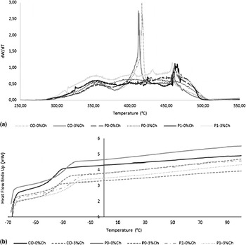
A TGA was performed using thermograms of PUs. The results for six matrices corresponding to the polymer matrices without Ch and those containing the highest percentage of Ch according to the type of polyol used are presented in Fig. 2(a).
(a) Derivative of the weight versus temperature of the PU thermograms. (b) DSC curves of PUs.

For PUs synthesized with polyols with 0 and 3% of Ch, the thermal behavior maintains the trend, indicating that there are no displacements of the degradation temperatures when modified polyols and Ch are used. In the derived curves from the TGA, several peaks were observed, which agrees with those of other authors who affirm that the mechanism of PU degradation is complex due to the formation of diverse compounds in the process. Reference Cakić, Ristić, Cincović, Nikolić, Nikolić and Cvetinov26
A thermogram was obtained for each polymer, allowing for determination of the stability of PUs synthesized in this study (complete results are not shown). The thermograms show that all PUs are stable at temperatures below 300 °C and exhibit complete degradation at temperatures close to 600 °C. These degradation temperatures coincide with those reported for PU synthesized with CO and isophorone diisocyanate that was proposed as a surgical adhesive. Reference Valero and Gonzalez22 In addition, the degradation temperatures do not affect the biomedical application because the physiological temperature is far lower (≈37 °C). Reference Ferreira, Pereira, Coelho, Silva and Gil42
Three degradation stages of PUs were determined using TGA thermograms. The first stage was between approximately 350–370 °C. This stage corresponds to the thermal degradation of the urethane bonds that are formed in the hard segments and are characterized by being thermally unstable. Reference Bakhshi, Yeganeh, Yari and Nezhad43 The second stage was approximately 390–430 °C, corresponding to the degradation of the soft segments. This is in agreement with Aung et al., Reference Aung, Yaakob, Kamarudin and Abdullah41 who synthesized Jatropha oil-based-PU adhesives and determined that the hard segments decompose before the soft segments (close to 400 °C). The last stage between 430 and 500 °C corresponds to the thermal degradation of the remaining fatty acid double bonds of CO. Reference Corcuera, Rueda, Fernandez d’Arlas, Arbelaiz, Marieta, Mondragon and Eceiza44
This is in agreement with a study of polymers synthesized with CO and isophorone diisocyanate with polycaprolactone and Ch at different concentrations in which T max 1 was between 250 and 370 °C, T max 2 was between 375 and 430 °C, and T max 3 was between 425 and 430 °C, Reference Arevalo, Uscategui, Diaz, Cobo and Valero25 indicating that the thermal stability of modified CO-polyol-based PUs was not affected by the type of diisocyanate and additives such as Ch used in synthesis.
In relation to the thermal behavior of chitosan, an isothermal analysis (110 °C for 12 h) and a dynamic analysis (25–600 °C) were performed (data not shown). With the isothermal analysis, no significant variation of the chitosan was observed indicating that the structure of chitosan is not affected during the curing process of the polymer. As for the dynamic analysis, it was observed that chitosan presents about 10% of weight loss that corresponds to the evaporation of physically adsorbed and strongly hydrogen bonded water to chitosan. Reference Moussout, Ahlafi, Aazza and Bourakhouadar45 The degradation of chitosan occurs between 245 and 350 °C which corresponds to the decomposition of polymer chains. Reference Moussout, Ahlafi, Aazza and Bourakhouadar45,Reference Gámiz-González, Correia, Lanceros-Mendez, Sencadas, Gómez Ribelles and Vidaurre46 Finally, a stage around 470 and 530 °C corresponds to residual decomposition reactions. Reference Gámiz-González, Correia, Lanceros-Mendez, Sencadas, Gómez Ribelles and Vidaurre46
3. Differential scanning calorimetry
Figure 2(b) shows the DSC curves obtained for the polymers synthesized with each polyol and with the minimum and maximum amounts of Ch addition.
By evaluating the DSC curves, the glass transition temperature of the PUs synthesized in this project was determined (complete data not shown). In the analysis in Fig. 2(b) it can be seen that polyols CO, P0, and P1 without Ch have glass transition temperature of −33.85 °C, −27.85 °C, and −21.87 °C, respectively. These results tend to be lower when the maximum concentration of Ch (3% of Ch) is used, that is, −37.86 °C, −31.87 °C, and −23.86 °C for polyols CO, P0, and P1, respectively. The glass transition temperature results are similar to those reported for PUs based on polyethylene glycol, poly (ε-caprolactone-co-D,L-lactide) and diurethane diisocyanate (based on hexamethylene diisocyanate and butanediol), for which the authors found values close to −33 °C. The authors did not find exothermic peaks because the materials were amorphous. Reference Hou, Zhang, Qu, Xu and Han34
4. Water absorption and contact angle
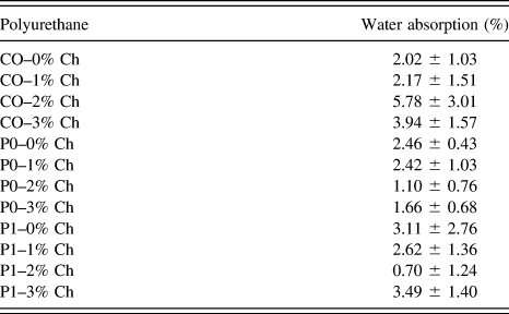
The hydrophilic character of PUs was determined by calculating the absorption percentage, and the results are presented in Table III.
Water absorption of PUs after 15 days.

Note: Water absorption is given as the mean ± SD (n = 3).
The swelling study was performed for 15 days. The water absorption was evaluated twice, at 5 and 15 days. The analysis of the results did not present statistical differences between the evaluation times. Table III shows the results obtained for the absorption of water at 15 days. PUs from polyol P1 presented lower water absorption than those obtained from CO and P0 when the concentration of Ch was 2–3%. The increase in hydrogen bonds due to the increase in the functionality of the polyol by the transesterification with pentaerythritol Reference Valero, Pulido, Ramírez and Cheng47 forms a barrier to the diffusion of fluid into the material. Reference Valero and Díaz24
An analysis of the concentration of Ch showed that PUs from polyol CO with Ch had greater swelling than those that do not contain it. Increasing the amount of Ch allows for more functional groups available to interact with the medium, which is also polar. However, when increasing the polyol functionality, the generated effect is inverse, as observed in PUs from polyol P1 that presented a significant reduction of swelling with respect to PUs without Ch. Internal interactions (hydrogen bonds) increased the barrier effect by preventing diffusion of the fluid. In addition, the filler function of Ch causes the chains to be reorganized, presenting a reduction of the volumetric defects or vacancies where water can be deposited. Reference Temenoff and Mikos48
Marques et al. Reference Marques, Santos, Ferreira, Correia, Correia, Gil and Baptista6 evaluated a bioadhesive synthesized from lactic acid in which water absorption was 10%. The authors noted that moderate percentages of water absorption improve the hemostatic character of the materials. Reference Marques, Santos, Ferreira, Correia, Correia, Gil and Baptista6 This is why PUs synthesized in this study can be suggested as bioadhesives.
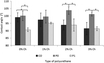
To evaluate the hydrophilic character of the polymer matrices the contact angle was determined and the results are presented in Fig. 3.
Contact angle of PUs. Notes: The contact angle is given as the mean ± SD (n = 10). * indicates statistical significance between polyol group type.

According to the statistical analysis in Fig. 3, there are significant differences between PUs synthesized with P1 and P0 and CO, with a reduction of the angle values. In evaluating the effect of Ch addition on contact angle values, it was observed that they did not present significant differences and therefore did not influence the hydrophilic character.
5. Scanning electron microscopy
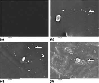
The morphology of PUs as a function of the type of polyol and Ch addition used in synthesis is presented in Fig. 4.
SEM micrographs of PUs at 500×. (a) P1–0% Ch, (b) P1–1% Ch, (c) P1–2% Ch, (d) P1–3% Ch.

In the micrographs presented in Fig. 4, deposition of Ch granules on the surface of the material was observed. Additionally, roughness can be observed that is related to cell adhesion. Reference Temenoff and Mikos48 It can also be inferred that by increasing of the amount of Ch, an incompatibility between the matrix and Ch can occur that results in phase separation. Ch on the surface can improve cell viability for biocompatible properties. Reference Depan, Surya, Girase and Misra49
B. In vitro cytotoxicity of PUs
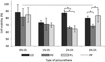
Cellular viability was evaluated using the mouse fibroblast cell line L-929 on PUs and the results are presented in Fig. 5.
Percentage of cellular viability of L-929 mouse fibroblasts at 24 h. Notes: The data are presented as the mean ± SD (n = 3). * indicates statistical significance between polyol group type.

Synthesized polymers generally have cell viability greater than 60%, which is desirable when evaluating materials are candidates for biomedical applications. The statistical analysis of the polyol types used showed significant differences (P < 0.05) between P0, CO, and P1 when the concentration of Ch was greater than 1%. As a positive control, a polypropylene biomaterial was used (98% cell viability under the assay conditions). As a negative control, doxorubicin at 50 ppm was used (37% cell viability under the assay conditions).
With the evaluation about the effect of Ch addition on cell viability, it can be inferred that Ch addition did not apparently affect the evaluated cells.
Guo et al. Reference Guo, Kim, Shan, Kim, Hu, Wang, Hamad, Qian, Rizk and Yang4 used a strategy to increase the cohesive strength of the adhesives to design crosslinked polymers with mechanical and biocompatible resistances. The cellular viability of cross-linked bioadhesives was similar to a commercial biomaterial [poly(lactic-co-glycolic acid)]. The results suggest that crosslinked bioadhesives can be used as bioadhesives. Reference Guo, Kim, Shan, Kim, Hu, Wang, Hamad, Qian, Rizk and Yang4
C. Cell adhesion testing of PUs
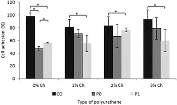
Mouse fibroblast cell line L-929 on PUs was using to evaluated cellular adhesion test and the quantitative analysis is presented in Fig. 6.
Cellular adhesion of L-929 mouse fibroblasts at 48 h on PUs. Notes: The data are presented as the mean ± SD (n = 3). * indicates statistical significance between polyol group type.

The statistical analysis of the polyol types used showed significant differences (P < 0.05) between CO, P0, and P1. The effect of Ch addition on cell adhesion was determined for each polyol, and significant differences in the results were observed for P0 and P1, indicating that Ch addition did not apparently affect the evaluated cell adhesion. As a positive control, a polystyrene standard tissue culture was used (104% cell viability under the assay conditions at 48 h). As a negative control, doxorubicin at 50 ppm was used (17% cell viability under the assay conditions at 48 h).
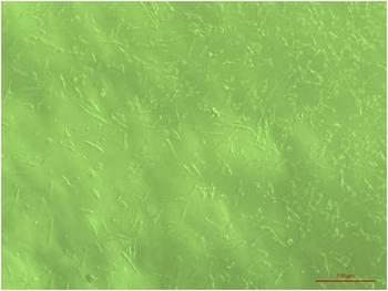
Similar behavior was observed for adhesion assessments at 6 and 24 h (data not shown). All PUs showed cell adhesion since 6 h. The minimum and maximum values at 24 h were between 28% for P0–0% Ch and 78% for CO–2% Ch, respectively. These results show that the effect of Ch addition tends to improve the adhesion of the L929 cells on the material. With the adhesion results, it can be inferred that PUs synthesized with polyols derived from the CO and chitosan can be used as bioadhesives. Figure 7 shows the cell morphology of L929 cells adhered to PU P0–1% Ch at 48 h.
Cell morphologies of L-929 mouse fibroblasts (10×) on P0–1% Ch at 48 h.

Sordel et al. Reference Sordel, Kermarec-Marcel, Garnier-Raveaud, Glade, Sauter-Starace, Pudda, Borella, Plissonnier, Chatelain, Bruckert and Picollet-D’hahan50 claim that cell adhesion on the surface of a material is an important factor to take into account when designing biomaterials. The adhesion of cells is important to fulfill a number of functions such as cell proliferation. Reference Sordel, Kermarec-Marcel, Garnier-Raveaud, Glade, Sauter-Starace, Pudda, Borella, Plissonnier, Chatelain, Bruckert and Picollet-D’hahan50,Reference Pehlivanova, Krasteva, Seifert, Lützow, Tsoneva, Becker, Richau, Lendlein and Tzoneva51 Zhu et al. Reference Zhu, Dong, Wejinya, Jin and Ye52 used human foreskin fibroblast cell line into collagen–chitosan hydrogel scaffolds and the authors suggest that chitosan addition (20–30%) with collagen can significantly improve cell attachment and proliferation. The authors affirm that chitosan is usually used to endow scaffolds with sufficient mechanical strengths required for cell growth. Reference Zhu, Dong, Wejinya, Jin and Ye52
D. Antibacterial activity of PUs
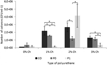
The antimicrobial activity of the polymeric materials synthesized was evaluated against gram-negative bacteria E. coli (ATCC® 2469), which is usually found in humans, Reference Marques, Santos, Ferreira, Correia, Correia, Gil and Baptista6 and the results are presented in Fig. 8.
Antibacterial activity of PUs against E. coli. Notes: The colony forming units per milliliter (CFU mL−1) are presented as the mean ± SD (n = 3). * indicates statistical significance between polyol group type.

The results in Fig. 8 show that PUs evaluated showed bacterial inhibition against E. coli, with percentages of inhibition between 60 and 90% compared to the control (1.17 × 10⁷ CFU mL−1). When performing the analysis for the polyols, P1 presents significant differences with respect to CO and P0. The statistical analysis of the results of PUs synthesized with CO did not present significant differences for the concentration of Ch, so an optimal concentration of Ch cannot be inferred. For P0 and P1, there were statistical differences related to the concentration of Ch. It was expected that increasing the Ch concentration would present bacterial inhibition, but Ch is apparently trapped in the polymer matrix, which prevents the release of Ch in 24 h.
Additional to this, Liu et al. Reference Liu, Chen, Park, Liu, Liu, Meng and Yu53 suggest that some factors affect the antibacterial activity of chitosan like molecular weight and concentration. Higher concentrations of chitosan are more effective used (higher than 10,000 Da, for example) for microorganism inhibition. Reference Liu, Chen, Park, Liu, Liu, Meng and Yu53 In this research, chitosan used was low-molecular-weight and low concentration (3%). Is possible that the results showed no significant differences for the molecular weight and concentration of chitosan used.
The polymers synthesized in this project generally have antibacterial activity against gram-negative bacteria, and this property is ideal for polymers used as bioadhesives.
IV. CONCLUSIONS
PUs were synthesized with polyols derived from CO, HDI, and Ch. The polyols were chemically modified with pentaerythritol to increase the hydroxyl groups that generate crosslinking. An evaluation of the mechanical, adhesive, thermal, and morphological properties, as well as the in vitro cytotoxicity, cellular adhesion, and in vitro antimicrobial activity, was performed. Chemical modification of the polyols increases the mechanical and adhesive properties due to an increase in the physical cross-links of the polymers. PUs were not toxic and revealed cellular adhesion for all polyols evaluated. PUs evaluated showed bacterial inhibition against E. coli, presenting inhibition between 60 and 90% compared to the control. In general, the mechanical, adhesive, and biological properties suggest that PUs synthetized in this research may be candidates for external biomedical tissue adhesives.
ACKNOWLEDGMENTS
This work was supported by the Universidad de La Sabana under grant number ING-176-2016 and by Colciencias under scholarship grant 617-2-2014. CIBER-BBN is an initiative funded by the VI National R&D&I Plan 2008–2011, Iniciativa Ingenio 2010, Consolider Program. CIBER Actions are financed by the Instituto de Salud Carlos III with assistance from the European Regional Development Fund. J.A.G.T. and A.V.L. acknowledge the support of the Spanish Ministry of Economy and Competitiveness (MINECO) through project DPI2015-65401-C3-2-R (including FEDER financial support). Finally, the authors thank the Universitat Politècnica de València for assistance and advice with the equipment.