In cases with severe or even subatretic valvar pulmonary stenosis and substantial right ventricular hypertrophy, diastolic dysfunction develops and right ventricle filling is impaired. Reference Medina, de Lezo, Delgado, Caballero, Segura and Romero1 Left alone, severe right ventricle outflow tract obstruction with severe cyanosis may cause biventricular dysfunction. Reference Kane, Kogon and Pernetz2 We present a case of a teenager with combined pulmonary stenosis and an atrial septal defect with severe cyanosis and secondary biventricular heart failure.
Case report
A 13-year-old boy came to the emergency room with severe breathlessness that worsened one day before admission. The blood pressure was 123/81 mmHg, and heart rate was 100 bpm with severe cyanotic (oxygen saturation 40–50%). Echocardiography examination revealed severe valvar pulmonary stenosis with minimal forward flow to the pulmonary artery and a pressure gradient of 110 mmHg, moderate tricuspid regurgitation, right to left shunt secundum atrial septal defect, and biventricular dysfunction (ejection fraction of 20% and TAPSE 1.0 cm). The patient was originally discussed to undergo surgical valvotomy, but was rejected due to the poor general condition and the estimated high perioperative risk. Therefore, pulmonary balloon valvuloplasty was deemed the only favourable approach.
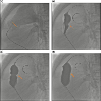
Right ventricular and pulmonary artery pressures were measured, then right ventriculography was performed. The pulmonary valve was found to be subatretic (Fig 1a) and could only be crossed with straight 0.035”RADIOFOCUS R guidewire (Terumo, Tokyo, Japan). As reperfusion oedema was deemed possible, a staged approach was chosen, and only incomplete balloon dilatation was performed, using a Tyshak R balloon catheter 12 × 30 cm (NuMed, Cornwall, Canada) (Fig 1b, c, d). The pressure gradient only decreased marginally, and peak-to-peak transvalvular pressure gradient decreased from 88 mmHg to 67 mmHg. The oxygen saturation, however, improved immediately, and therefore, the initial target of the procedure was achieved despite the significant resting gradient.
Pulmonary Balloon Valvuloplasty Procedure: A. Right ventriculography in lateral projection showed sub-atretic pulmonary valve with pulmonary valve annulus size of 25 mm. B, C and D showed balloon (TyshakR balloon catheter 12 × 30 cm, NuMed,Cornwall,Canada) dilated progressively to maximum until balloon silhouette disappear, as indicated by the arrow.

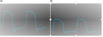
Following the initial valve dilation, normal saturation, and oxygen delivery to the body, marked improvement in peripheral oedema, regression of hepatomegaly, and onset of massive diuresis were observed. His haemodynamic was improving, and he was discharged on the seventh day. After 6 months, we performed the scheduled re-dilation of the pulmonary valve. Peak-to-peak transvalvular pressure gradient decreased, and the right ventricular end diastolic pressure decreased to 12 mmHg compared to 23 mmHg at the initial intervention (Fig 2). Evaluation in the following year showed left to right atrial septal defect; therefore, it was closed percutaneously.
End-diastolic pressures of the RV at initial intervention and after the second procedure. RVEDp decreased from 23 mmHg (a) to 10 mmHg (b).

Discussion
In severe cases of pulmonary valve stenosis, severe right ventricular hypertrophy will cause diastolic dysfunction. Reference Mitchell and Mhlongo3 This patient’s right ventricle was exposed to high pressures from the obstructed outflow tract and volume overload due to the atrial septal defect. The coronary blood flow may be impaired, causing ischaemia and cardiomyocyte apoptosis and ultimately leads to dilatation and stiffening of right ventricle, further impairing the stroke volume. Finally, the profound cyanosis will aggravate the deterioration of biventricular myocardial impairment. Reference Iyer, Moreno, Fernando, Faiz, Shekerdemian and Iyer4
This case presented a clinical challenge due to the presence of biventricular failure. A study by Pieri et al mentioned the higher risk of complications in patient with low left ventricle ejection fraction. Reference Pieri, Belletti and Monaco5 Thus, staged valvuloplasty is the preferred treatment of choice. Reference Tefera, Qureshi, Bermudez-Cañete and Rubio6 Following percutaneous balloon valvuloplasty, there was a high incidence of pulmonary oedema, and pulmonary haemorrhage in severe cases. Relieving the right ventricular obstruction at once may result in a high preload to the left heart for which the left ventricle may not be prepared, also the reperfusion damage may occur within the pulmonary vascular bed due to a great haemodynamic change. Many literatures said none of the patients who underwent graded dilation developed evidence of reperfusion injury/pulmonary oedema. Reference Tefera, Qureshi, Bermudez-Cañete and Rubio6 The recovery of left and right ventricles function in this patient proved that the dramatic increase in saturation was the key factor in the improvement of myocardial function and subsequent cardiac output, organ perfusion, and recovery. This could be achieved by submaximal dilatation of the pulmonary valve alone. In addition, the improvement of right ventricle function reversibly increased the performance of left ventricular function. Reference Medina, de Lezo, Delgado, Caballero, Segura and Romero1,Reference Tefera, Qureshi, Bermudez-Cañete and Rubio6,Reference Cho and Ma7
Supplementary material
To view supplementary material for this article, please visit https://doi.org/10.1017/S1047951121001967
Acknowledgements
We would like to thank Oktavia Lilyasari, MD, and Bayushi Eka Putra, MD for their valuable guidance and support.
Financial support
The author(s) received no financial support for the research, authorship, and/or publication of this article.
Conflicts of interest
None.
Ethical standards
No human and/or animal experimentation was conducted in this study.