Introduction
The poultry oviduct is a highly complex and dynamic organ where the ovulated yolk from the ovary is developed into egg. Unlike other mammalian species, hens have a single set of functional reproductive organs, the left ovary and the oviduct. When a chick hatches, the development of the right ovary and oviduct cease and gradually regress. However, during the formation of the egg, oviduct tissues undergo extensive proliferation of luminal and glandular epithelial cell, extracellular matrix remodelling, and neovascularisation (Jeong et al., Reference JEONG, LIM, KIM, AHN, LEE, JEONG, HAN, SONG and BAZER2012). Therefore, it is important to understand the oviductal proteins/genes involved in the egg formation.
The oviduct itself is a long tubular structure consisting of five functionally distinct parts namely the infundibulum (site of fertilisation), magnum (production of the components of egg white), isthmus (formation of the soft shell-membrane), shell gland or uterus (formation of calcified egg shell) and vagina (oviposition or egg laying). Following ovulation, the ovum passes through the entire length of the oviduct, where the constituents of the egg are secreted and deposited from respective parts of the oviduct.
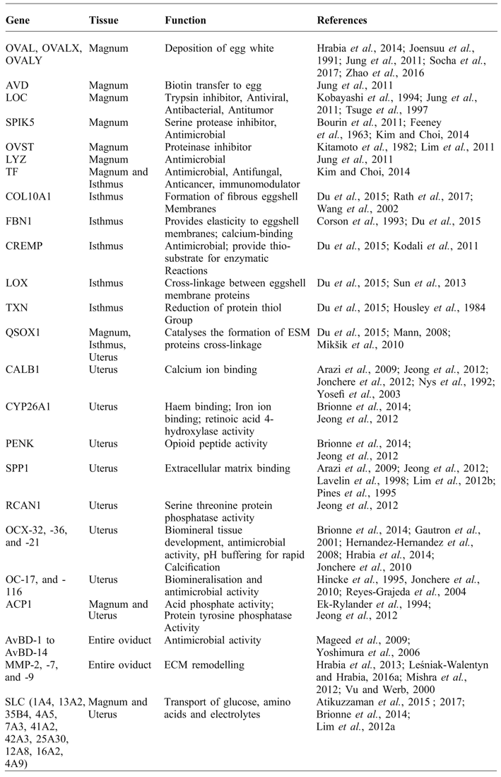
Two recent studies reported numerous genes associated with albumin deposition, eggshell membrane formation and shell calcification (Du et al., Reference DU, HINCKE, ROSE-MARTEL, HENNEQUET-ANTIER, BRIONNE, COGBURN, NYS and GAUTRON2015; Willems et al., Reference WILLEMS, DECUYPERE, BUYSE and EVERAERT2014). As shown in Table 1, gene expression and protein secretion with the oviduct varies in accordance with different functional roles in the regions of the oviduct. Together the oviduct completes egg formation by creating the middle albumin portion and the outer protective eggshell that surrounds the central yolk. This review highlights the current knowledge of the oviductal proteins/genes and their biological functions in the egg formation.
Summary of the expression, localisation and functions of the genes in the laying hen oviduct.

Functional role of the oviduct
role of infundibulum
The infundibulum is a muscular funnel-shaped structure marking the beginning of the oviduct. It is in close proximity to the growing follicles and surrounds them so that the ovulated follicle is trapped and directed to descending parts of the oviduct. The infundibulum has no secretory functions and the egg remains here for a period of 15 to 17 minutes. However, if the infundibulum malfunctions, the follicle may ovulate outside of oviduct into the internal peritoneal cavity. A study conducted on Brown-Leghorn pullets revealed that when this ‘internal laying’ was evident there was a loss of 12% of potential eggs (Wood-Gush and Gilbert, Reference WOOD-GUSH and GILBERT1970).
role of magnum
The magnum is the longest part of the oviduct and contributes the egg white protein to the developing egg during its 3 h of passage through this segment. Synthesis of albumin largely occurs continuously in the tubular gland cells of magnum; however, deposition rate is heightened when the egg is present in this region (Muramatsu et al., Reference MURAMATSU, HIRAMOTO and OKUMURA1991). Albumin provides an antimicrobial function prior to egg incubation, and its proteins are the major source of nutrition for the growing embryo during incubation (Willems et al., Reference WILLEMS, DECUYPERE, BUYSE and EVERAERT2014). Among the several egg white proteins, including ovalbumin, ovomucin and conalbumin, the latter constitutes the major share.
role of isthmus
The isthmus bridges the magnum and the shell gland in the chicken oviduct. When the inchoate egg passes through the isthmus, the eggshell membranes (ESM) are secreted and assemble around the egg white. Besides covering the inner soft structures, the ESM provides a structural foundation for eggshell calcification. The ESM is a clear film primarily composed of fibrous proteins such as collagen. The expression of COL10A1, FBN1, CREMP, LOX, TXN, QSOX1 in the isthmus are involved in the formation of ESM (Corson et al., Reference CORSON, CHALBERG, DIETZ, CHARBONNEAU and SAKAI1993; Kodali et al., Reference KODALI, GANNON, PARAMASIVAM, RAJE, POLENOVA and THORPE2011; Du et al., Reference DU, HINCKE, ROSE-MARTEL, HENNEQUET-ANTIER, BRIONNE, COGBURN, NYS and GAUTRON2015,) as listed in Table 1. However, the mechanism of formation of ESM in the isthmus is not completely understood.
role of shell gland
The uterus in chickens is commonly known as the shell gland, and the glandular secretions function to provide the outer calcified, protective layer for the chicken embryo. The egg remains in the uterus for the longest period during formation, for a duration of 20 h, during which eggshell calcification occurs in three stages; initiation, growth, and arrest. Calcification initiates from nucleation sites on the ESM. During the initiation stage, the rate of calcification is slow; however, it gets accentuated over 15 hours and ebbs down to a nadir in the last two hours before oviposition. Pigments (protoporphyrin and biliverdin) are synthesised in the uterus and deposited by ciliated cells during the last 3 h before egg laying (Zhao et al., Reference ZHAO, XU, LIU, LI and YANG2006). Apart from providing physical protection to the chicken embryo, the eggshell functions effectively and efficiently in offering protection against bacterial ingression, providing a source of calcium and sites for respiration of the growing embryo. The mineral component and the organic matrix proteins interact to give the eggshell its physical and chemical protective function. Several genes and proteins are responsible for the eggshell formation. Identification and functional annotation of the uterine genes have potential to serve as biological markers for genetic improvement of eggshell quality and egg self-sustainability (Jonchere et al., Reference JONCHERE, REHAULT-GODBERT, HENNEQUET-ANTIER, CABAU, SIBUT, COGBURN, NYS and GAUTRON2010).
role of vagina
The vagina is the caudal portion of the chicken oviduct and does not play a role in egg formation. However, this is where the outer cuticle forms and pigmentation occurs. The muscles of the vagina help to turn the egg longitudinally to the blunt end and push the egg out of the oviduct.
Functions of oviduct genes/proteins in the egg formation
ovalbumin
Ovalbumin (OVL) describes a group of glycoproteins making up approximately 55% of total egg proteins and is found in the egg white. Presence of the ovum in the magnum increases OVL synthesis and its expression is upregulated in chickens with high laying rate (Zhao et al., Reference ZHAO, ZHANG, JIAO, WANG, JIANG, LUO and LIN2016). The ovalbumin protein family comprises three sequential homologous proteins: ovalbumin (OVAL), ovalbumin-related protein X (OVALX) and ovalbumin-related protein Y (OVALY). Their expressions in the chicken oviduct are regulated through oestrogen, but the hormonal responsiveness is in the order of OV:Y:X=100:10:1 (Colbert et al., Reference COLBERT, KNOLL, WOO, MACE, TSAI and O'MALLEY1980). Progesterone induces OVL production in the magnum mucosa of laying hens (Joensuu et al., Reference JOENSUU, TUOHIMAA and VILJA1991), whereas OVAL expression decreases in moulting birds (Zhao et al., Reference ZHAO, ZHANG, JIAO, WANG, JIANG, LUO and LIN2016). The expression of OVL is also increased in the magnum of juvenile, adult and moulting hens treated with recombinant chicken growth hormone (Hrabia et al., Reference HRABIA, LEŚNIAK-WALENTYN, SECHMAN and GERTLER2014; Socha et al., Reference SOCHA, SECHMAN, MIKA and HRABIA2017). OVALX is effective against Listeria monocytogenes and Salmonella enterica subsp. enterica whereas OVAL is not. Since OVL is mainly important as a constituent of the albumin, the antimicrobial activity adds specificity to the function of OVALX (Réhault-Godbert et al., Reference RÉHAULT-GODBERT, LABAS, HELLOIN, HERVÉ-GRÉPINET, SLUGOCKI, BERGES, BOURIN, BRIONNE, POIRIER, GAUTRON, COSTE and NYS2013).
avidin
Avidin is secreted by the goblet cells of the magnum. Additionally, avidin is expressed in the intestine and skeletal muscles of the developing embryo. Expression in the magnum is very high, 549-fold greater in adult chickens than in juveniles (Jung et al., Reference JUNG, LIM, PARK, KIM, HAN, SONG and HAN2011). It has been reported that avidin in the oviduct scavenges unbound biotin and transfers it to albumin. And it is only when avidin becomes saturated by high dietary intakes that unbound biotin gets accumulated to yolk and plasma (White and Whitehead, Reference WHITE and WHITEHEAD1987). Avidin production is induced in the magnum at a dose-dependent rate of progesterone administration (Joensuu et al., Reference JOENSUU, TUOHIMAA and VILJA1991).
ovomucin
Ovomucin is a mucoprotein found in egg white and responsible for the gel-like property in fresh egg whites. Adult chickens show very high (4,931-fold) expression of ovomucoid in the magnum, more than juveniles (Jung et al., Reference JUNG, LIM, PARK, KIM, HAN, SONG and HAN2011). It represents the major allergen found in egg whites and is heat-stable, which accounts for around 3.5% of the total egg white protein. Because of its inhibitory effect on trypsin, it has been commercially available as a trypsin inhibitor, and can imparts anti-viral (Tsuge et al., Reference TSUGE, SHIMOYAMADA and WATANABE1997) and anti-bacterial (Kobayashi et al., Reference KOBAYASHI, HATTORI, HARA-KUDO, OKUBO, YAMAMOTO, TAKITA and SUGITA-KONISHI2004) activities.
ovoinhibitor
Ovoinhibitor (OIH) has been found to be highly expressed in the magnum and liver (Kim and Choi, Reference KIM and CHOI2014) and in the uterus to a lesser amount (Bourin et al., Reference BOURIN, GAUTRON, BERGES, ATTUCCI, LE BLAY, LABAS, NYS and REHAULT-GODBERT2011). A study showed that OIH expression increased in the liver of pre-laying hen and gradually decreased in matured birds (Kim and Choi, Reference KIM and CHOI2014). OIH has been shown to have serine protease inhibition activity (Matsushima, Reference MATSUSHIMA1958) and provides defence against bacterial ingression of Bacillus spp. (Bourin et al., Reference BOURIN, GAUTRON, BERGES, ATTUCCI, LE BLAY, LABAS, NYS and REHAULT-GODBERT2011). The inhibitory activity of OIH is more effective than ovomucoid (Feeney et al., Reference FEENEY, STEVENS and OSUGA1963).
ovostatin
One of the four proteinase inhibitors found in egg white is ovostatin (OVST) (Kitamoto et al.,1982). Because of its similar composition to plasma α2 macroglobulin, it has been earlier referred to as ovomacroglobulin. Unlike ovoinhibitor, its expression is limited to the glandular and luminal epithelium of the chicken oviduct and is induced by oestrogen (Lim et al., Reference LIM, JEONG, KIM, LEE, KIM, BAZER, HAN and SONG2011).
ovotransferrin
Ovotransferrin, previously known as conalbumin, is expressed predominantly in the magnum and the isthmus. This protein is found in the uterine fluid during the early mineralisation stage (Gautron et al., Reference GAUTRON, HINCKE, PANHELEUX, GARCIA-RUIZ, BOLDICKE and NYS2001). The expression of ovotransferrin mRNA in the magnum is down-regulated with dietary corticosterone, but there is increased ovotransferrin protein in the egg white. Therefore, it can be used as a marker to estimate the exposure of the laying birds to environmental stress condition (Kim and Choi, Reference KIM and CHOI2014). Ovotransferrin has been reported to have an antimicrobial function (Baron et al., Reference BARON, JAN, GONNET, PASCO, JARDIN, GIUDICI, GAUTIER, GUÉRIN-DUBIARD and NAU2014; Giansanti et al., Reference GIANSANTI, LEBOFFE and ANTONINI2012).
cystatin
Chicken cystatin is a cysteine proteinase inhibitor (Anastasi et al., Reference ANASTASI, BROWN, KEMBHAVI, NICKLIN, SAYERS, SUNTER and BARRETT1983) and makes up 0.05% of the total egg white protein. Cystatin has antimycotic activity (Kolaczkowska et al., Reference KOLACZKOWSKA, KOLACZKOWSKI, SOKOLOWSKA, MIECZNIKOWSKA, KUBIAK, ROLKA and POLANOWSKI2010) and is expressed in various tissues including lung, gizzard brain, heart, muscle, liver, and oviduct of chicken. Its expression in magnum is not directly affected by oestrogen (Colella et al., Reference COLELLA, JOHNSON and BIRD1991).
lysozyme
Lysozyme (LYZ) is an antimicrobial enzyme. The expression of LYZ in magnum is very much higher (37,557-fold) in adult chickens compared to juveniles (Jung et al., Reference JUNG, LIM, PARK, KIM, HAN, SONG and HAN2011).
collagen x
Collagenous fibrous proteins are the primary components of eggshell membranes. It gives the membrane its insoluble and stable nature. Collagen X (COL10A1) expression is localised in the tubular gland cells of the white isthmus (Wang et al., Reference WANG, FORD, PRAUL and LEACH2002). Microarray analysis has revealed that COL10A1 mRNA expression in the isthmus is 155-fold greater in the magnum and 111-fold greater in the uterus (Du et al., Reference DU, HINCKE, ROSE-MARTEL, HENNEQUET-ANTIER, BRIONNE, COGBURN, NYS and GAUTRON2015).
fibrillin-1
Fibrillin-1 (FBN1) is a microfibrillar glycoprotein rich in cysteine and is abundant in the extracellular matrix throughout the body. It is responsible for the elastic nature of chicken ESM and has 43 calcium-binding EGF-like repeats, which are the sites of nucleation for calcium deposition (Corson et al., Reference CORSON, CHALBERG, DIETZ, CHARBONNEAU and SAKAI1993). Expression of FBN 1 mRNA in white isthmus is 2.5-fold higher than in the magnum and 14-fold higher than in the uterus (Du et al., Reference DU, HINCKE, ROSE-MARTEL, HENNEQUET-ANTIER, BRIONNE, COGBURN, NYS and GAUTRON2015). However, a proteomics study based on protein extraction with methanol failed to detect FBN1 protein from ESM (Rath et al., Reference RATH, LIYANAGE, MAKKAR and LAY2017). This indicated that FBN1 protein was highly insoluble in nature (Du et al., Reference DU, HINCKE, ROSE-MARTEL, HENNEQUET-ANTIER, BRIONNE, COGBURN, NYS and GAUTRON2015).
cysteine-rich eggshell membrane protein
These eggshell membrane proteins (CREMPs) are very rich (13.8%) in cysteine, similar to fibrillin, and are major contributors of the cysteine content in chicken ESM. The isthmus shows the strongest expression of CREMP relative to other oviduct segments (Du et al., Reference DU, HINCKE, ROSE-MARTEL, HENNEQUET-ANTIER, BRIONNE, COGBURN, NYS and GAUTRON2015). CREMP proteins also provide protection against microbial contamination in the egg (Kodali et al., Reference KODALI, GANNON, PARAMASIVAM, RAJE, POLENOVA and THORPE2011). The CREMP proteins contain multiple disulphide crosslinks that are formed through the actions of any of several enzymes, including sulphydryl oxidase, thioredoxin, protein disulphide isomerase (Kodali et al., Reference KODALI, GANNON, PARAMASIVAM, RAJE, POLENOVA and THORPE2011; Du et al., Reference DU, HINCKE, ROSE-MARTEL, HENNEQUET-ANTIER, BRIONNE, COGBURN, NYS and GAUTRON2015).
lysyl oxidases
The chicken lysyl oxidase (LOX) gene family consists of five gene members, namely lysyl oxidase (LOX) and lysyl oxidase homologue-1, -2, -3, and -4, (LOXL1, LOXL2, LOXL3, LOXL4). The members of this gene family contribute to the formation of crosslinks between eggshell membrane proteins (Harris et al., Reference HARRIS, BLOUNT and LEACH1980). They are copper-dependent, and chickens fed a deficient diet have been shown to produce abnormal eggshell membranes (Harris et al., Reference HARRIS, BLOUNT and LEACH1980). Among this enzyme family members, LOXL2, LOXL3 have been identified in the eggshell membrane by proteomics analysis (Sun et al., Reference SUN, XU and YANG2013). LOXL2 and LOXL3 are up-regulated in the isthmus relative to magnum and uterus respectively (Du et al., Reference DU, HINCKE, ROSE-MARTEL, HENNEQUET-ANTIER, BRIONNE, COGBURN, NYS and GAUTRON2015), indicating that these homologues have a crucial role in forming crosslinks between collagen and ESM fibrillar proteins.
quiescin q6 sulfhydryl oxidase 1
Quiescin Q6 sulphydryl oxidase 1 (QSOX1) is an enzyme having flavin-linked sulphydryl oxidase activity and protein disulphide isomerase activity. It catalyses the formation of ESM proteins cross-linkages to impart the insolubility and stability of the ESM. Proteomic studies have confirmed the presence of this enzyme in ESM (Mann, Reference MANN2008) and eggshell matrix (Mikšik et al., 2010). Du et al. (Reference DU, HINCKE, ROSE-MARTEL, HENNEQUET-ANTIER, BRIONNE, COGBURN, NYS and GAUTRON2015) reported an equivalent level of expression of QSOX1 in magnum, isthmus and uterus of chickens.
thioredoxin
In chicken oviduct tissues, thioredoxin (TXN) is differentially expressed in the isthmus relative to both magnum and uterus (Du et al., Reference DU, HINCKE, ROSE-MARTEL, HENNEQUET-ANTIER, BRIONNE, COGBURN, NYS and GAUTRON2015). As the name suggests, it helps to maintain the reduced state of protein thiol groups. It functions as an antioxidant by scavenging hydrogen peroxides. NADPH-dependent TXN has been identified as an activating factor to propitiate hormone-receptor binding (Housley et al., Reference HOUSLEY, GRIPPO, DAHMER and PRATT1984).
calbindin 1
Calbindin-1 (CALB1) is a protein-coding gene having a mass of 28 kDa found in the intestine, kidney and oviduct of chickens and mammals. Expression and function of CALB1 are more evident and crucial in the oviduct (Nys et al., Reference NYS, BAKER and LAWSON1992) than in other tissues. During the calcification stage, its expression is highly increased in the uterus to accommodate the augmented need of calcium ions for mineralisation of the eggshell (Jeong et al., Reference JEONG, LIM, KIM, AHN, LEE, JEONG, HAN, SONG and BAZER2012). Further, its expression and concentration in the shell glands are abruptly downregulated in the absence of an egg (Nys et al., Reference NYS, BAKER and LAWSON1992). Expression of CALB1 in uterine gland cells is related to abnormal eggshells due to intensified expression under such conditions (Arazi et al., Reference ARAZI, YOSELEWITZ, MALKA, KELNER, GENIN and PINES2009). Low dietary calcium intake negatively affects CALB1 uterine concentration, but not its mRNA expression, as this is related to disrupted eggshells (Nys et al., Reference NYS, BAKER and LAWSON1992). Though older birds have lower eggshell weights and densities, senescence has no effect on uterine CALB1 (Yosefi et al., Reference YOSEFI, BRAW-TAL and BAR2003). However, this is relatively undetectable during the moulting stage and readjusts to normal levels when laying resumes. (Yosefi et al., Reference YOSEFI, BRAW-TAL and BAR2003). Expression of CALB1 in eggshell gland is oestrogen-dependent (Nys et al., Reference NYS, BAKER and LAWSON1992).
cytochrome p450
Cytochrome P450 is a member of the superfamily of haemoproteins and a reducing enzyme. Expression of CYP26A1, a member of the cytochrome P450 family, is upregulated in uterine tissue and is abundantly localised in the glandular epithelium of the uterus during the calcification stage, suggesting its role in tissue remodelling (Jeong et al., Reference JEONG, LIM, KIM, AHN, LEE, JEONG, HAN, SONG and BAZER2012; Brionne et al., Reference BRIONNE, NYS, HENNEQUET-ANTIER and GAUTRON2014).
proenkephalin
Proenkephalin (PENK) is a member of the opioid polypeptide hormone found in mammals, rodents and avian species. Beside its dominance in the CNS, it is expressed in the oviducts (Jeong et al., Reference JEONG, LIM, KIM, AHN, LEE, JEONG, HAN, SONG and BAZER2012). Two independent studies have reported increased expression of PENK mRNA in the shell gland during the calcification stage, indicating that PENK is required for eggshell (Jeong et al., Reference JEONG, LIM, KIM, AHN, LEE, JEONG, HAN, SONG and BAZER2012; Brionne et al., Reference BRIONNE, NYS, HENNEQUET-ANTIER and GAUTRON2014). However, its mechanistic role in the uterus has not been established.
acid phosphatase-1
Acid phosphatase-1 (ACP1) has the highest homology to mammalian ACP5 and is tartrate resistant. It dephosphorylates phosphoproteins and enhances osteoclastic activity (Ek-Rylander et al., Reference EK-RYLANDER, FLORES, WENDEL, HEINEGARD and ANDERSSON1994). In the chicken oviduct, ACP1 expression is induced when an egg is present in the magnum or the shell gland (Jeong et al., Reference JEONG, LIM, KIM, AHN, LEE, JEONG, HAN, SONG and BAZER2012). Along with the over-expression of SPP1 in the shell gland, it is believed to phosphorylate SPP1 and enhance eggshell mineralisation.
secreted phosphoprotein-1
Secreted phosphoprotein-1 (SPP1) gene, encoding for the protein osteopontin (OPN), is a potent inhibitor of calcification and helps in giving the shell its form and structure (Chien et al., Reference CHIEN, HINCKE and MCKEE2008). Its secretion is induced by the mechanical distention of the uterine lumen, and unlike calbindin, is independent of calcium flux in the uterus (Lavelin et al., Reference LAVELIN, YARDEN, BEN-BASSAT, BAR and PINES1998). Expression of OPN in pre-laying chickens and hens with a premature expulsion of the egg (before calcification) is null (Pines et al., Reference PINES, KNOPOV and BAR1995), which suggests that non-layers and moulting birds lack OPN expression in the shell gland. OPN expression is weak in the magnum but high in the shell gland with the presence of an egg (Jeong et al., Reference JEONG, LIM, KIM, AHN, LEE, JEONG, HAN, SONG and BAZER2012). A localised expression is seen in the eggshell matrix and eggshell (Pines et al., Reference PINES, KNOPOV and BAR1995). Sometimes, secretion of osteopontin is irregular in the shell gland and correlates to abnormal eggshells like pimpled, corrugated, and cracked eggs (Arazi et al., Reference ARAZI, YOSELEWITZ, MALKA, KELNER, GENIN and PINES2009). Oestrogen has been shown to suppress the expression of SPP1 in the oviduct (Lim et al., Reference LIM, JEONG, KIM, KA, BAZER, HAN and SONG2012b).
regulator of calcineurin-1
The gene that regulates calcineurin-1 (RCAN1) in humans is annotated to regulate calcineurin-NFTA, signalling a cascade in a negative feedback mechanism (Minami et al., Reference MINAMI, HORIUCHI, MIURA, ABID, TAKABE, NOGUCHI, KOHRO, GE, ABURATANI, HAMAKUBO, KODAMA and AIRD2004). Calcineurin is a protein (serine/threonine) phosphatase activated by calcium and calmodulin. Its function is correlated with angiogenesis (Minami et al., Reference MINAMI, HORIUCHI, MIURA, ABID, TAKABE, NOGUCHI, KOHRO, GE, ABURATANI, HAMAKUBO, KODAMA and AIRD2004). RCAN1 regulates the expression of calcineurin to prevent these conditions. In the dearth of established mechanisms, its function is obscure in the chicken oviduct. However, an amplified expression level of RCAN1 transcripts in the shell gland during the eggshell calcification period is indicative of its signalling role on calcineurin, and thus shell calcification and oviposition (Jeong et al., Reference JEONG, LIM, KIM, AHN, LEE, JEONG, HAN, SONG and BAZER2012).
ovocalyxin
It has been well established that the hard, calcified chicken eggshell is a result of the interaction between organic matrix and inorganic minerals present in the uterine fluid. Ovocalyxin (OCX) - 32 and -36 are major constituents of the organic matrix (Jonchere et al., Reference JONCHERE, REHAULT-GODBERT, HENNEQUET-ANTIER, CABAU, SIBUT, COGBURN, NYS and GAUTRON2010; Brionne et al., Reference BRIONNE, NYS, HENNEQUET-ANTIER and GAUTRON2014). Ovocalyxin-36 is an antimicrobial protein incorporated into the eggshell while ovocalyxin-32 is a major determinant of eggshell quality (Cordeiro et al., Reference CORDEIRO, ESMAILI, ANSAH and HINCKE2013). OCX-32 is secreted from the glandular epithelium of the shell gland and localised in the outermost cuticle layer of the eggshell, with higher expression in the distal oviduct (isthmus and uterus) of chickens (Gautron et al., Reference GAUTRON, HINCKE, PANHELEUX, GARCIA-RUIZ, BOLDICKE and NYS2001; Jonchere et al., Reference JONCHERE, REHAULT-GODBERT, HENNEQUET-ANTIER, CABAU, SIBUT, COGBURN, NYS and GAUTRON2010). The OCX-36 is shell gland specific and is upregulated during calcification of eggshell (Jonchere et al., Reference JONCHERE, REHAULT-GODBERT, HENNEQUET-ANTIER, CABAU, SIBUT, COGBURN, NYS and GAUTRON2010; Brionne et al., Reference BRIONNE, NYS, HENNEQUET-ANTIER and GAUTRON2014). This is localised mainly to the base of mammillary cores and the palisade layer of the shell (Hernández-Hernández et al., Reference HERNÁNDEZ-HERNÁNDEZ, GÓMEZ-MORALES, RODRÍGUEZ-NAVARRO, GAUTRON, NYS and GARCÍA-RUIZ2008). Expression of OCX-32 and -36 increases under the influence of growth hormones (Hrabia et al., Reference HRABIA, LEŚNIAK-WALENTYN, SECHMAN and GERTLER2014). Ovocalyxin-21 is another haplotype of the ovocalyxin gene family. It has been identified as uterus specific and expressed at stage I (initiation) and stage II (active growth) calcification (Hernández-Hernández et al., Reference HERNÁNDEZ-HERNÁNDEZ, GÓMEZ-MORALES, RODRÍGUEZ-NAVARRO, GAUTRON, NYS and GARCÍA-RUIZ2008). Presence of OCX-21 in stage I uterine fluid buffers the pH and create a conducive environment for rapid calcification during stage II.
ovocleidin
Chicken ovocleidin 17 (OC-17) is expressed in the shell gland and has been identified as one of the eggshells soluble matrix proteins. It functions as an antimicrobial and biomineralisation regulator (Hincke et al., Reference HINCKE, TSANG, COURTNEY, HILL and NARBAITZ1995; Reyes-Grajeda et al., Reference REYES-GRAJEDA, MORENO and ROMERO2004). Reyes-Grajeda et al. (Reference REYES-GRAJEDA, MORENO and ROMERO2004) observed a strong concentration-dependent correlation between OC-17 and aggregation of calcite crystals. Further, OC-17 induces calcite crystal formation by binding to amorphous calcium carbonate in a catalytic manner (Freeman et al., Reference FREEMAN, HARDING, QUIGLEY and RODGER2010). Ovocleidin-116 is another member of the ovocleidin gene but is characterised as an insoluble eggshell matrix protein. Its distribution in the eggshell is similar to OC-17 (Hincke et al., Reference HINCKE, GAUTRON, TSANG, MCKEE and NYS1999). Expression of OC-116 is higher in the uterus tissues compared to those in which the egg is expelled (Brionne et al., Reference BRIONNE, NYS, HENNEQUET-ANTIER and GAUTRON2014).
avian beta defensins
Avian β defensins (AvBD) possesses antimicrobial activity in the chicken oviduct (Yacoub et al., Reference YACOUB, ELAZZAZY, ABUZINADAH, AL-HEJIN, MAHMOUD and HARAKEH2015) and are incorporated into the eggshell membrane and eggshells (Mageed et al., Reference MAGEED, ISOBE and YOSHIMURA2009). Expression analysis of all 14 AvBD in the oviduct epithelium revealed that these defensins can be categorised into three groups, based on their expression levels under uninfected conditions: Group I (AvBD-4, -5, -9, -10, -11, and -12) with high expression levels; group II (AvBD-1, -3, -13, and -14) with moderate expression and group III (AvBD-2, -6, -7, and -8) with low expression (Ebers et al., Reference EBERS, ZHANG, ZHANG, BAILEY and ZHANG2009). Expression of AvBDs has been found in vaginal epithelium and these genes are upregulated by lipopolysaccharides (LPS) treatment in laying hens (Yoshimura et al., Reference YOSHIMURA, OHASHI, SUBEDI, NISHIBORI and ISOBE2006; Mageed et al., Reference MAGEED, ISOBE and YOSHIMURA2008). Recently, a new member of defensin, called gallin, was identified in egg white and found to be over thousand times more highly expressed in the magnum than in the shell gland (Gong et al., Reference GONG, WILSON, BAIN, MCDADE, KALINA, HERVÉ-GRÉPINET, NYS and DUNN2010).
solute carrier genes
Solute carrier (SLC) genes are mostly located in the cell membrane and code for membrane transport proteins. They transport glucose, electrolytes, and amino acids. The SLC superfamily is very wide and consists of 52 families. Since a large number of nutrients and electrolytes need to be transported from the liver and blood to the uterine environment for the proper composition of the chicken egg, SLC genes have an important role in the oviduct.
Currently, the identified SLC genes in chicken oviduct are SLC1A4, SLC13A2, SLC35B4, SLC4A5, SLC7A3, SLC41A2, SLC41A3, SLC25A30, SLC12A8, SLC16A2 and SLC4A9. SLC1A4 transports L-forms of serine, cysteine, alanine, and threonine. Glandular epithelium in the magnum expresses messenger RNA for this gene when the egg is present, allowing neutral amino acids to be incorporated into and support the developing egg mass (Lim et al., Reference LIM, JEONG, LIM, KIM, SONG and BAZER2012a). Lim et al. (Reference LIM, JEONG, LIM, KIM, SONG and BAZER2012a) reported the expression of SLC4A5 (a co-transporter of sodium bicarbonate), SLC13A2 (dicarboxylate transporter), SLC35B4 (transporter of UDP-galactose) and SLC7A3 (transporter of cationic amino acids) genes in the magnum and shell gland, suggesting that these coded proteins play a significant role in incorporating nutrients in chicken eggs before they are laid. SLC41A2 and SLC41A3, associated with transportation of Mg-ion, and SLC25A30, associated with the mitochondrial carrier, are differentially expressed in the shell gland during eggshell calcification (Brionne et al., Reference BRIONNE, NYS, HENNEQUET-ANTIER and GAUTRON2014). Expression of SLC12A8, which is a pH regulator gene, is increased at the utero-vaginal junction (UVJ) in mated hens 24 h after natural mating compare to unmated layers. The increased expression of SLC12A8 at the UVJ suggests that mating and/or exposure of sperm is required for SLC12A8 up-regulation. (Atikuzzaman et al., Reference ATIKUZZAMAN, MEHTA BHAI, FOGELHOLM, WRIGHT and RODRIGUEZ-MARTINEZ2015). Other SLC family members, SLC16A2 and SLC4A9 (pH regulators) are over-expressed in the UVJ after mating (Atikuzzaman et al., Reference ATIKUZZAMAN, ALVAREZ-RODRIGUEZ, VICENTE-CARRILLO, JOHNSSON, WRIGHT and RODRIGUEZ-MARTINEZ2017).
Role of steroid hormones in egg formation
Similar to hormonal regulation of ovulation in mammals, the hypothalamus-pituitary-ovarian axis is the regulatory pathway of follicle development and ova formation in chickens. Photoreceptors in the hypothalamus are stimulated by the exposed photoperiod in a hen and GnRH is released. The GnRH induces the synthesis and secretion of FSH and LH from the anterior pituitary. These hormones travel through the blood-stream and reach the ovary where FSH stimulates follicular growth, while LH stimulates steroidogenesis by the follicles (Johnson and Bridgham, Reference JOHNSON and BRIDGHAM2001). Steroid hormones such as oestradiol, progesterone, and growth hormones along with their receptors play a critical role in the development and functions of the chick oviduct (Kawashima et al., Reference KAWASHIMA, TAKAHASHI, KAMIYOSHI and TANAKA1996). Concentrations of progesterone and oestrogen receptors in the magnum and shell gland are regulated by growth hormones, and their expression is decreased during the regression phase of induced moulting (Hrabia et al., Reference HRABIA, LEŚNIAK-WALENTYN, SECHMAN and GERTLER2014; Socha et al., Reference SOCHA, SECHMAN, MIKA and HRABIA2017). Oestrogen induces expression of pleiotrophin, ovalbumin Y, and ovostatin, responsible for the development of the oviduct and the formation of the egg (Lim et al., Reference LIM, JEONG, KIM, LEE, KIM, BAZER, HAN and SONG2011; Lee et al., Reference LEE, JEONG, LIM, KIM, BAZER, HAN and SONG2012). Progesterone induces ovalbumin and avidin expression that codes for proteins of the egg white. Growth hormones regulate the expression of ovocalyxins, ovalbumin, and ovocleidin, whose coded proteins are important constituents of an egg (Hrabia et al., Reference HRABIA, LEŚNIAK-WALENTYN, SECHMAN and GERTLER2014; Socha et al., Reference SOCHA, SECHMAN, MIKA and HRABIA2017).
Extracellular matrix remodelling and matrix metalloproteinases family
As the egg passes through the oviduct segments, a high amount of extra-cellular matrix remodelling (ECM) and cellular remodelling occur in the concomitant segment. Matrix metalloproteinases (MMP) are known to degrade the ECM for cellular proliferation, differentiation, migration, and apoptosis (Vu and Werb, Reference VU and WERB2000). MMP activities are induced by extracellular matrix metalloproteinase inducer (EMMPRIN) and inhibited by tissue inhibitors (TIMPs) (Mishra et al., Reference MISHRA, KIZAKI, SATO, ITO and HASHIZUME2012; Vu and Werb, Reference VU and WERB2000). In the chicken oviduct, the genes MMP-2 and -9 degrade gelatine and induce angiogenesis (Leśniak-Walentyn and Hrabia, Reference LEŚNIAK-WALENTYN and HRABIA2016a). It was recently proposed that expression of MMPs in an age-specific and segment-specific manner in the avian oviduct is highly correlated to the undergoing cellular growth and differentiation for the required physiological needs (Leśniak-Walentyn and Hrabia, Reference LEŚNIAK-WALENTYN and HRABIA2016a). In another study, involving forced moulting of chicken, it was reported that MMP had a further role in rejuvenating the oviduct (Leśniak-Walentyn and Hrabia, Reference LEŚNIAK-WALENTYN and HRABIA2016b).
Conclusion and future directions
Formation of poultry eggs requires the orchestration of genes/proteins expression in a spatial and- temporal manner in the oviduct, along with the optimal synthesis of reproductive hormones. Further studies are needed to identify the novel genes and biological pathways using RNA-sequencing and their precise role in the egg formation.