Introduction
Endoscopic dacryocystorhinostomy was first performed by McDonogh and Meiring in 1989,Reference McDonogh and Meiring1 and subsequently mastered by Sprekelson and Barberan (1996).Reference Sprekelsen and Barberan2 Endoscopic dacryocystorhinostomy is currently the treatment of choice for saccal and infrasaccal obstruction of the lacrimal pathway. Idiopathic stenosis and trauma are the commonest causes of obstruction. In one series of nasolacrimal duct obstruction cases, endoscopic dacryocystorhinostomy had a significantly higher success rate than external dacryocystorhinostomy.Reference Ben Simon, Joseph, Lee, Schwarcz, McCann and Goldberg3
During dacryocystorhinostomy, surgeons may rarely encounter a chronic granulomatous lesion or tumour in the drainage pathway. A tumour of the lacrimal drainage system often presents late or inadvertently (43 per cent) because it is surrounded by the lacrimal bone and the frontal process of the maxilla.Reference Flanagan and Stokes4 Epiphora is one of the common presentations; this condition is persistent and socially debilitating. Recurrent or chronic dacryocystitis can be another presenting feature. A bloody discharge from the punctum is a symptom which warrants investigation to rule out malignancy. Proptosis or non-axial globe displacement occurs late with sac tumours.Reference Flanagan and Stokes4
Tumours may arise from the lumen wall itself or may extend from the surrounding nasal tissue, causing extramural pressure. Most intramural tumours arise from the lacrimal sac.Reference Stefanyszyn, Hidayat, Pe'er and Flanagan5 They comprise a large and variable spectrum of entities.Reference Heindl, Jünemann, Kruse and Holbach6 Malignant tumours are more common. Inverted papilloma is the most common benign tumour, but it can undergo malignant transformation. Benign and small tumours limited to the nasolacrimal sac can be managed endoscopically.
Here we present a literature review of nasolacrimal duct tumours and our experience of these tumours in the era of endoscopic dacryocystorhinostomy. The purpose of this paper was to alert ENT surgeons to the possibility of chronic granulomatous diseases and tumours affecting the lacrimal duct and sac.
Materials and methods
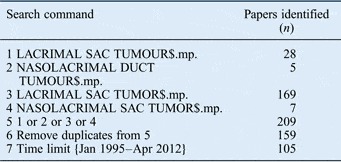
An extensive web-based search (using Pubmed, Medline, Embase and Google Scholar) was conducted by the first and second author. This focused on case reports and series or review articles on primary nasolacrimal duct and lacrimal sac tumours, and their endoscopic management, which had been published in the last 16 years (June 1995 to May 2011). Table I shows the terms used in the systematic review, which were limited in terms of their number and spelling variation. For the literature search, a combination of Medical Subject Headings and text words were used to generate the list of citations (nasolacrimal duct tumour, lacrimal duct tumour, lacrimal sac tumour, nasolacrimal sac tumour and endoscopic dacryocystorhinostomy). A total of 105 articles were identified in the systematic review. Articles were carefully reviewed by the first and second authors independently, and references were cross checked for any relevant papers. The exclusion criteria were: metastatic nasolacrimal duct tumours or tumour extension from the surrounding area, and non-English literature (Abstracts not available in English). In total, 77 papers were identified, comprising a total of 118 cases.
Systematic search on medline and embase

[mp = ti, ab, ot, nm, hw, ps, rs, ui, an, tx, bt, sh, ct, tn, dm, mf, dv, kw]
A retrospective chart review of the senior author's database was conducted for the patients who underwent endoscopic dacryocystorhinostomy from February 2001 to February 2012.
Results
Literature review
The literature search revealed paucity of articles published in ENT journals. Most of the case reports had been published in ophthalmology journals. This may be because epiphora used to be managed by ophthalmologists. The search revealed 16 large series publications (with 3 or more cases) and 61 individual case reports related to nasolacrimal tumours.
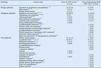
In 1994, Stefanyszyn et al. compiled an extensive review of nasolacrimal sac tumours in 115 patients.Reference Stefanyszyn, Hidayat, Pe'er and Flanagan5 We compared their findings with more recent reports (since 1995) on nasolacrimal tumours found during endoscopic dacryocystorhinostomy (Table II).Reference Tucker, Chow, Stockl, Codère and Burnier7–Reference Khan and Dhillon63
Primary nasolacrimal duct tumours before and after 1994 review

*Total n=115. †Total n = 118. DCR = dacryocystorhinostomy
Retrospective chart review
The review of the senior author's database (of a dedicated epiphora clinic) identified 4 nasolacrimal duct tumours in 411 patients (around 1 per cent); these tumours were exposed during a total of 525 endoscopic dacryocystorhinostomy procedures over the last 10 years. All benign cases were successfully managed by day-case endoscopic dacryocystorhinostomy carried out by the senior author, with a median follow up of three years (ranging from two months to five years). Most tumours were coincidental findings. No mortality was noted. Malignant cases were discussed in multidisciplinary team meetings and managed accordingly.
Patient 1, a 41-year-old man, presented with epiphora. A lump had been removed from his lacrimal sac 12 years previously and 10 years later he developed a lacrimal cystocoele. Examination revealed an opaque and smooth swelling near the puncta. Endoscopic dacryocystorhinostomy was performed and biopsy indicated an inverted papilloma. There was no sign of recurrence at the latest annual follow up.
Patient 2, a 38-year-old man, was found to have a small nodule in his right lacrimal duct. Biopsy demonstrated a solitary fibrous tumour. There was no recurrence at three months' follow up.
Patient 3, a 67-year-old man, presented with epiphora and a small swelling near the medial canthus. Pre-operative scans (computed tomography (CT) and magnetic resonance imaging (MRI)) and dacryocystography revealed a cystic lesion. Histological examination of the obstructive lesion showed a benign oncocytoma. There were no signs of disease recurrence in the first three months of follow up.
Patient 4, an 80-year-old female, presented with bilateral acute dacryocystitis and epiphora. Past medical history included chronic lymphocytic leukaemia, and cervical and axillary lymphadenopathy known to her haematologist (presumably lymphoma). Bilateral endoscopic dacryocystorhinostomy procedures and biopsy of suspicious sac tissue revealed a low-grade B-cell lymphoma of the right nasolacrimal duct. No active treatment was proposed by the haematologist. The stent was left in place with instructions to the patient to manually empty the sac daily.
Discussion
More than 350 primary lacrimal sac tumours have been reported in the English literature, most of which were primary epithelial tumours.Reference Flanagan and Stokes4, Reference Stefanyszyn, Hidayat, Pe'er and Flanagan5, Reference Ryan and Font64–Reference Hornblass, Jakobiec, Bosniak and Flanagan67 Papilloma was the most frequently reported benign tumour and lymphoma was the most common malignant tumour.Reference Stefanyszyn, Hidayat, Pe'er and Flanagan5, Reference Pe'er, Stefanyszyn and Hidayat68–Reference Kheterpal, Chan, Batch and Kirkby70 Adult males are most affected, and the condition is more common in the sixth decade of life.Reference Stefanyszyn, Hidayat, Pe'er and Flanagan5 The aetiology of nasolacrimal duct tumours is unknown. Lymphoma of the nasolacrimal duct may be associated with systemic lymphoma or leukaemia. Diffuse, large B-cell lymphoma or mucosa-associated lymphoid tissue lymphoma are common (33 per cent).Reference Sjö, Ralfkiaer, Juhl, Prause, Kivelä and Auw-Haedrich71 Metastatic spread has also been reported, with the breast and prostate being primary sites.
Primary malignant tumours of the nasolacrimal duct are relatively less common than adjacent spread from nearby tissues such as eyelid cancers or tumours of the nasal cavity. Radiological evaluation (CT and/or MRI) is recommended (Figure 1) if there is clinical suspicion of malignancy on endoscopic examination or on the dacryocystogram. Imaging techniques can provide information regarding the three-dimensional relationships with the surrounding bony and soft tissue structures.Reference Weber, Rodriguez-DeVelasquez, Lucarelli and Cheng72 Pre-operative incisional biopsy is only recommended in cases of obvious exophytic or ulcerative lesions.
Axial magnetic resonance image of a left nasolacrimal duct tumour

In the current series, the incidence of nasolacrimal duct tumours found during endoscopic dacryocystorhinostomy (4 out of 411 cases; 1 per cent) was similar to that reported by other studies published in the pre-endoscopic era (2 out of 150 cases; 1.3 per cent).Reference Tucker, Chow, Stockl, Codère and Burnier7 However, when we compared the case reports published after 1994, we found that there was a slight increase in the incidence of oncocytoma and lymphoma, but there was a decrease in cases of squamous cell carcinoma and papilloma (Table II). It is difficult to ascertain the exact reasons for the discrepancy, but it may be because of the rarity (anecdotal publications) and varied nature of pathology.
In another series of 377 dacryocystorhinostomy biopsies, the incidence of nasolacrimal duct tumours was as follows: lymphoma, 1.9 per cent; papilloma, 1.1 per cent; lymphoplasmacytic infiltrate, 1.1 per cent; transitional cell carcinoma, 0.5 per cent; and single cases of adenocarcinoma, undifferentiated carcinoma, granular cell tumour, plasmacytoma and leukaemic infiltrate.Reference Anderson, Wojno and Grossniklaus73 In a different series of 166 patients undergoing dacryocystorhinostomy, no malignancy was reported.Reference Lee-Wing and Ashenhurst8
• Nasolacrimal duct tumours are rare
• They present with obstructive symptoms
• Biopsy of the sac or duct mucosa is mandatory if there is any suspicion of tumour
• Inverted papilloma is the most common benign nasolacrimal duct tumour
• Malignant tumours are relatively common; they can be mutilating and are challenging to manage
In an elaborate study of cases by the Lacrimal Clinic (Moorfields Eye Hospital, London, UK) published in 2003, the most common tumour was non-Hodgkin's B-cell lymphoma (5 out of 15 cases).Reference Parmar and Rose19 At a median follow up of 30 months (ranging from 2 months to 17 years), 2 patients had died from metastatic disease, but 9 patients remained without evidence of recurrent tumour (i.e. recurrence was about 26 per cent). Mortality rates for malignant tumours depend on tumour stage and type, with a mean rate of 38 per cent (as reported by a study published in 1978).Reference Flanagan and Stokes4
In a 15-year retrospective chart review published in 2010 by an institute in France, 7 malignant tumours were found in the lacrimal sac.Reference Montalban, Liétin, Louvrier, Russier, Kemeny and Mom74 The commonest tumour was squamous cell carcinoma (five cases). Surgery was performed in six cases, with additional radiotherapy in four cases. The mean follow up was 68 months (with a range of 6 months to 17 years). Two patients developed recurrences (approximately 30 per cent), 3 were disease-free at the time of the study, 1 had persistent disease and another died after 30 months.
Malignant lesions of the nasolacrimal duct can be quite mutilating and potentially lethal. Complete surgical removal including the surrounding periosteum (if possible) is the treatment of choice.Reference Heindl, Jünemann, Kruse and Holbach6, Reference Schenck, Ogura and Pratt65, Reference Nakamura, Uehara, Omagari, Kunitake, Kimura and Makino69 Malignant cases should be discussed in the head and neck multidisciplinary team meeting. Patients with lymphoma and other haematological malignancies should be referred to the haematologist. In most of the literature reports, relapse occurred in 50 per cent of patients within 5 years.Reference De Stefani, Lerda, Usai, Cavalot, Riella and Bussi75 Malignant melanomas of the lacrimal sac have a poor prognosis, with an average survival of 18 months.Reference Lloyd and Leone76–Reference Carnevali, Trimarchi, Rosso and Stringa78
Conclusion
Nasolacrimal duct tumours are rare and present with obstructive symptoms. Incidental findings of such tumours have become more common in the endoscopic dacryocystorhinostomy era. We found that there has been a slight increase in the incidence of oncocytoma and lymphoma, and a decrease in papilloma and squamous cell carcinoma. Papilloma remains the most common benign tumour, while lymphoma is the most common malignant tumour.
Since the advent of endoscopic dacryocystorhinostomy, tumours are being diagnosed relatively early when smaller in size. Every attempt should be made to remove the whole tumour during the primary surgery. Endoscopic dacryocystorhinostomy provides immediate symptomatic relief. Decisions regarding any further treatment should be made on the basis of histology and tumour extent.
Long-term follow up is required, particularly for locally invasive or potentially malignant tumours, in order to rule out the possibility of any recurrence or malignant transformation. Because of the rarity of the tumours, it is advisable that such cases are managed in a multidisciplinary setting through a dedicated epiphora service framework.