
Introduction
Nanomedicine strategies for diagnosis and treatment of human diseases are of growing interest in recent years, owing to their unique and appealing properties, such as prolonged circulation lifetimes, reduced side effects of the drugs.[Reference Shi, Kantoff, Wooster and Farokhzad1, Reference Adiseshaiah, Crist, Hook and McNeil2] Until now, various nanoparticles (NPs) platforms have been developed including liposomes, micelles, dendrimers, gold, silica, and other inorganic NPs, etc.[Reference Torchilin3] Although some of the NPs have been used in clinical trials, few of them have been proved by the US Food and Drug Administration (FDA) for diagnosis or treatment of human diseases so far.[Reference Torchilin3, Reference Chauhan and Jain4] One of the main barriers stems from the safety problems of the materials and undesirable therapeutic efficiencies.[Reference Chen, Zhang, Zhu, Xie and Chen5] Therefore, it is highly desired to develop safer and more effective NP platforms.
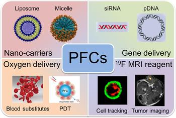
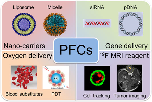
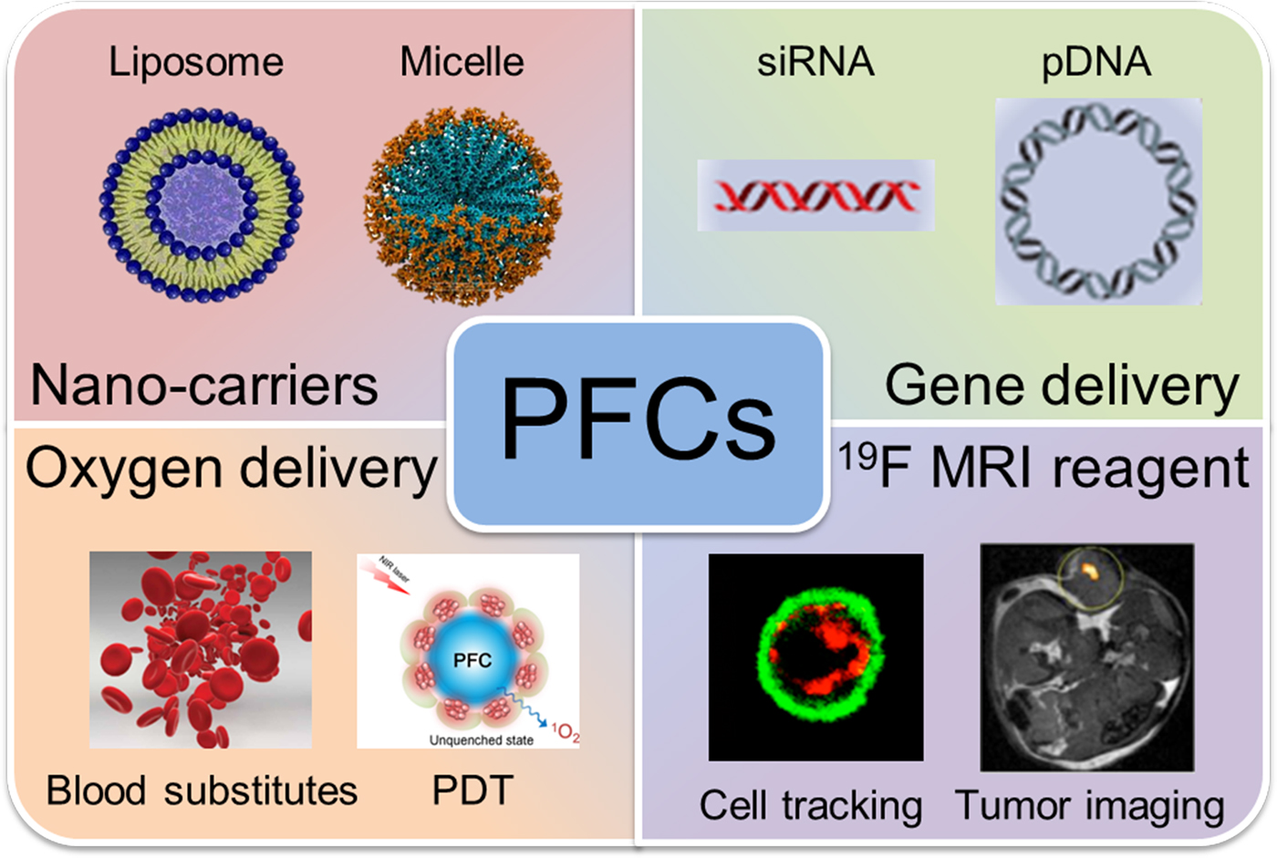
Perfluorocarbons (PFCs) show biologic inertness and low toxicity even at high doses.[Reference Krafft and Riess6] In contrast to the hydrocarbons, PFCs are more hydrophobic as well as capable of dissolving oxygen, detection and diagnosis of diseases by 19F magnetic resonance imaging (MRI).[Reference Janjic and Ahrens7] In addition, PFCs are also lipophobic and have a strong tendency to self-aggregation regardless of the solvent polarity.[Reference Riess8] Initially, the PFCs were mostly fabricated as the oxygen carriers because of their superior oxygen-dissolving capability.[Reference Riess and Krafft9, Reference Riess10] They thereby have been widely explored for lung injury, emergency transfusion, and traumatic brain injury.[Reference Castro and Briceno11] Due to the extremely favorable nuclear magnetic resonance (NMR) properties and virtual absence in the human body of fluorine, the PFCs can also be used for 19F MRI.[Reference Tirotta, Dichiarante, Pigliacelli, Cavallo, Terraneo, Bombelli, Metrangolo and Resnati12] In recent years, the PFCs were also used for gene/drug vectors, displaying excellent serum-resistant capability for gene delivery and good stability for drug delivery.[Reference Wang, Wu, Xu and You13, Reference Wang, Liu, Li and Cheng14] This review aims to give an overview of recent achievements on PFC-based nanomedicine for diagnosis and treatment of diseases (Fig. 1). This review is expected to provide inspiration for the design of PFC-based carriers and prompt the application of PFCs in nanomedicine.
Schematic illustration of the typical applications of perfluorocarbons (PFCs) in the fabrication of nano-carriers, gene delivery, oxygen delivery, and 19F MRI.
PFC-based nano-carriers
Nanotechnology has been considered as an effective strategy to improve aqueous solubility, decrease side effects, and enhance bioavailability of the conventional drugs.[Reference Petros and DeSimone15] According to the physicochemical properties of the drugs, the specific types of the nano-carriers can be chosen.[Reference Doane and Burda16] Until now various NPs have been developed, including liposome, micelle, dendrimer, metal NPs, etc.[Reference Zhang, Zhang, Xing, Xu, Li, Wang, Liang, Keservani, Sharma and Kesharwani17]
Liposomes, as a kind of conventional nano-carrier, have proven advantageous for chemotherapeutic drugs or biopharmaceutical drugs delivery to treat the diseases.[Reference Mitragotri, Burke and Langer18, Reference Ozpolat, Sood and Lopez-Berestein19] One paper reported by the FDA described that liposomal drug formulations were the most prevalent category (33% of the submissions) in nanomaterial-based drug products from 1973 to 2015, and more than ten of them have been approved by the FDA.[Reference D'Mello, Cruz, Chen, Kapoor, Lee and Tyner20, Reference Pattni, Chupin and Torchilin21] For example, the first FDA-approved nano-drug, Doxil®, is mainly composed of doxorubicin and PEGylated liposomes.[Reference Barenholz22] The Doxil® has been used for the treatment of Kaposi's sarcoma, ovarian, and breast cancer. Typically, liposomes are formed by the self-assembly of the phospholipids such as phosphatidylethanolamine, phosphatidylcholine, and phosphatidylglycerol, as well as cholesterol, displaying the hydrophilic core and hydrophobic lipid bilayer. Thus, liposomes can encapsulate hydrophilic, hydrophobic, and even amphiphilic drugs. In addition, the surface of liposomes can also be modified with the target-specific ligands or antibodies to improve their accumulation in the target tissue. Compared with the conventional drugs, the liposomes display reduced toxicity and higher accumulation efficiency of drugs that can reach the targeted tissue or pathology. However, delivery efficiency of the liposomes is still of depression. A latest statistical work reported by Chan and co-workers demonstrated that the average delivery efficiency of liposome-based drug delivery systems was only 0.5%.[Reference Wilhelm, Tavares, Dai, Ohta, Audet, Dvorak and Chan23] This suggests that just 0.5% of administered liposomes are able to reach the targeted diseased tissue. The low delivery efficiency of the liposomes is mainly attributed to their short circulation lifetimes.[Reference Allen and Cullis24]
In the aspect of the enhancement on the circulation times of the liposomes, the most widely used strategy is to construct the PEGylated liposome.[Reference Caponigro, Cornelia, Budillon, Bryce, Avallone, De Rosa, Ionna and Cornelia25, Reference Yang, Cui, Choi, Cho, Chung, Shim and Kim26] It is reported that half-life of the conventional liposome increased from 30 min to 5 h via the introduction of PEG-based amphiphilic materials.[Reference Klibanov, Maruyama, Torchilin and Huang27] Alternatively, the construction of fluorinated liposomes has also emerged as an attractive strategy to prolong the circulation lifetimes of liposomes with PFCs as hydrophobic component.[Reference Riess8] The fluorinated liposomes were more stable and displayed longer circulation lifetimes, due to their unique properties of both hydrophobicity and lipophobicity, high phase-separation tendency, and biomolecules adsorption resistance.[Reference Vierling, Santaella and Greiner28] As early as 1993, Riess and co-workers have demonstrated that the fluorinated liposomes have much longer circulation lifetimes compared with the conventional liposomes composed of 1,2-distearoyl-sn-glycero-3-phosphocholine (DSPC) and cholesterol.[Reference Santaella, Frézard, Vierling and Riess29]
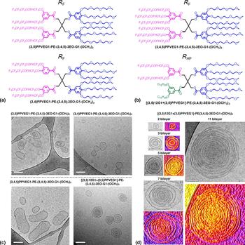
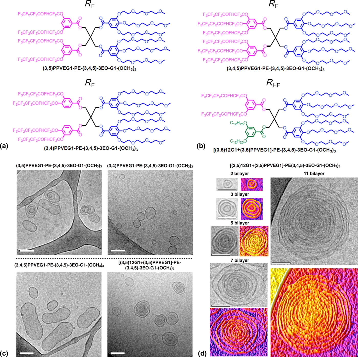
The researchers also developed a series of liposome-like fluorinated nano-carriers to pursue the efficient drug/gene vectors.[Reference Klein, Ciobanu, Klein, Machi, Leborgne, Vandamme, Frisch, Pons, Kichler, Zuber and Lebeau30] Via the self-assembly of the amphiphilic fluorinated dendrimers, a library of liposome-like nano-assemblies named dendrimersomes were developed by Percec and co-workers (Fig. 2).[Reference Xiao, Rubien, Wang, Reed, Hammer, Sahoo, Heiney, Yadavalli, Goulian, Wilner, Baumgart, Vinogradov, Klein and Percec31] The morphology of the dendrimersomes could be unilamellar or onion-like depending on the composition of the hydrophobic chains [Figs. 2(b) and 2(c)]. Additionally, the potential imaging properties of dendrimersomes for 19F MRI and fluorescence imaging make them interesting tools for nanomedicine. With the aim of developing novel nano-carriers that can co-deliver therapeutic genes and fluorinated drugs, Cheng and co-workers synthesized a library of amphiphilic materials composed of heptafluorobutyric anhydride and polyamidoamine (PAMAM) dendrimers with low generation.[Reference Wang, Hu, Cai, Xiao and Cheng32, Reference Wang, Wang, Wang, Hu, Li, Liu, Zhang and Cheng33] The fluorodendrimer-based nano-assemblies with the features of both lipids and polymers can readily encapsulate the fluorinated drugs regardless of hydrophilicity and hydrophobicity as well as efficiently deliver therapeutic genes.
(a) Library containing three janus dendrimers with fluorinated dendrons in their hydrophobic part (RF), one hybrid with a combination of chiral-racemic fluorinated and achiral hydrogenated dendrons (RHF). (b) Cryo-transmission electron microscopy (TEM) images of dendrimersomes assembled by RF and RHF Janus dendrimers. (c) Selected cryo-TEM images of onion-like dendrimersomes self-assemblies and their three-dimensional intensity-plotting images with different numbers of bilayers and diameters. Scale bar, 100 nm. Adapted with permission from Ref. Reference Xiao, Rubien, Wang, Reed, Hammer, Sahoo, Heiney, Yadavalli, Goulian, Wilner, Baumgart, Vinogradov, Klein and Percec31 (American Chemical Society, 2016).
The fluorinated catansomes were also developed by Mouritsen and co-workers aimed at obtaining alternative vectors to liposomes.[Reference Rosholm, Arouri, Hansen, González-Pérez and Mouritsen34] The fluorinated catansomes were constructed by the self-assembly of the oppositely charged surfactants with the diameter of 100–200 nm. The catansomes were pretty stable for over 5 months and could efficiently load the fluorescent molecule calcein as a model drug.
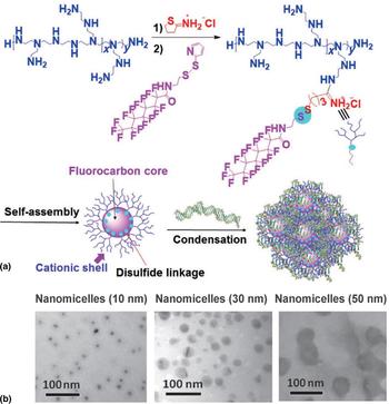
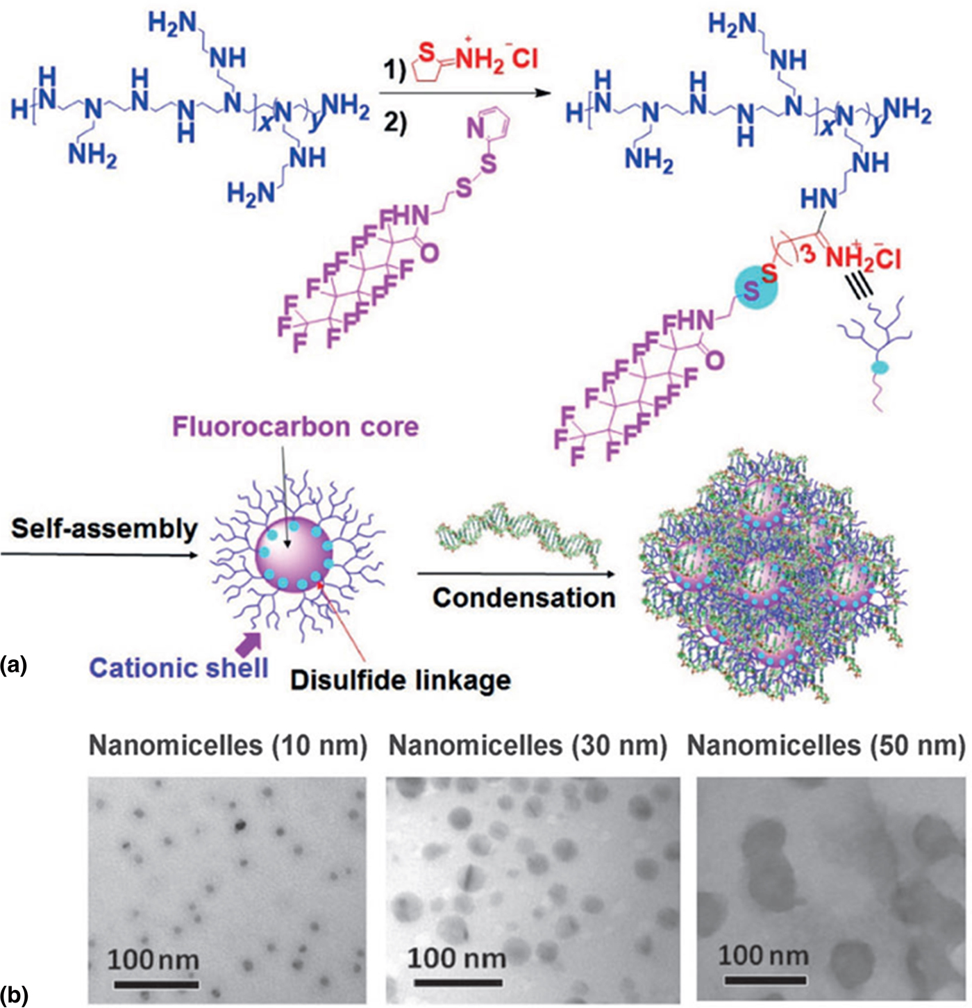
Micelles, another traditional nano-carrier, have been widely used for drugs and genes delivery.[Reference Kataoka, Harada and Nagasaki35, Reference Kakizawa and Kataoka36] The micelles are typically constructed via the self-assembly of the amphiphilic molecules into the core–shell architectures. The hydrophilic shell can impart them steric stability and prolong their circulation lifetimes, while the hydrophobic core can load the cargoes and further enhance their stability.[Reference Wei, Liu, Ma, Cheng, Huang, Zhao, Huo, Xue, Liang and Liang37] Until now, various kinds of micelles have been developed, such as PEG-b-PLGA, PEG-b-PCL, and PVP-b-PDLLA, for the drugs or genes delivery.[Reference Gaucher, Dufresne, Sant, Kang, Maysinger and Leroux38–Reference Gong, Chen, Zheng, Wang and Wang40] But few of them have been approved by the FDA. One of the major problems is the poor stability of the micelles in blood.[Reference Feiner-Gracia, Buzhor, Fuentes, Pujals, Amir and Albertazzi41, Reference Owen, Chan and Shoichet42] In comparison with hydrocarbons, the PFCs are more hydrophobic with lower van der Waals interactions,[Reference Krafft43] which will enhance the cohesion of the hydrophobic core and make the micelles more stable.[Reference Vierling, Santaella and Greiner28, Reference Matsuoka and Moroi44] Thus, the development of fluorinated micelles will be a promising strategy to combat the stability problems of micelles. A kind of bioreducible fluorinated micelle was reported by You and co-workers for gene delivery (Fig. 3).[Reference Wang, Wu, Xu and You13] The reported micelles were prepared via the self-assembly of PFC-modified poly(ethylenimine) (Mn = 10,000 g/mol). The introduction of the PFCs vastly enhanced the stability of the vectors and DNA complexes. Surprisingly, they can maintain high transfection efficiencies even in the presence of 50% fetal bovine serum (FBS).
(a) Scheme outlining the conjugation of PFCs to PEI, nanomicelle formation, nanomicelle@DNA complexes. (b) TEM images of the nanomicelles. Adapted with permission from Ref. Reference Wang, Wu, Xu and You13 (Wiley, 2016).
PFC-based gene delivery
Gene-based therapy has been recognized as the promising approach to treat the diseases by the modulation of gene expression in the targeted cells.[Reference Zuckerman and Davis45, Reference Stewart, Sharei, Ding, Sahay, Langer and Jensen46] The modulation can be realized by using various exogenous nucleic acids, including DNA, mRNA, siRNA, and CRISPR.[Reference Zhang, Zhang, Jin, Xue, Yang, Gong, Zhang, Wang, Tian, Xing and Liang47–Reference Xue, Chen, Yin, Tammela, Papagiannakopoulos, Joshi, Cai, Yang, Bronson, Crowley, Zhang, Anderson, Sharp and Jacks49] Although the mechanisms of these nucleic acids are different, it is essential to realize efficient gene transfection with the aid of gene vectors.[Reference Kanasty, Dorkin, Vegas and Anderson50, Reference Wang, Liu, Chung, Wang, Gaffey, Lu, Cavanaugh, Zhou, Kanade, Atluri, Morrisey and Burdick51] Viral-based vectors are able to escape the sequestration by the mononuclear phagocyte system and display high gene transfection efficiency both in vitro and in vivo.[Reference Truong, Gu, Prasadam, Jia, Crawford, Xiao and Monteiro52, Reference Hernandez-Garcia, Kraft, Janssen, Bomans, Sommerdijk, Thies-Weesie, Favretto, Brock, de Wolf and Werten53] However, the potential immunogenicity and carcinogenesis limit the application of them in gene delivery.[Reference Mingozzi and High54, Reference Naldini55] Unlike viral-based vectors, non-viral vectors, such as polycations and liposomes, are safer and more feasible to prepare.[Reference Ozpolat, Sood and Lopez-Berestein19, Reference Samal, Dash, Van Vlierberghe, Kaplan, Chiellini, van Blitterswijk, Moroni and Dubruel56] While the low gene transfection efficiency in the presence of serum is undesirable, due to the attack of serum proteins leading to the disassembly or aggregation of the vectors.[Reference Oishi, Nagasaki, Itaka, Nishiyama and Kataoka57, Reference Gary, Puri and Won58]
To overcome the poor delivery efficiency of the non-viral vectors in the presence of serum, researchers have developed various strategies, for example, the construction of virus-like vectors and the development of charge-reversal nanosystems.[Reference He, Nie, Cheng, Xie, Shen and Gu59–Reference Zhang, Guo, Zhang, Yu, Xu, Li, Tian, Wang, Xing and Liang62] Although these strategies can improve their delivery efficiency in serum-containing medium, the complex synthetic procedures and cytotoxicity issue limit their clinical use.[Reference Guo, Huang, Jiang, Sun, Deng, Liang, Du, Xing, Zhao, Wang, Dong and Liang63–Reference Noble, De Santis, Ravi, Lamarre, Castelletto, Mantell, Ray and Ryadnov65] PFCs are synthetically and biologically inert, displaying special properties of both hydrophobicity and lipophobicity, low surface energy, and high phase-separation tendency.[Reference Krafft and Riess6] In recent years, researchers have found that the PFC-modified vectors could well maintain their gene transfection efficiency in serum-containing medium.[Reference Wang, Liu, Li and Cheng14, Reference Yang, Zhang, Chang and Cheng66]
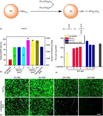
The strategy of functionalizing generation 5 (G5) PAMAM dendrimers with perfluoro acid to construct the pDNA vectors was explored by Cheng and co-workers [Fig. 4(a)].[Reference Wang, Liu, Li and Cheng14] Upon the fluorination of the dendrimers by heptafluorobutyric acid (G5-F768), both the cellular uptake and endosomal escape were enhanced. The gene transfection efficiency of the fluorinated dendrimer was improved from 21.8% to 97.1% in HEK293 cells [Fig. 4(b)]. Moreover, the G5-F768 achieved 55.4% in HeLa cells even in the presence of 50% FBS, whereas Lipo 2000, a commercialized vector, almost lost its gene delivery capability under the same conditions, indicating the good serum-resistant capability of fluorinated vectors [Fig. 4(c)]. The fluorination strategy also worked on polypropylenimine dendrimer and other cationic polymers such as bPEI 25k.[Reference Liu, Wang, Wang, Xiao and Cheng67, Reference Lv, Chang, Wang, Wang, Xiao, Zhang and Cheng68] Considering the cytotoxicity problems of the polycations and their complexes with nucleic acids in vivo environment, the biodegradable fluorinated peptide dendrimers with low generation were developed by Gu and co-workers for pDNA/siRNA delivery.[Reference Cai, Jin, Wang, Yue, Jiang, Wu and Gu69, Reference Cai, Zhu, Zhang and Gu70] The biodegradable and biocompatible G2 poly(L-lysine) dendrimer with 32 peripheral amino groups is served as positive charged component. Fluorination of the dendrimer by heptafluorobutyric acid can create an inactive surface to suppress protein adsorption and improve cellular delivery efficiency. The reversible crosslinking of the vectors by disulfide-contained linker is able to decrease the cytotoxicity of the vectors, and simultaneously enhance their stability in vivo.[Reference Chen, Wang, Hu, Ding, Yu, Zhou, Zhou, Li, Sun and Oupický71] The vectors also well preserved the gene transfection efficiency in HEK293 and HepG2 cells in the presence of 30% FBS and achieved considerable gene transfection efficiency in HepG2 tumor xenografts. As discussed before, the fluorinated micelles/liposomes with the PFCs as hydrophobic component were also prepared for gene delivery.[Reference Wang, Wu, Xu and You13, Reference Wang, Wang, Wang, Hu, Li, Liu, Zhang and Cheng33] These carriers can well maintain their gene delivery efficiency in vivo, and simultaneously encapsulate chemotherapy drugs such as 5-fluorouracil, displaying high efficiency in tumor inhibition.[Reference Wang, Hu, Cai, Xiao and Cheng32]
(a) Schematic illustration of the synthetic process of fluorinated dendrimers. (b) Comparison of the ability of G5-F749, G5-F768, G5, and Lipofectamine 2000 to mediate in vitro enhanced green fluorescent protein (EGFP) (b) and luciferase (c) gene transfection in HEK293 cells after 24 h. (d) Serum-resistance capabilities of fluorinated dendrimers on EGFP gene transfection in HeLa cells. Scale bar, 200 µm. Adapted with permission from Ref. Reference Wang, Liu, Li and Cheng14 (Springer Nature, 2014).

PFC-based oxygen storage and transport
The PFCs have also attracted much attention due to their properties in dissolving oxygen.[Reference Riess and Krafft9, Reference Wesseler, Iltis and Clark72] As early as 1966, researchers have found that PFC liquids could support respiration of mice and cats.[Reference Clark and Gollan73] Afterward, a series of PFC-based oxygen carriers have been developed as blood substitutes, organ preservation solutions, and oxygen suppliers for photodynamic therapy (PDT).[Reference Riess10, Reference Maluf, Mas, Yanek, Stone, Weis, Massey, Spiess, Posner and Fisher74, Reference Cheng, Cheng, Jiang, Qiu, Wang, Huan, Yuan, Wu and Hu75]
The PFCs are initially explored for artificial blood fabrications.[Reference Riess76, Reference Lowe77] There is increasing demand of blood throughout the world because of wars, natural disasters, and aging population issues.[Reference Williamson and Devine78] In hospitals, especially for the emergency services, the patients are urgently needed the matched blood to live.[Reference Shehata, Forster, Lawrence, Rothwell, Fergusson, Tinmouth and Wilson79] However, the amount of blood supply is not sufficient to meet the demand of blood, and the chasm is continued to increase.[Reference Greinacher, Fendrich and Hoffmann80] In addition, complex process of blood transfusion and strict storage condition of blood further threaten the human life.[Reference Hovav, Yedgar, Manny and Barshtein81] Thus, it is highly desirable to develop the available and safe blood substitutes. PFCs with comparable oxygen solubility capability to hemoglobin are promising candidates to overcome the drawbacks of human blood.[Reference Riess and Krafft9, Reference Fraker, Mendez and Stabler82] Until now, many PFC-based blood substitutes have been developed, for example, Fluosol-DA as a blood substitute in hemorrhage and emergency transfusion, and Oxygent as a blood substitute in hemorrhagic shock and cerebral ischemia.[Reference Riess10, Reference Castro and Briceno11] However, there is no FDA-approved blood substitute to date because of the safety issue.[Reference Tao and Ghoroghchian83] Therefore, it is a prerequisite to develop the next generation of the blood substitutes with the biocompatibility as the first priority consideration.
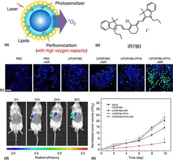
In recent years, researchers have also extended the applications of the PFCs onto PDT therapy as an oxygen supplier.[Reference Que, Liu, Tan, Feng, Shi, Li and Xiaoyu84, Reference Day, Estabrook, Logan and Sletten85] The therapeutic efficacy of PDT therapy is mainly determined by the 1O2 generation rate.[Reference Shen, Shuhendler, Ye, Xu and Chen86] The oxygen concentration is pretty low in some biophysical circumstances, for example, hypoxia tumors microenvironment, leading to low 1O2 generation rate and poor therapeutic efficacy.[Reference Lucky, Soo and Zhang87] PFCs with highly dissolved oxygen capability are promising candidates to overcome the limitation of PDT.[Reference Riess88] Hu and co-workers designed a kind of oxygen self-enriching nanosystem with PFCs and photosensitizer coencapsulated by lipids (Fig. 5).[Reference Cheng, Cheng, Jiang, Qiu, Wang, Huan, Yuan, Wu and Hu75] The nanosystem can not only enhance the 1O2 generation rate, but also prolong the half-life of 1O2. In contrast to the traditional PDT, the oxygen self-enriching nanosystem showed higher therapeutic efficacy on the treatment of hypoxia tumors (Fig. 5). Another effective strategy was explored by Liu and co-workers, wherein the oxygen carriers composed of albumin and PFCs were administered to relieve the tumor hypoxia microenvironment firstly, and then the PDT treatment was performed.[Reference Song, Feng, Liang, Yang and Liu89] The carriers can efficiently adsorb oxygen in the lung and the adsorbed oxygen can be rapidly released into the tumor upon the stimulation of ultrasound. In comparison with the conventional PDT treatment using liposomal chlorin e6, the combination of oxygen carriers and PDT treatment remarkably inhibited the growth of tumors.
(a) Schematic illustration of the structure and design of the Oxy-PDT agent. PFCs as a hydrophobic core are coencapsulated by lipids. Photosensitizer is uniformly dispersed inside the nanoparticle. (b) The chemical structure of IR780. (c) Enhanced 1O2 production by Oxy-PDT in cells. 1O2 generation was detected using carboxy-H2DCFDA, showing green fluorescence indicate positive staining for ROS; the cells are stained with Hoechst 33342. Scale bar, 50 mm. (d) Near-infrared imaging of tumor accumulation of IR780 in tumor-bearing mice after intravenous injection of LIP (IR780 + PFH). Images were taken at 0, 12, 24, and 32 h postinjection. Tumors are circled with white dashed lines. (e) Tumor volumes were measured after treated with LIP (IR780 + PFH) and laser irradiation. Values are the means ± SEM. Adapted with permission from Ref. Reference Cheng, Cheng, Jiang, Qiu, Wang, Huan, Yuan, Wu and Hu75 (Springer Nature, 2015).

PFC-based diagnostic reagent
Owing to the negligible fluorine concentration in the human body and extremely favorable NMR properties of fluorine, the 19F MRI techniques have also been employed for various biomedical applications, such as cell tracking and diseases diagnosis.[Reference Tirotta, Dichiarante, Pigliacelli, Cavallo, Terraneo, Bombelli, Metrangolo and Resnati12, Reference Flögel, Ding, Hardung, Jander, Reichmann, Jacoby, Schubert and Schrader90, Reference Srinivas, Heerschap, Ahrens, Figdor and de Vries91] To meet the requirements of 19F MRI applications, kinds of PFC-based contrast agents have been developed, including the aromatic PFCs, saturated linear PFCs, perfluoroethers, and polyethers.[Reference Janjic and Ahrens7] The properties of conventional PFC-based MRI agents have been well discussed in the previous reviews.[Reference Tirotta, Dichiarante, Pigliacelli, Cavallo, Terraneo, Bombelli, Metrangolo and Resnati12] Herein, this part will focus on the introduction of the recent works related to PFC-based MRI agents.
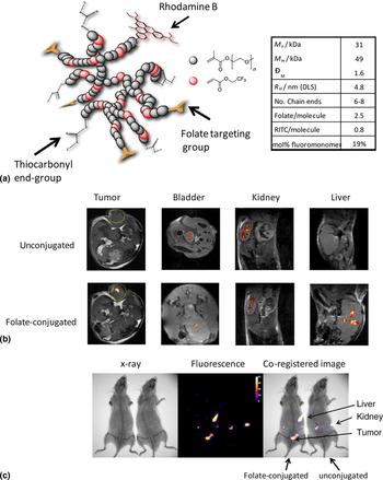
One of the main barriers of the 19F MRI lies on the low sensitivity. To improve the sensitivity of the probes, Ahrens and co-workers developed a library of paramagnetic fluorinated nanoemulsions with perfluoropolyether (PFPE) as metal chelators, which could incorporate various paramagnetic metal ions in the fluorous phase.[Reference Kislukhin, Xu, Adams, Narsinh, Tsien and Ahrens92] The formed nanoemulsions were stable in the intracellular milieu with pretty low rates of metal leakage. Additionally, this formation of the 19F MRI probes had a reduction in spin–lattice relaxation time (T 1), which contributed to improving the cell detection sensitivity. For the iron (III) tris-β-diketonate nanoemulsions with PFPE, their 19F MRI detection sensitivity was improved more than eightfold at clinical field strengths. The strategy to develop the multimodal imaging agents was also adopted to compensate for the low sensitivity of 19F MRI.[Reference Guo, Gao, Song, Li, Zhang, Xu, You, Wang, Zhuang, Su, Liu, Du and Zhang93] Thurecht and co-workers developed a multimodal polymer NPs system by combining high-resolution 19F MRI and sensitive fluorescence imaging for diagnosis of disease in vivo [Fig. 6(a)].[Reference Rolfe, Blakey, Squires, Peng, Boase, Alexander, Parsons, Boyle, Whittaker and Thurecht94] The hyperbranched polymer (HBP) was synthesized by the reversible addition-fragmentation chain transfer (RAFT) polymerization technique, comprising stealth component (PEG), tumor targeting agents (folate), and two imaging modalities (19F MRI and fluorescence). The polymer NPs were capable of fluorescence imaging of individual cells and detection of B16 melanoma tumors by the combination of 19F MRI and fluorescence in vivo [Figs. 6(b) and 6(c)].
(a) Schematic illustration of the multifunctional hyperbranched polymer used in this work. (b) The application of the HBP for imaging of the mouse subcutaneous B16 melanoma model. MRI images of the tumor (circled in image) bladder, kidney, or liver in the tumor-bearing mice 1 h following intravenous injection of 100 µL of unconjugated or folate-conjugated HBP (20 mg/mL in PBS). The 1H MR image is overlaid with the 19F image. (c) Fluorescence images of mice after injection of two compounds at the same concentration. The fluorescence images are co-registered with x-ray images of the mice 1 h following subcutaneous injection. Adapted with permission from Ref. Reference Rolfe, Blakey, Squires, Peng, Boase, Alexander, Parsons, Boyle, Whittaker and Thurecht94 (American Chemical Society, 2014).

Conclusion
In this review, we have systematically described the recent achievements of PFC-based nanomedicine. Compared with hydrocarbons, the PFCs are more hydrophobic and stable because of their high tendency to segregate from the surrounding environment. Besides, the biologic inert PFCs are good candidates for oxygen delivery and 19F MRI because of their great oxygen-dissolving ability, negligible background signal, and extremely favorable NMR properties of fluorine. In recent years, the PFC-based gene/drug carriers have also been developed. These works well demonstrated that fluorination of the carriers, such as liposomes or micelles, was able to vastly improve their stability and serum-resistant capability both in vitro and in vivo. Additionally, the work reported by Riess and co-workers also demonstrated that fluorination of the liposome vastly prolonged the circulation lifetimes. Thus, the construction of fluorinate carriers will be a promising strategy to overcome the limitations of systemic delivery for conventional nano-carriers.
The strategy of combining the properties of PFCs with other imaging agents or drugs may be a rational choice to achieve more definitive diagnosis and effective treatment of diseases. For example, to achieve accurate early detection of diseases, the multimodal imaging platforms can be constructed by the combination of 19F MRI with other imaging modalities with a good sensitivity such as fluorescence, single-photon emission computed tomography, or positron emission tomography. The highly dissolved oxygen of PFCs can also be applied to promote the efficacy of radiotherapy and PDT for cancer therapies. In addition, the PFC-based theranostics platforms can also be constructed by applying the diagnostic functions of PFC-based 19F MRI and the therapeutic functions of drugs/genes. Therefore, the PFC-based nanomedicines hold appealing potentials as a new strategy to develop stable and efficient nano-carriers for definitive diagnosis and efficacious treatment of diseases.
Acknowledgments
This work was supported by the National Natural Science Foundation of China (grant numbers 31771094 and 31371014). This work was also supported by the Natural Science Foundation key projects (grant numbers 31630027 and 31430031) and NSFC-DFG project (grant no. 31761133013). The authors also appreciate the support by the “Strategic Priority Research Program” of the Chinese Academy of Sciences (grant no. XDA09030301).