Introduction
Neoplasms of the lymphoid tissues represent a large, heterogeneous group of diseases that vary in clinical severity and their response to treatment. In Europe, the incidence of lymphoma is around 18–20 cases per 100,000 people per year.Reference Sant, Allemani and Tereanu1 In the past decade, the median age at diagnosis has increased to 65–70 years.Reference Morton, Wang and Devesa2 Over the past century, the population older than 65 years has more than doubled and is expected to double again over the next 50 years, whilst the population older than 85 years is likely to quadruple.Reference Jemal, Murray, Samuels, Ghafoor, Ward and Thun3 As a result, further increases are expected in the incidence of lymphoma in older patients. The incidence of lymphoma in the 80–84 year age group is 50 times higher than in the 20–24 year age group.Reference Groves, Linet, Travis and Devesa4 Outcomes in older patients are worse than those in younger patients, but numerous studies have shown that this difference cannot be explained by age alone.Reference Hasselblom, Ridell, Nilsson-Ehle and Andersson5–Reference Peyrade, Gastaud and Ré7 Lymphoma in older patients may be histologically and biologically different from lymphomas in younger patients and older people are significantly under-represented in clinical trials, so these worse outcomes may be due to undertreatment. This review considers the optimal strategy for lymphoma diagnosis and treatment in older patients.
General concepts
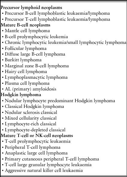
Lymphomas are a diverse group of malignant solid tumours of the lymphoid tissues. There are two types of lymphomas: Hodgkin lymphoma (HL) and non-Hodgkin lymphoma (NHL). HL, formerly called Hodgkin's disease, arises from germinal centre or post-germinal centre B cells. It has a unique cellular composition, containing a minority of neoplastic cells (Reed Sternberg cells and their variants) in an inflammatory background. NHLs are derived from B cells progenitor, T cells progenitor, mature B cells, mature T cells, or (rarely) natural killer cells. There are many typesReference Swerdlow, Campo and Harris8 (Table 1).
Classification of lymphoid neoplasms (adapted from Swerdlow et al.Reference Swerdlow, Campo and Harris8)

Clinical presentation
The majority of patients with classical HL present with overt symptoms, most often with an asymptomatic enlarged lymph node or a mass on chest X-ray. A minority present with non-specific symptoms and signs that are more compatible with infection than malignant disease. The clinical presentation of NHL varies depending on the type of lymphoma and the areas of involvement. Some lymphomas behave indolently with lymphadenopathy waxing and waning over years. Others are highly aggressive, resulting in death within weeks if left untreated.
Aggressive lymphomas commonly present acutely or sub-acutely with a rapidly growing mass, systemic ‘B’ symptoms (fever, night sweats, weight loss), and/or elevated levels of lactate dehydrogenase and uric acid. Indolent lymphomas are often insidious, presenting only with slow-growing lymphadenopathy, hepatomegaly, splenomegaly or cytopenias. Less common presentations include skin rash, pruritus, generalized fatigue, malaise, fever of unknown origin, ascites and effusions.
Approximately 50% of patients develop extranodal disease (secondary extranodal disease) during the course of their illness, while between 10 and 35% of patients have primary extranodal lymphoma at diagnosis.Reference Anderson, Chabner and Young9 The most common site of primary extranodal lymphomas is the gastrointestinal tract, followed by skin. The initial evaluation of patients with newly diagnosed lymphoma must establish the precise histological subtype, the extent and the sites of disease (located or advanced; nodal or extranodal) and the general health status of the patient. These studies help to determine the appropriate therapy and the patient's prognosis
Clinical evaluation
The evaluation of the patient with suspected lymphoma starts with a complete history and physical examination. The patient history must include systemic complaints (‘B’ symptoms): fever (temperature >38ºC), weight loss (unexplained loss of >10% of body weight over the past 6 months) and sweats (the presence of drenching night sweats). The physical examination needs to be directed to all potentially involved lymphoid sites. These include: Waldeyer's ring (tonsils, base of the tongue, nasopharynx), standard lymph node sites (e.g. cervical, supraclavicular, axillary, inguinal, femoral), liver and spleen, abdominal nodal sites (mesenteric, retroperitoneal), and less commonly involved nodal sites (e.g. occipital, preauricular, epitrochlear).
A lymph node biopsy of involved tissue is required for the diagnosis and classification of lymphoma. More than two-thirds of patients present with peripheral lymphadenopathy that is generally painless. The decision to biopsy a lymph node is dependent upon the clinical situation and characteristics of the patient, as well as the location of the involved lymph node(s). Biopsy should be considered if one or more of the following characteristics are present: significant enlargement, persistence for more than 4–6 weeks, or progressive increase in size. Enlarged peripheral lymph nodes are generally preferred over other lymph nodes for diagnostic biopsy because they are easily accessed and have a relatively high diagnostic yield. However, when enlarged peripheral lymph nodes are absent, computed tomography (CT)-guided core biopsiesReference Metzgeroth, Schneider and Walz10 or laparoscopic biopsiesReference Asoglu and Porter al, Donohue11 can be used to obtain diagnostic material in a majority of cases.
The diagnosis of lymphoma is based upon the evaluation of histological, immunophenotypic and genetic studies interpreted in the clinical context. After the initial tissue biopsy provides a diagnosis of lymphoma, the following baseline laboratory tests are appropriate for most cases:
• complete blood count, white blood cell differential, platelet count, and examination of the peripheral smear for the presence of atypical cells, suggesting peripheral blood and bone marrow involvement, and if it is possible an immunophenotype using flow cytometry;
• biochemical test including blood urea nitrogen (BUN), creatinine, alkaline amino transferase (AST), alanine amino transferase (ALT), lactate dehydrogenase (LDH) and albumin;
• serum calcium, electrolytes and uric acid;
• HIV, hepatitis B and C serology;
• beta-2 microglobulin levels.
These studies may not only contribute to staging but also influence the type of therapy chosen and its timing and guide further investigations to other potential sites of disease or complications.
Imaging studies
Routine radiological studies in lymphoma should include contrast enhanced CT imaging of chest, abdomen and pelvis. These serve to help determine disease stage at diagnosis and provide a baseline for comparison to determine response to treatment. Integrated positron emissions tomography (PET)/CT scanning is more sensitive and specific than CT in diffuse large B cell lymphoma and HL, so PET/CT scanning is recommended in treatment assessment for these lymphomas.Reference Cheson, Pfistner and Juweid12
Staging and prognosis
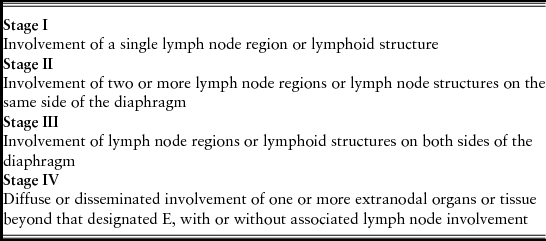
The Ann Arbor staging system with Cotswold modification, originally developed for HL, has been adapted for NHLReference Lister, Crowther and Sutcliffe13 (Table 2). This staging system focuses on the number of tumour sites (nodal and extranodal), their location, and presence or absence of systemic symptoms. The prognosis depends on histopathology and the influence of clinical parameters, including age, presence of extranodal disease, performance status and stage (I/II versus III/IV). There are different prognostic indices for each type of lymphoma.
The Ann Arbor staging system with Cotswolds modifications (adapted from Lister et al.Reference Lister, Crowther and Sutcliffe13)
All cases are subclassified to indicate the absence (A) or presence (B) of the systemic symptoms.
The designation ‘E’ refers to extranodal contiguous extension. More extensive extranodal disease is designated stage IV.
The subscript ‘X’ is used if bulky disease is present. This is defined as a mediastinal mass with a maximum width that is equal to or greater than one-third of the internal transverse diameter of the thorax at the level of T5/6 interspace or >10 cm maximum dimension of a nodal mass. No subscripts are used in the absence of bulk.
Characteristics of older patients
The definition of ‘elderly’ has been arbitrarily established as people above 60 years of age, but this is inadequate. In a German study of older patients with lymphoma, the investigators suggest that over 75 years should be the age cut-off for ‘elderly’ because outcome in those older than 75 was significantly inferior to patients younger than 75 years.Reference Pfreundschuh, Schubert and Ziepert14 The National Comprehensive Cancer Network (NCCN) classifies older patients into ‘young old’ (aged between 65 and 75 years), ‘old’ patients (aged 76 to 85 years) and ‘oldest old’ patients (aged >85 years).Reference Balducci15 Currently the definition of ‘elderly’ or preferably ‘older’ patients should be based not only on chronological age, but on a thorough evaluation of each patient individually to decide best therapy.Reference Latta, Cygan and Fried16
Performance status
The Karnosky score and the Eastern Cooperative Oncology Group (ECOG) scales have been used routinely in clinical trials to assess performance status in individual patients. Despite their limitation, they have been incorporated in most prognostic indices.
Retrospective analysis of patients with lymphoma over the age of 80 years demonstrates that a patient's ability to perform activities of daily living (ADLs) (instrumental ADLs as opposed to their ECOG-performance status), is more predictive of overall survival and progression free survival.Reference Nabhan, Smith and Helenowski17
Comprehensive geriatric assessment
The decision to choose a treatment based on performance status without conducting a comprehensive geriatric assessment (CGA) may underestimate older patients’ ability to tolerate treatment, and might, in part, explain why they are offered less therapy.Reference Latta, Cygan and Fried16 CGA is a multidisciplinary evaluation of physical and psychological conditions that affect the choice of therapy for older patients with malignant disease. The CGA evaluates the nutritional status, cognitive skills, functional status and co-morbidities of older patients and helps decide the intensity and density of chemotherapy. Whilst it has resulted in excellent outcomes in patients with various cancers,Reference Merli, Luminari and Rossi18 few trials were specifically designed for lymphoma patients.
Co-morbidities
Co-morbidities are defined as chronic medical conditions that can affect a patient's life. The modified Charlson co-morbidity index has been used to estimate risk of mortality, based on pre-existing morbid conditions. Reference Charlson, Pompei and Ales19 Several studies have demonstrated how co-morbid conditions adversely affect outcomes in patients with different cancers and suggest a similar effect in lymphoma patients.Reference Pfreundschuh20
Organ dysfunction
In older patients, organ dysfunction becomes more likely. Dysfunction may affect the reserve of the kidney, the liver, the heart and bone marrow.Reference Pal, Katheria and Hurria21 Thus older patients with lymphoma need a detailed evaluation that includes all these factors in order to offer the best treatment tailored to the baseline situation and illness.
Main types of lymphoma in older patients: clinical and therapeutic characteristics
Diffuse large B-cell non-Hodgkin lymphoma (DLBCL)
DLBCL is the most commonly diagnosed NHL in western countries, accounting for approximately 25% of all NHLs. In Europe, its incidence is approximately 3.81 cases per 100,000 person years.Reference Sant, Allemani and Tereanu1 Like most other NHLs, there is a male predominance. Incidence increases with age, with the median age at presentation being 70.4 years.Reference Smith, Howell, Patmore, Jack and Roman22 Almost one-third of newly diagnosed patients are over the age of 75 years.
DLBCL is a heterogeneous group of tumours. A number of clinicopathological entities are now recognized that are sufficiently distinct to be considered separate diagnostic categories: T-cell-rich large B-cell lymphoma, primary mediastinal large B-cell lymphoma, intravascular lymphoma and Epstein-Barr virus positive large B-cell lymphoma. DLBCL arises from a mature B-cell, usually a centroblast or immunoblast with a diffuse pattern and high proliferation fraction. Tumour cells in DLBCL generally express pan-B cell antigens (CD19, CD20, CD22, CD79a). The majority have genetic abnormalities, but there is no single cytogenetic change that is typical or diagnostic.Reference Swerdlow, Campo and Harris8
It can occur de novo, but it can also arise through the transformation of many different types of low grade B-cell lymphomas.
Patients with DLBCL typically present with a rapidly enlarging symptomatic mass, most usually nodal enlargement in the neck or abdomen. Systematic ‘B’ symptoms are observed in approximately 30% of patients and the serum LDH is elevated in over half. Approximately 60% of patients present with advanced stage (III or IV). Extranodal disease occurs in up to 40% of cases. The most common site of extranodal involvement is the stomach/gastrointestinal tract.
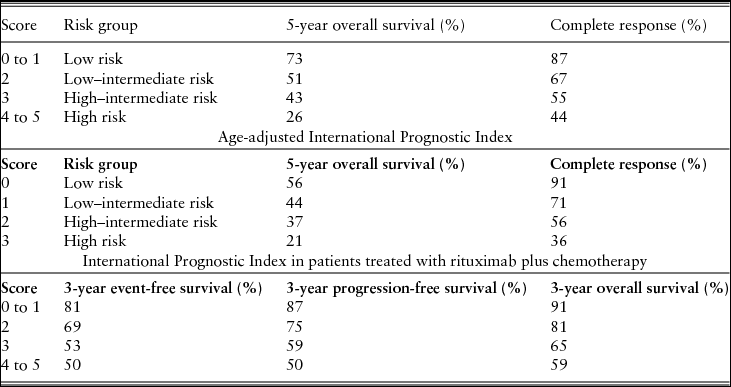
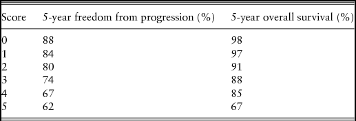
The International Prognostic Index (IPI) and its modifications are the main prognostic tools used in patients with DLBCL23,Reference Ziepert, Hasenclever and Kuhnt24 (Table 3). Molecular pathology also influences the prognosis. DLBCL is classified according to expression of genes into three groups: the germinal centre B-cell-like (GCB), the activated B-cell-like (ABC) and a not-otherwise specified type 3. Patients in the GCB group have the highest 5-year survival rate.Reference Rosenwald, Wright and Chan25,Reference Klapper, Kreuz and Kohler26
International Prognostic Index and its modifications (adapted from the International Non-Hodgkin's Lymphoma Prognostic Factors Project 23and Ziepert et al.Reference Ziepert, Hasenclever and Kuhnt24)
One point is given for each characteristic present: age >60 years, serum lactate dehydrogenase concentration above normal, ECOG performance status >2, Ann Arbor stage III or IV, number of extranodal disease sites >1
For all types of lymphoma, one important clinical decision is whether treatment may be curative. Thus distinction can be made between older, ‘fit’ patients who can be treated as adult patients, ‘frail’ patients handled mostly with palliation, and patients in an intermediate state, or ‘vulnerable’, who are likely to be suited to adjusted or reduced chemotherapy regimens. DLBCL is a potentially curable malignancy. For patients up to 80 years, rituximab plus cyclophosphamide, doxorubicin, vincristine and prednisone (R-CHOP) is the standard regimen for CD20-positive DLBCL.Reference Coiffier, Thieblemont and Van den Neste27 The benefit of the addition of rituximab has been shown for all age groups to 80 years.Reference Pfreundschuh, Schubert and Ziepert14 However, patients who are frail or over 80 years of age have been excluded from clinical trials, and guidelines on how these patients should be treated are lacking. There is a need for such patients to be enrolled in prospective clinical trials specifically designed for this group. In the absence of clinical studies, the decision of how to treat is usually left to the physician's discretion.Reference Van der Poel, Mulder and Ossenkoppele28 It would seem reasonable for DLBCL in stage I or II disease to be treated with an abbreviated course of chemo-immunotherapy using R-CHOP followed by involved-field radiation.Reference Odejide, Cronin, Davidoff, LaCasce and Abel29 In fit patients with adequate cardiac reserve, full dose R-CHOP is recommended. In those with compromised cardiac function or other co-morbidity, a dose reduced R-CHOP (mini-R-CHOP) or non-anthracycline-based programme is recommended (e.g. rituximab plus cyclophosphamide, vincristine and prednisone (R-CVP) or rituximab plus bendamustine (R-B).Reference Peyrade, Jardin and Thiblemont30,Reference Weidmann, Neumann and Fauth31
Some older patients present with a decline in their performance status that is due only to their lymphoma. In such instances, starting with a pre-phase treatment might be appropriate, to improve the patient's functional status. The German Lymphoma Study Group suggested treating patients who present with ECOG performance status of 2 or more with 7 days of prednisone and 1 mg of vincristine before the first chemotherapy administration and, if a patient's status improves, the goal of therapy may then be modified.Reference Pfreundschuh, Trumper and Kloess32
Follicular lymphoma
Follicular lymphoma (FL) is the second most common subtype of NHL and the most common of indolent NHL. The estimated incidence of FL in Europe is 3.1 cases per 100,000 people per year.Reference Sant, Allemani and Tereanu1 Median age at diagnosis is 60 yearsReference Smith, Howell, Patmore, Jack and Roman22 and the incidence increases with age. Most patients present with asymptomatic lymphadenopathy, whilst other frequently involved sites are spleen, bone marrow, peripheral blood, Waldeyer's tonsillar ring and extranodal sites such as the gastrointestinal tract.
FL is derived from germinal centre B-cells and morphologically is composed of centrocytes and centroblasts. The grade (1 to 3) depends on the proportion of centroblasts per high power field. Grades 1 and 2 (0 to 15 centroblasts per high power field) are indolent disease. Grade 3 is divided into grade 3a (with centrocytes present) and 3b (with a solid sheet of centroblasts). Grade 3b is more aggressive and the management of this entity must be like that for diffuse large B cell lymphoma.Reference Swerdlow, Campo and Harris8 FL cells express surface immunoglobulin, CD19, CD20, CD22 and are bcl-2+, bcl6+ and CD10+. The genetic characteristic of FL is the translocation t (14; 18) (q32; q21) contributing to overexpression of bcl-2.Reference Swerdlow, Campo and Harris8 The prognosis is calculated using the Follicular Lymphoma International Prognostic Index (FLIPI) and its modificationsReference Solal-Céligny, Roy and Colombat33,Reference Federico, Bellei and Marcheselli34 (Table 4).
Follicular lymphoma International prognostic index (FLIPI) (adapted from Solal-Celigny et al.Reference Solal-Céligny, Roy and Colombat33)
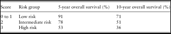
One point is given for each characteristic present: age >60 years, serum lactate dehydrogenase concentration above normal, haemoglobin level <12 g/dl, Ann Arbor stage III or IV, number of involved nodal areas >4
Treatment options for FL depend on the disease stage, age and co-morbidities of the patient and if there is symptomatology associated with the disease. In early stages (I/II), radiotherapy is a treatment option that may increase survival, but one that produces toxicity, so that the observation is a reasonable option if there are co-morbidities and expected toxicity of radiotherapy is considered likely to be significant.Reference Pugh, Ballonoff, Newman and Rabinovitch35 In advanced stages (III/IV) that are still asymptomatic, observation is a reasonable option until there is disease progression and does not worsen survival.Reference Ardeshna, Smith and Norton36 For patients with symptomatic advanced stage or high-volume disease, treatment is chemoimmunotherapy: R-CHOP or bendamustine-rituximab (B-R). B-R can be considered as a first-line treatment because of increased progression-free survival (PFS) and fewer side-effects.Reference Rummel, Niederle and Maschmeyer37 R-CVP can be used in patients with heart disease, but the response rate is lower.Reference Marcus, Imrie and Solal-Celigny38 For ‘oldest old’ or unfit patients, rituximab 375 mg/m2 weekly for four doses, or treatment with single agents (e.g. chlorambucil or cyclophosphamide) with or without rituximab, can be options.39 Patients who have achieved a good response can be treated with rituximab maintenance therapy to improve PFS.Reference Salles, Seymour and Offner40 Currently, there are clinical trials of new drugs with promising results, such as 90Y-ibritumomab tiuxetan.Reference Scholz, Pinto and Linkesch41
Mantle cell lymphoma
Mantle cell lymphoma (MCL) is a mature B-cell NHL that accounts for approximately 7% of adult NHL. In Europe the incidence is 0.45 cases per 100,000 people per year.Reference Sant, Allemani and Tereanu1 It is more prevalent in males than females. Median age at diagnosis is around 74 years and the incidence increases with age.Reference Smith, Howell, Patmore, Jack and Roman22 Frequently, MCL patients present at an advanced stage of disease (III or IV), with lymphadenopathy, hepato-splenomegaly and bone marrow infiltration. The diagnosis is made by lymph node biopsy, bone marrow aspiration, or even with identification of an immunophenotype in peripheral blood. The lymphoid cells are positive for CD20, CD5 and FMC7. Cyclin D1 and translocation t (11:14) (q13:32) identifies MCL.Reference Swerdlow, Campo and Harris8
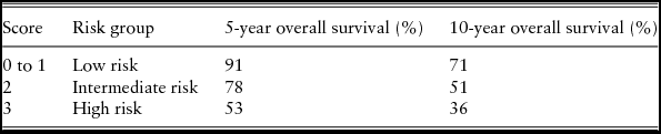
The clinical course of MCL is very heterogeneous. Most cases have an aggressive course and poor prognosis, with short duration of remission following standard therapies. However, there is a small percentage of patients that have an indolent course and do not need treatment for several years. The prognostic index for MCL is the Mantle Cell International Prognostic Index (MIPI)Reference Hoster, Dreyling and Klapper42 (Table 5). Other factors associated with worse prognosis in MCL are high proliferative activity or increased Ki-67 staining, blastoid variant, inactivation of p53 and secondary chromosomal aberrations.
Mantle cell lymphoma international prognostic index (MIPI) (adapted from Hoster et al.Reference Hoster, Dreyling and Klapper42)

One to three points is given for each characteristic present: age (<50: 0; 50 to 59: 1; 60 to 69: 2; >70: 3), ECOG performance status (0 to 1: 0; 2 to 4: 2), lactate dehydrogenase:upper limit of normal (LDH:ULN) ratio (<0.67: 0; 0.67 to 0.99: 1; 1.00 to 1.49: 2; >1.50: 3), white blood cells 109/l (<6.7: 0, 6.7 to 9.9: 1; 10.0 to 14.9: 2; >15.0: 3)
The choice of treatment in MCL older patients is difficult. For patients with asymptomatic low MIPI, older MCL and indolent MCL, the strategy of ‘watch and wait’ should be considered.Reference Martin, Chadbum and Christos43 R-CHOP should remain an important consideration in older patients,Reference Kluin-Nelemans, Hoster and Hermine44 despite the cardiotoxicity associated with anthracycline. B-R has been associated with a longer PFS as it is less toxic and is not associated with cardiac toxicity.Reference Rummel, Niederle and Maschmeyer37 Therefore, B-R is a reasonable first option. Purine analogues have been used for the treatment of MCL in older patients, but the outcomes are poorer than with R-CHOP.Reference Kluin-Nelemans, Hoster and Hermine44 Rituximab maintenance after induction chemotherapy may improve the overall survival.Reference Kluin-Nelemans, Hoster and Hermine44 More intensive treatments, such as hyper-CVAD regimens, have significantly more toxicity in older patients than in younger patients. Other recent studies have combined bendamustine and rituximab with other agents such as bortezomib, lenalidomide and cytarabine.Reference Visco, Finotto and Zambello45 However, further clinical trials including older patients and the monitoring of existing studies are needed before these treatments can be recommended outside the context of clinical trials. New treatments such as ibrutinib are being investigated with promising results.Reference Zucca and Bertoni46
Hodgkin lymphoma
Hodgkin lymphoma is a B-cell neoplasm that represents approximately 10% of all lymphomas. The incidence in Europe is around 2.49 cases per 100,000 people.Reference Sant, Allemani and Tereanu1 HL has a bimodal age distribution with one peak in young adults around 20 years and one peak in older age around 65 years. Histologically, HL consists of neoplastic cells called Reed-Sternberg cells and their variants on an inflammatory background. There are two groups: nodular lymphocyte predominant HL (NLPHL) and classical HL (CHL). CHL is divided into four groups based on the appearance of the tumour cells and the composition of the reactive background: nodular sclerosis, mixed cellularity, lymphocyte rich and lymphocyte depleted. Usually CHLs express CD15 and CD30, variably express CD20 and do not express CD3 and CD45.Reference Swerdlow, Campo and Harris8 The main prognostic index for HL is the International Prognostic Score for Hodgkin LymphomaReference Hasenclever and Diehl47,Reference Moccia, Donaldson and Chhanabhai48 (Table 6).
The International Prognostic Score for Hodgkin lymphoma (adapted from Hasenclever et al.Reference Hasenclever and Diehl47 and Moccia et al.Reference Moccia, Donaldson and Chhanabhai48)
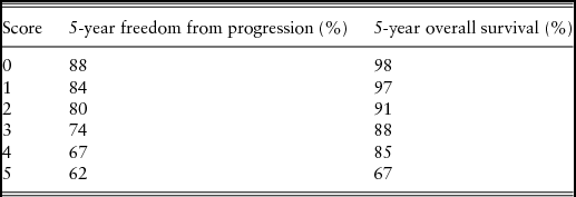
One point is given for each characteristic present: serum albumin <4 g/dl; haemoglobin<10.5 g/dl; male gender; age >45 years; stage IV disease; white blood cell count >15,000/µl; absolute lymphocyte count <600/µl and/or <8% of the total white blood cell count
The percentage of HL patients aged over 60 years is approximately 15–35%. Older patients have biological and histological features associated with poor prognosis. Mixed cellularity, EBV positive HL, B symptoms, infra-diaphragmatic disease and advanced stage of disease are other features more commonly observed in older than younger patients. However, a bulky mediastinal mass is infrequent in older patients.Reference Björkholm, Svedmyr and Sjöberg49,Reference Halbsguth, Böll, Borchmann and Diehl50
The most common treatment for HL is ABVD (adriamycin, bleomycin, vinblastine and dacarbazine) with or without radiotherapy, but this regimen can be very toxic in older patients. Patients aged 60–70 years with a good performance status and without co-morbidities can receive ABVD. The number of courses depends on stage of the disease and presence of favourable or unfavourable features. Use of granulocyte colony stimulation factor (GCS-F) with bleomycin regimens increases the risk of bleomycin toxicity,Reference Martin, Ristow, Habermann, Colgan, Witzig and Ansell51 so GCS-F should be avoided in these cases. In patients aged 60–70 years with co-morbidities and in patients older than 70 years, ABVD may be poorly tolerated and adjustments of doses could be needed.Reference Björkholm, Svedmyr and Sjöberg49 The Stanford V or BEACOPP regimens are very toxic in HL older patients. There are other regimens – PVAG (prednisone, vinblastine, adriamycin and gemcitabine)Reference Böll, Bredenfeld and Görgen52 and VEPEMB (vinblastine, cyclophosphamide, procarbazine, prednisone, etoposide, mitoxantrone and bleomycinReference Proctor, Wilkinson and Jones53 – that provide satisfactory disease control in early and advanced stages of disease with acceptable toxicity.
Nodular lymphocyte predominant HL (NLPHL) constitutes less than 5% of all HL cases, express CD20, are often localized, have a male predominance and the median age just over 30 years.Reference Swerdlow, Campo and Harris8 Thus NLPHL is rarely seen among older patients. Moreover, the treatment of this disease differs from CHL.39 PET/CT is used for the early evaluation of response in order to adapt the subsequent intensity and duration of treatment.Reference Ansell54
Brentuximab-vedotin is an anti-CD30 antibody that has been used in patients with relapsing or refractory disease and has a favourable safety profile and long term response.Reference Younes, Gopal and Smith55,Reference Younes, Bartlett and Leonard56
Conclusions
The incidence of lymphoma in older patients is growing and their treatment is a challenge. Specific guidelines are lacking. Moreover they are excluded from most clinical trials and management decisions usually need to be based on data obtained in younger patients. The choice of the best treatment for each patient should be individualized with an overall assessment that includes PS, co-morbidities and CGA. These patients should be included in clinical trials when available. Furthermore, the development of clinical trials for older patients with lymphoma could be help to determine the optimal treatment.
Conflicts of interest
None