I. INTRODUCTION
Since the first report by Widawski et al. in 1994,Reference Widawski, Rawiso and Francois1 the breath figure method has been widely used to prepare honeycomb-patterned porous films because of the fascinating advantages of simplicity and cost-efficiency.Reference Bunz2, Reference Escale, Rubatat, Billon and Save3 Functional honeycomb films showed great potential as advanced materials including microarrays of biomoleculesReference Zhang and Wang4–Reference Wan, Li, Chen and Xu7 or particles,Reference Sun, Li, Bu, Xu and Wu8–Reference Jiang, Zhou, Zhang, Zhang, Guo and Gu14 templates,Reference Li, Zhong, Ma, Li, Chen, Zhang, Tang, Xie and Ma15 superhydrophobic surface,Reference Yabu and Shimomura16 biomaterials,Reference Nishikawa, Nishida, Ookura, Nishimura, Wada, Karino and Shimomura17–Reference Du, Zhu, Yan, Su, Song and Li19 optical and electronic devices,Reference Liu, Gao and Yan20, Reference Yin, Zhang, Kong, Zou, Li, Lu, Ma, Boey and Chen21 and separation membranes with narrow distribution of pore size.Reference Wan, Li, Ke and Xu22 There are two typical methods to fabricate functional honeycomb films, i.e., the bottom–up method and the top–down method. The former is often based on a self-assembly processReference Wan, Li, Chen and Xu7–Reference Jiang, Zhou, Zhang, Zhang, Guo and Gu14 and is very desirable for the breath figure method because segregation of hydrophilic moieties or particles can take place to decrease the interfacial tension between polymer solution and condensed water droplets.Reference Chen, Wan, Ke and Xu23 The top–down strategies such as vapor depositionReference Hirai, Yabu, Matsuo, Ijiro and Shimomura24 and surface modificationReference Ke, Wan and Xu6, Reference Hernandez-Guerrero, Min, Barner-Kowollik, Muller and Stenzel25, Reference Nystrom, Malmstrom, Hult, Blakey, Boyer, Davis and Whittaker26 provide diverse functional surface compared to the bottom–up method. In previous work, we reported an approach to functional honeycomb films based on facilitated and site-specific assembly of microspheres.Reference Wan, Lv, Ke and Xu27 The surface wettability greatly affects the assembly of microspheres in honeycomb films, on which there is a Cassie–Wenzel wetting transition.Reference Ke, Wan, Li, Xu and Xu28That approach may be versatile to various functionalities but depends on the microspheres.
There exist many methods to prepare functional microspheres.Reference Kawaguchi29 Seeded polymerization can form a skin layer to introduce new functions.Reference Okubo, Izumi and Takekoh30 On existing microspheres, new functional layer can also be introduced by methods such as adsorption,Reference Carmonaribeiro and Midmore31 deposition,Reference Dokoutchaev, James, Koene, Pathak, Prakash and Thompson32 surface grafting,Reference Chen, Tao, Mantovani, Geng, Nystrom and Haddleton33 and surface reaction.Reference Chen, Tao, Mantovani, Geng, Nystrom and Haddleton33–Reference Alvarez-Paino, Munoz-Bonilla, Marcelo, Rodriguez-Hernandez and Fernandez-Garcia37 For example, Chen et al.Reference Chen, Tao, Mantovani, Geng, Nystrom and Haddleton33 introduced alkyne groups to the surface of the Wang resin beads and used a copper-catalyzed Huisgen reaction to attach mannose-containing azides to the bead surface. Most recently, Alvarez-Paino et al.Reference Alvarez-Paino, Munoz-Bonilla, Marcelo, Rodriguez-Hernandez and Fernandez-Garcia37 reported glycosylated polystyrene (PS) microspheres functionalized via thiol–para-fluorine “click” reaction. It is well known that PS microspheres are most commonly used and often cross-linked using divinylbenzene (DVB) or ethylene glycol dimethacrylate (EGDMA) as the cross-linkers that lead to a number of free surface vinyl groups. The residual vinyl groups can be used for further surface functionalization by thiol–ene reaction. This thiol–ene reaction is important as it can provide a straightforward method to prepare functional cross-linked PS microspheres by avoiding the procedures for introducing other reactive groups such as alkyne groups.Reference Chen, Tao, Mantovani, Geng, Nystrom and Haddleton33 While the reactivity difference of different vinyl monomers in bulk thiol–ene reaction was elucidatedReference Hoyle, Lee and Roper38 and Gu et al.Reference Gu, Chen and Stenzel36demonstrated the thiol–ene chemistry on poly(EGDMA) microspheres, it is important to investigate the difference of two typical cross-linkers, i.e., DVB and EGDMA, during the thiol–ene reaction performed at PS microspheres surface, as the reactivity may greatly affect the functionalities of the microspheres.
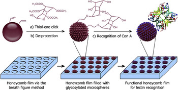
In this study, we synthesized cross-linked PS microspheres that contain surface vinyl groups using DVB and EGDMA as the cross-linkers. We expected to compare the reactivity of surface vinyl groups introduced by DVB and EGDMA in the thiol–ene reaction and then achieve surface functionalization of the microspheres via the thiol–ene reaction. Finally, the functionalized microspheres were assembled in honeycomb porous films to demonstrate a new pathway to fabricate functional patterned films (Scheme 1).
Illustration of synthesis of glycosylated microspheres and formation of functional patterned films. This scheme is not to scale.

II. EXPERIMENTAL
A. Materials
Styrene (St) and DVB were commercially obtained from Sinopharm Chemical Reagent, Co. (Shanghai, China), and distilled under reduced pressure before use. Azobis(isobutyronitrile) (AIBN) was recrystallized from ethanol. Triton X-305 was kindly obtained from Dow Chemical Co. (Shanghai, China). 2-[4-(2-Hydroxyethyl)-1-piperazinyl]ethanesulfonic acid (HEPES) was a commercial product of Nanjing Robiot (Nanjing, China). Fluorescein-labeled concanavalin A (FL-Con A) (Vector, Shanghai, China) was used as received. Polyethylene terephthalate film was kindly provided by Hangzhou Tape Factory (Hangzhou, China) and cleaned with acetone for 2 h before use. Water used in all experiments was deionized and ultrafiltrated to 18.2 MΩ with an ELGA LabWater system (Shanghai, China). All other reagents, such as EGDMA and poly(N-vinyl-2-pyrrolidone) (PVP), were analytical grade and used without further purification.
B. Characterizations
Field-emission scanning electron microscope (SEM Hitachi S4800, Tokyo, Japan) was used to observe the surface morphology of films. The diameter of microspheres was analyzed using ImageJ (v1.42q; Wayne Rasband), and the size and polydispersity were determined by dynamic light scattering (DLS; Malvern Instruments, Worcestershire, UK). Fourier transform infrared (FTIR) spectra were recorded on a Nicolet FTIR/Nexus470 spectrometer (Thermo Fisher Scientific Inc., Waltham, MA). All spectra were taken by 32 scans at a nominal resolution of 1 cm−1. X-ray photoelectron spectroscopy (XPS) was recorded on a PHI-5000C ESCA system (Perkin-Elmer, Waltham, MA) with Al Kαexcitation radiation (1486.6 eV). The take-off angle is 45°. The water contact angles were measured by a DropMeter A-200 contact angle system (MAIST Vision Inspection & Measurement Ltd Co., Ningbo, China) at room temperature. Before measurements, the microspheres were deposited on a clean glass surface to form a uniform film, which was measured after drying in the air. Confocal laser scanning microscopy (CLSM) was performed on a Leica TCS SP5 confocal setup mounted on a Leica DMI 6000 CS inverted microscope (Leica Microsystems, Wetzlar, Germany) and was operated under the Leica Application Suite Advanced Fluorescence program.
C. Synthesis of cross-linked PS microspheres by two-stage dispersion polymerization
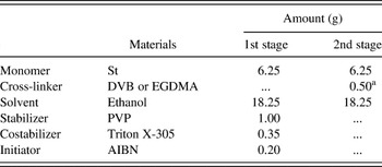
The synthesis was performed according to a slightly modified procedure proposed by Song and Winnik.Reference Song and Winnik39 The recipe for the two-stage dispersion polymerization of St and DVB, or St and EGDMA, in ethanol is listed in Table I. In the first stage, St, ethanol, stabilizer (PVP), costabilizer (Triton X-305), and initiator (AIBN) were added to a 50 mL three-necked bottle equipped with a condenser and a constant pressure drop funnel. After a homogeneous solution formed at room temperature, the solution was deoxygenated by bubbling nitrogen gas for 30 min at room temperature. After that, the flask was placed in a 70 °C oil bath and stirred mechanically at 100 rpm. DVB (or EGDMA) and the remaining St were then dissolved in ethanol at 70 °C under nitrogen flow. When the polymerization had run for 1 h, the hot dissolved DVB (or EGDMA) solution was slowly added into the reaction flask through constant pressure drop funnel in 1 h. After the polymerization run for 18 h, the resulting dispersion was separated by centrifugation, washed with ethanol and followed with water both in triplicate, and then dried under reduced pressure. The amount of DVB was controlled at various proportions to St of 1%, 2%, 3%, 4%, and 5% by weight. The amount of EGDMA was the same as DVB.
Typical recipes for the two-stage dispersion copolymerization of St with DVB or EGDMA.

a The ratio of DVB or EGDMA to St is 4 wt%. Other ratios including 1%, 2%, 3%, and 5% were also used.
D. Measurement of amounts of vinyl groups at microsphere surface
Potentiometric titration was carried out according to the following procedure. 50 mL of 0.1 M KBrO3–KBr (5:1) water solution was added into a 250 mL conical flask, in which 0.2 g PS microspheres and 10 mL water solution including 0.5 mL glacial acetic acid and 0.5 mL methanol had been added. After the conical flask was immersed into the ice water bath, 2 mL of concentrated hydrochloric acid was dropped in and the flask was shaken up for 0.5 h in dark. Then, 1.5 g KI was added and kept in dark place until the formation of homogeneous solution. The solution was titrated by 0.1 M Na2S2O3and turned into pale yellow at the end point. At this moment, 1 mL of 1% amylum was added and then continued to titrate until the blue disappeared. Other samples were operated in the same way, and each sample was repeated 3 times to calculate the mean value.
E. Immobilization of glucose via the thiol–ene reaction
To explore the effects of solvents on the morphology of microspheres, 10 mL ethanol, 0.17 mL 3-mercaptopropionic acid (1.96 mmol, 212 mg), 3.1 mg PS microspheres with 2 wt% DVB (1.96 mmol double bond), and 10 mg AIBN (0.06 mmol) were added to a round bottom flask. The solution was degassed with nitrogen for 20 min, and the flask was then sealed and kept at 70 °C for 24 h. Acetonitrile was also used as solvent for comparison with ethanol.
To introduce glucose, 2,3,4,6-tetra-O-acetyl-β-D-glucopyranosyl thiol and cross-linked PS microspheres in different ratios were added to 10 mL ethanol with 10 mg AIBN (0.06 mmol). The solution was degassed with nitrogen for 20 min. Then, the flask was sealed and kept at 70 °C for 24 h.
F. Filling of microspheres into the pores of honeycomb-patterned films
The simplest method to fill microspheres into pores of patterned porous films is direct deposition by gravity. It should be noted that the film must be prewetted using ethanol before filling. Microspheres dispersed in ethanol were prepared at a given concentration. Patterned porous films were then immersed into the dispersions for 5 min with gentle shaking. Afterward, the film was rinsed with ethanol to remove the microspheres adhered on the surface and dried in air. This process can be repeated for several times to achieve high filling ratio. For lectin recognition, we deprotected the glucose on the surface by dispersing microspheres in MeOH/MeONa for 30 min. Afterward, the microspheres were rinsed with methanol and water.
G. Recognition of lectin
After being fully wetted in HEPES buffer solution [pH 7.5, containing 10 mM HEPES, 0.15 M NaCl, 0.1 mM Ca2+, 0.01 mM Mn2+(not for peanut agglutinin, PNA), 0.08% sodium azide], a piece of film assembled with glucose-functionalized microspheres was dipped into 200 mL of FL-Con A HEPES buffer solution (0.1 mg/mL) and incubated at 25 °C for 2 h. The film was then washed with HEPES buffer solution 6 times. After being dried under reduced pressure at room temperature, images of the lectin-adsorbed film were recorded by CLSM.
III. RESULTS AND DISCUSSION
A. Synthesis and characterization of cross-linked PS microspheres with surface vinyl groups
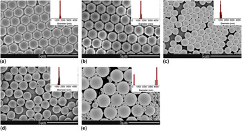
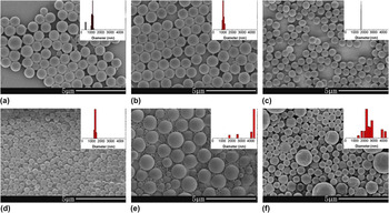
Microspheres show many features such as large specific surface area, high diffusibility and mobility, stable dispersions, high uniformity, and variety in surface chemistry and texture. They have been widely used in the fields of photonics, electronics, and biotechnology.Reference Kawaguchi29 Cross-linked PS microspheres contain surface vinyl groups, which are useful for postfunctionalization. However, the introduction of cross-linker may distort the morphology of microspheres. In this work, cross-linker such as DVB was added 1 h after the initiation of polymerization to avoid its negative influence on the nucleation. As shown in Fig. 1, the addition of DVB at the second stage affects the diameter and polydispersity of the microspheres. Monodisperse microspheres can be obtained when the content of DVB was less than 4 wt%. In this range, the average diameter changes little. As the content of DVB increases to 5 wt%, double size distribution appears. SEM images suggest that the surface of the microspheres is smooth. At the same time, results of DLS (insets in Fig. 1) are consistent with those from SEM observation. When DVB was replaced by EGDMA, similar results were obtained (Fig. 2). It is clear that there are limitations to the content of cross-linker in this two-stage dispersion polymerization when compared with precipitation polymerization, which yields highly cross-linked microspheres but generally low productivity.Reference Wang and Pan40
SEM images and DLS results of PS microspheres with (a) 1 wt%, (b) 2 wt%, (c) 3 wt%, (d) 4 wt%, and (e) 5 wt% DVB as the cross-linker. (a)–(d) 10000x; (e) 5000x.

SEM images and DLS results of PS microspheres with (a) 1 wt%, (b) 2 wt%, (c) 3 wt%, (d) 4 wt%, (e) 5 wt%, and (f) 6 wt% EGDMA as the cross-linker. (a) and (b) 10000x; (c), (d), and (f) 5000x; (e) 2000x.

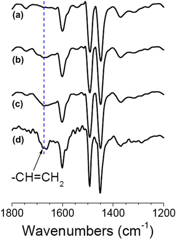
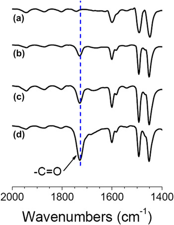
The microspheres cross-linked with DVB were characterized by FTIR (Fig. 3). The absorption peak at 1675 cm−1 is assigned to the residual vinyl groups. The intensity of the absorption peak increases with the feed of DVB, demonstrating the increase of amount of vinyl groups in the microspheres. But it is well known that the molar extinction coefficient of vinyl groups is small, and hence, the intensity of the peak is relatively very weak. For microspheres cross-linked using EGDMA, besides the vinyl groups, there are typical carbonyl groups that show a very strong absorption peak at 1725 cm−1. As shown in Fig. 4, the intensity of peak at 1725 cm−1increases with EGDMA.
FTIR spectra of PS microspheres containing (a) 0 wt%, (b) 1 wt%, (c) 3 wt%, and (d) 5 wt% DVB as the cross-linker.

FTIR spectra of PS microspheres containing (a) 0 wt%, (b) 2 wt%, (c) 4 wt%, and (d) 6 wt% EGDMA as the cross-linker.

It is necessary to determine the amount of vinyl groups on microsphere surface for the subsequent surface functionalization. Amounts of vinyl groups at microsphere surface were further quantitatively measured by potentiometric titration. The bromine index (BI) was calculated from the following expression:
 $${\rm{BI}} = 7790(V_1 - V_2 )c/m\quad,$$
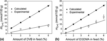
where BI is the quality of bromine consumed in 100 g sample, V 1 and V 2 denote the volume of Na2S2O3 that has been titrated to the blank and sample solutions, respectively, c is the concentration of the standard solution of Na2S2O3, and m is the mass of the sample. The content of the residual vinyl groups can be calculated from the BI by converting the unit of BI (mg/100 g) to mol/100 g, which is the amount of residual vinyl groups. Figure 5shows the calculated and experimental amounts of residual vinyl groups for microspheres with DVB and EGDMA as the cross-linkers. Results reveal that the amounts of surface vinyl groups, about 10 mmol/100 g, are less than that theoretically calculated. It is reasonable because the conversion of cross-linkers cannot reach 100%, and some of the residual vinyl groups are imbedded into the bulk, although the cross-linker was added at the second stage. Besides, for EGDMA system, the amount of surface vinyl groups is slightly less than that of DVB system. As a result, microspheres with 2 wt% DVB and those with 4 wt% EGDMA were used in the following reaction, which have similar surface vinyl groups of about 7 mmol/100 g.
Calculated and experimental amounts of residual vinyl groups on PS microspheres with (a) DVB and (b) EGDMA as the cross-linkers.

B. Attachment of glucose onto the microspheres via the thiol–ene reaction
The thiol–ene reaction was used for surface functionalization of the microspheres. We tried two solvents for the thiol–ene reaction and found that the microspheres distort in acetonitrile because of serious swelling, whereas the morphology changes little when ethanol was used. In addition, the microspheres disperse well in ethanol. Therefore, sulfhydryl glucose was reacted with vinyl groups at the cross-linked microspheres surface using ethanol as the solvent.
We investigated the effects of thiol–ene ratio and reaction time on the reaction degree of vinyl groups at the microspheres surface. The reaction degree fis defined as follows:
 $$f = (n_0 - n_1 )/n_0 \quad,$$
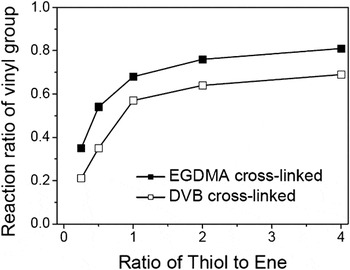
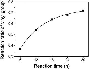
where n 0 and n 1 are the amounts of surface vinyl groups measured before and after the reaction, respectively. It has been reported that the ratio of thiol to ene has great influence on the reaction.Reference Hoyle and Bowman34 Our results also confirm that the reaction degree of vinyl groups greatly depends upon the ratio of thiol to ene. The reaction can be proceeded sufficiently only when the ratio of thiol to ene is larger than 1.0 (Fig. 6). Moreover, the reaction degrees are different between the two cross-linker systems. Microspheres cross-linked with EGDMA show higher reactivity. Hoyle et al.Reference Hoyle, Lee and Roper38 summarized typical enes used in thiol–ene polymerization and pointed out that the reaction rate of methacrylate (EGDMA) is larger than that of St (DVB) because EGDMA is an activated alkene and DVB is not. Another reason is the longer spacer of EGDMA, which decreases steric hindrance for the surface reaction. As shown in Fig. 7, the reaction degree increases first and then levels off with reaction time. It reaches about 65% at 24 h. This reaction degree is relatively low when compared with other bulk thiol–ene reaction, which may be due to steric hindrance of microspheres surface.
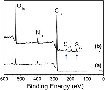
Microspheres bound with protected glucose were characterized by XPS (Fig. 8). For microspheres cross-linked with EGDMA, there are two peaks at binding energy of 281.45 and 529.35 eV, which are attributed to C1s and O1s, respectively. A peak at 396.55 eV that belongs to N1s also appears. The nitrogen element is introduced by the stabilizer PVP added in the polymerization. After the thiol–ene reaction, the peak arising from O1s shows an increased intensity because protected glucose contains a large number of oxygen atoms. Furthermore, peaks at 161.70 and 220.00 eV, arising from S2p and S2s, respectively, can also be clearly observed, which are introduced by sulfhydryl glucose.
Effects of ratios of thiol to ene on the reaction degree of vinyl groups on PS microspheres cross-linked with 4 wt% DVB or EGDMA. The reaction time was 24 h.

Effects of reaction time on the reaction degree of vinyl groups on PS microspheres cross-linked with 4 wt% EGDMA. The ratio of thiol to ene is 1.0.

XPS results of 4 wt% EGDMA cross-linked PS microspheres (a) before and (b) after modified with glucose via thiol–ene reaction.

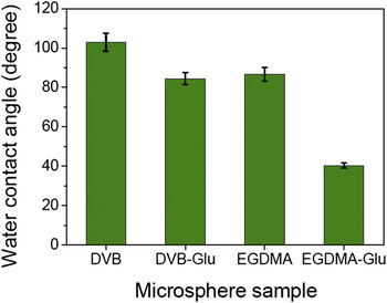
As we know, glycosylated surface is generally hydrophilic, and hence, water contact angle measurements were performed to evaluate the surface hydrophilicity of the modified microspheres.Reference Wang, Ren, Huang, Wu and Xu41 To this end, deprotected microspheres were dispersed in ethanol and deposited on a clean glass to form a uniform film packed with microspheres. Although the film surface is not smooth from the micro point of view, the water contact angles decrease after binding with hydrophilic glucose (Fig. 9). The water contact angles are about 103.0° and 86.8° for PS microspheres cross-linked with DVB and EGDMA, respectively. The introduction of EGDMA endows a slightly more hydrophilic surface. For those bound with glucose, the contact angles sharply decrease to 84.5° and 40.4°, respectively. The decrease is more prominent for EGDMA microspheres than for DVB microspheres, which is consistent with the results of reaction degree of vinyl groups (Fig. 6). The improved surface hydrophilicity also indicates the surface coverage of the microspheres by glucose.
Water contact angles of microsphere films deposited on glass surface. DVB and DVB-Glu represent microspheres cross-linked with DVB before and after immobilization of glucose, respectively, whereas EGDMA and EGDMA-Glu are those cross-linked with EGDMA.

C. Recognition of Con A by honeycomb film filled with glucose-functionalized microspheres
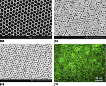
Glucose can specifically interact with lectins such as Con A. Patterned carbohydrate arrays are useful in sensing of biomolecules, investigating carbohydrate–protein interactions, and high throughout screening.Reference Kiessling, Gestwicki and Strong42 According to our previously reported procedure, PS-based honeycomb-patterned porous films were prepared by the breath figure method.Reference Ke, Wan, Chen, Zhang and Xu11 The average pore size can be modulated in the range of 0.8–3.6 μm when the polymer concentration changes from 3 to 1.5 mg/mL. In this work, PS microspheres cross-linked with 4 wt% EGDMA have an average diameter of 1.35 μm. Therefore, patterned film with a pore diameter of about 2 μm was used [Fig. 10(a)]. The microspheres were dispersed in ethanol that can effectively wet the porous film to give a dispersion of 2 mg/mL and then were assembled into the pores of the film forming a functional microspheres array [Fig. 10(b)]. This filling process is more convenient compared with other methods such as chemical attachment. It can be seen that most pores have been filled with one microsphere. Fluorescence results elucidate that the glucose-functionalized film can recognize Con A [Fig. 10(d)]. However, the control sample of film filled with blank microspheres does not recognize Con A, and the glucose-functionalized film cannot recognize fluorescein-labeled PNA, both of which resulted in a totally black image under the same observation conditions.Reference Ke, Wan and Xu6, Reference Ke, Wan, Zhang and Xu43
SEM images of (a) free honeycomb-patterned film, (b) film filled with EGDMA-Glu microspheres, and (c) film adsorbed with fluorescein isothiocyanate-labeled Con A. (d) Fluorescence image of sample (c).

IV. CONCLUSIONS AND PERSPECTIVE
PS microspheres cross-linked with DVB or EGDMA have been synthesized by a two-stage dispersion polymerization and functionalized with glucose via thiol–ene reaction. Up to 4 wt% DVB or EGDMA can be introduced into the microspheres without increasing the polydispersity. DVB or EGDMA leads to residual vinyl groups of about 7 mmol/100 g at the microsphere surface, on which glucose can be bound via the thiol–ene reaction. Vinyl groups of EGDMA show relatively higher reactivity than that of DVB. The microspheres with deprotected glucose were then assembled into the pores of honeycomb-patterned porous films prepared by the breath figure method, forming functional patterned films that can recognize lectins such as Con A. This work opens a new top–down approach to the preparation of functional patterned films, showing an advantage of easy and diverse functionalities based on the microspheres.
ACKNOWLEDGMENTS
Financial support from the National Natural Science Foundation of China (Grant No. 51173161) and the Zhejiang Provincial Natural Science Foundation of China (Grant No. Y4110076) is gratefully acknowledged.