1. Introduction
Thrombolites are benthic microbial carbonates recognized by their patchy mottled/clotted structures with no internal lamination (Aiken, Reference Aiken1967; Tang et al. Reference Tang, Shi, Jiang, Pei and Zhang2012) and are composed of calcimicrobes, micrite and peloids (Riding, Reference Riding2008). Kershaw et al. (Reference Kershaw, Li, Crasquin-Soleau, Feng, Mu, Collin, Reynolds and Guo2007) applied the term digitate dendrolite to Permian–Triassic boundary microbialites in Sichuan, China. However, Riding (Reference Riding, Reitner, Quéric and Arp2011) presented a plausible argument that a dendrolitic form seen in vertical section may also be classified as a thrombolite because in transverse section the branched structure appears as clots on a cut surface (Riding, Reference Riding, Reitner, Quéric and Arp2011; Kershaw et al. Reference Kershaw, Zhang and Li2021). Thrombolites are found globally in various modern depositional environments such as open subtidal settings (Planavsky & Ginsburg, Reference Planavsky and Ginsburg2009; Myshrall et al. Reference Myshrall, Mobberley, Green, Visscher, Havemann, Reid and Foster2010; Mobberley et al. Reference Mobberley, Ortega and Foster2012), freshwater (Laval et al. Reference Laval, Cady, Pollack, McKay, Bird, Grotzinger, Ford and Bohm2000; Gischler et al. Reference Gischler, Gibson and Oschmann2008), alkaline (Arp et al. Reference Arp, Reimer and Reitner2003) and hypersaline (Puckett et al. Reference Puckett, McNeal, Kirkland, Corley and Ezell2011) lakes, and hot springs (Campbell et al. Reference Campbell, Francis, Collins, Gregory, Campbell, Greinert and Aharon2008). Early works suggested that the clotted fabrics of thrombolites may have resulted from the destruction of stromatolitic laminae by metazoans or diagenesis (Hofmann, Reference Hofmann1973; Walter & Heys, Reference Walter and Heys1985). Research on modern thrombolites in the Bahamas (Highborne Cay; Myshrall et al. Reference Myshrall, Mobberley, Green, Visscher, Havemann, Reid and Foster2010), Australia (Shark Bay; Jahnert & Collins, Reference Jahnert and Collins2012) and Kuwait (Arabian Gulf; AlShuaibi et al. Reference AlShuaibi, Khalaf and Al-Zamel2015) have improved our knowledge of these kinds of calcareous microbialites and also confirmed their primary biogenic origin. Similar to other microbialites, thrombolites are formed from complex interactions between microbial communities and the surrounding waters, whereas microbially induced calcification and/or trapping and binding of sediments are the most important processes involved in their growth and expansion (Burne & Moore, Reference Burne and Moore1987; Riding, Reference Riding2000; Planavsky & Ginsburg, Reference Planavsky and Ginsburg2009; Jahnert & Collins, Reference Jahnert and Collins2012).
The lower Cambrian deposits in Morocco are widely exposed in the Anti-Atlas belt and High Atlas Mountains (Álvaro et al. Reference Álvaro, Benziane, Thomas, Walsh and Yazidi2014). During this period, the marine platform of the Anti-Atlas was part of the northern margin of the Gondwana supercontinent. Microbial reefs (dominated by stromatolites) remained unaffected until the early Cambrian Stage 3 (Atdabanian). During the formation of the Tiout Member (upper unit of the Igoudine Formation) an important palaeoecological event took place, characterized by the replacement of the microbial consortium (stromatolite-dominated) by thrombolites and shelly metazoans (Hupé, Reference Hupé1960; Schmitt & Monninger, Reference Schmitt, Monninger and Flügel1977; Schmitt, Reference Schmitt1979; Destombes et al. Reference Destombes, Hollard, Willefert and Holland1985; Debrenne & Debrenne, Reference Debrenne and Debrenne1995; Álvaro & Clausen, Reference Álvaro and Clausen2006; Álvaro & Debrenne, Reference Álvaro and Debrenne2010; Clausen et al. Reference Clausen, Álvaro and Zamora2014). Several authors have mentioned the first episode of thrombolite reefs with archaeocyaths in the lower Cambrian (Series 2) Tiout Member in the western Anti-Atlas (Benssaou & Hamoumi, Reference Benssaou and Hamoumi2004; Álvaro & Debrenne, Reference Álvaro and Debrenne2010; Álvaro et al. Reference Álvaro, Benziane, Thomas, Walsh and Yazidi2014); however, the detailed study and interpretation of this event is still lacking. The current work is focused on the details of the structures, mineralogy and geochemistry, providing a genesis model for the first thrombolite reef complex containing archaeocyaths in the lower Cambrian of Morocco.
2. Geological setting and stratigraphy
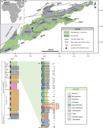
The Anti-Atlas Mountains are a c. 1300 km long NE–SW-trending belt in the central part of Morocco (Fig. 1a, b). The southern slope of the Anti-Atlas contains well-exposed Palaeozoic sedimentary successions resting on the Precambrian Pan-African orogen (Destombes et al. Reference Destombes, Hollard, Willefert and Holland1985; Geyer & Landing, Reference Geyer and Landing1995). It comprises a ∼1800 m thick mixed siliciclastic-carbonate deposit on the north Gondwana margin (western Anti-Atlas, Morocco) directly deposited on the ∼2000 m thick volcanic and volcaniclastic rocks of the Ediacaran Ouarzazate Supergroup (577–560 Ma) (Thomas et al. Reference Thomas, Fekkak, Ennih, Errami, Loughlin, Gresse, Chevallier and Liégeois2004; Gasquet et al. Reference Gasquet, Ennih, Liégéois, Soulaimani, Michard, Michard, Saddiqi, Chalouan and Frizon de Lamotte2008; Walsh et al. Reference Walsh, Benziane, Aleinikoff, Harrison, Yazidi, Burton, Quick and Saadane2012; Fig. 1b). This Ediacaran–Cambrian succession accumulated on the thermally subsiding continental shelf (Maloof et al. Reference Maloof, Schrag, Crowley and Bowring2005) in the upper part of the eastern Anti-Atlas platform. The Tata Group is well exposed in the Taroudant province, and has previously been described in the Tiout, Amouslek and Tazemmourt areas (Álvaro et al. Reference Álvaro, Ezzouhairi, Vennin, Ribeiro, Clausen, Charif, Ait Ayad and Moreira2006), and within the Issafen and Fouanou synclines. The ∼1000 m thick Tata Group consists of four formations: Igoudine, Amouslek, Issafen and Asrir (Fig. 1c). The basal Igoudine and Amouslek deposits represent carbonate-dominated environments such as peritidal flats and ooid shoals. The upper part of the Igoudine Formation has been described only in the Tiout area as the Tiout Member (Álvaro et al. Reference Álvaro, Ezzouhairi, Vennin, Ribeiro, Clausen, Charif, Ait Ayad and Moreira2006), containing the boundary interval where the microbial consortium (stromatolite-dominated) sediments were first replaced by thrombolite reefs with archaeocyaths and shelly metazoans (Álvaro et al. Reference Álvaro, Benziane, Thomas, Walsh and Yazidi2014). The thickness of the overlying Amouslek Formation ranges between 20 and 220 m and consists of variegated shales with interbedded limestones.
(a) Location map of the Anti-Atlas of Morocco. (b) Simplified geological sketch showing the distribution of the Precambrian–Cambrian outcrops in the Anti-Atlas Mountains (drawn after Saadi et al. Reference Saadi, Hilali, Bensaïd, Boudda and Dahmani1983). (c) Stratigraphic section through the lower Cambrian of the Fouanou syncline. (d) Detailed lithostratigraphic section of the studied horizon in the Fouanou syncline (the diameters of archaeocyath cups were measured within the levels L1 and L2).

The Tommotian–Atdabanian boundary interval is widely exposed in the western Anti-Atlas (Fouanou syncline). One characteristic section comprising the Igoudine and Amouslek formations has been logged by us (Fig. 1d). The stratigraphic succession in this area comprises similar sedimentary facies to those described within the type sections of the Tiout and Amouslek areas (Álvaro et al. Reference Álvaro, Ezzouhairi, Vennin, Ribeiro, Clausen, Charif, Ait Ayad and Moreira2006; Álvaro & Debrenne, Reference Álvaro and Debrenne2010).
The lowermost unit of Igoudine Formation (∼40 m thick) consists of massive black oolithic limestones, with rare dome-shaped stromatolites, symmetric ripples and cross-stratifications. In the middle unit (∼35 m thick) dominated by stratified dolostones, displaying crinkled dome-shaped and stratiform stromatolites, wave ripples and rare cross-stratifications, horizons shows slumped and faulted stromatolites. Based on the sedimentary facies, both previous units were deposited in upper subtidal to intertidal environments. The upper unit of the Igoudine Formation formed by massive bedded oolitic limestone (‘black oolitic limestone facies’ of Monninger, Reference Monninger1979 and Schmitt, Reference Schmitt1979) is characterized by the emergence of small-sized archaeocyaths, Adtabanian in age, including Dictyocyathus, Erismacoscinus and Agastrocyathus. Some beds exhibit cross-stratifications and rare wave ripples and erosion surfaces. The latter unit is overlain by the Tiout Member dominated by dendritic thrombolites with archaeocyaths, which is the first microbial archaeocyathan reef barrier of the Anti-Atlas. Thrombolite–archaeocyathan reefs are characterized by successive reef-growth phases delimited by surfaces of reef-growth interruption, which suggest that this reef barrier may have formed under low-energy conditions with occasional high-energy events, generating erosion surfaces. The latter conditions can occur between the middle to lower subtidal zone (Jahnert & Collins, Reference Jahnert and Collins2012). The Amouslek Formation consists of variegated shales and siltstones, strongly bioturbated, recording a significant sea-level change from shallow water restricted conditions to deeper open sea conditions (Álvaro & Debrenne, Reference Álvaro and Debrenne2010). The lower part of this unit displays numerous archaeocyathid-bioherms and patch reefs. The upper part consists mainly of shales and fine-grained sandstones with interbedded oolitic limestones, cross-stratifications and wave ripples, recording rhythmic transgressive–regressive depositional sequences.
3. Materials and study methods
Thrombolites were macroscopically examined in the field and in polished slabs in the laboratory. About 70 thin-sections were made for microfacies analyses using an Olympus BH2 polarizing binocular microscope. Platinum-coated and uncoated freshly broken sample fragments were observed for micro/nanostructures using an FEI Teneo Volume Scope scanning electron microscope (SEM), operated at a 5 to 20 kV accelerating voltage and a varying working distance of between 9 and 11 mm, at UFR SFA Pole Biology University of Poitiers, France. Semi-quantitative element analyses of sub-micron-sized spots were determined using an EDAX energy-dispersive X-ray spectrometer (EDS) connected to the SEM at 20 kV and a working distance of 9 mm. The bulk mineral compositions of some thrombolite fragments from the bottom and upper part of the lower reef complex of the Igoudine Formation were determined using a Bruker D8 ADVANCE X-ray diffractometer (CuKα radiation) with operating conditions of 40 kV and 40 mA and a 0.025/s step size.
4. Results
4.a. Thrombolytic reefs with archaeocyaths
4.a.1. Morphology and macrofabric
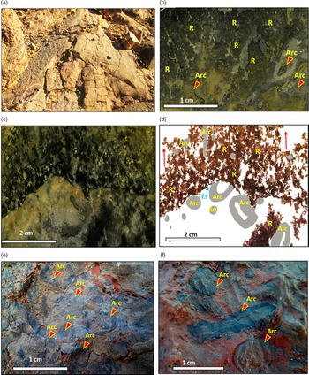
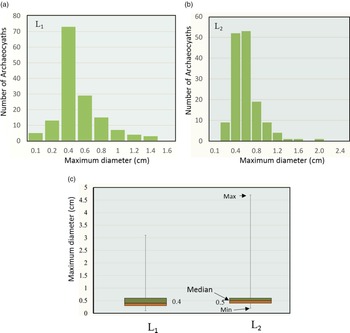
Thrombolite–archaeocyathan reefs of the Igoudine Formation are characterized by successive reef-growth phases delimited by surfaces of reef-growth interruption (Fig. 2a). The Atdabanian thrombolites of the Igoudine Formation exhibit dark micritic mesoclots in varying sizes and shapes and have maximum diameters ranging between 5 and 20 mm (Fig. 2a, b). The mesoclots are interlaced to form upward-growing dendritic structures (Fig. 2b, c, d). Scattered small-size archaeocyaths are apparent within the studied materials (Fig. 2b, c, d, e, f), but they never exceed 20 % of the total rock volume. Three genera have been identified in the studied horizon including Dictyocyathus, Erismacoscinus and Agastrocyathus. They are usually less than 20 mm in height and preserved in their growth position. The maximum diameters of 150 cups of archaeocyaths measured within two spaced reef cores (Fig. 2b) vary from 1 to 31 mm with a mean of 4.9 mm in the lower level (L1) and from 2 to 47 mm with a mean of 5.6 mm in the upper level (L2) (Fig. 3). Dendritic thrombolites in the lower Cambrian Series 2 of the Founou syncline show well-preserved calcimicrobes such as Renalcis, Epiphyton and Girvanella.
Macroscopic features of the thrombolite reef with archaeocyaths. (a) Outcrop photograph of thrombolite–archaeocyathan reefs characterized by successive reef-growth phases delimited by surfaces of reef-growth interruption (length of hammer scale is 29.5 cm). (b) Sample slab showing dark grey dendritic mesoclots and light grey matrix with scarce archaeocyaths (Arc). (c, d) Photographs and sketches of longitudinal section showing dendritic clotted fabric growing upwards (red arrows) (Arc – archaeocyath; Es – erosion surface). (e, f) Photographs of transverse sections showing small-sized regular and irregular archaeocyaths (Arc).

Distribution patterns of the maximum diameter of 150 cups of archaeocyaths measured within two spaced levels (a) L1 and (b) L2 (shown in Fig. 1d). (c) Box-plots of cup diameters in both L1 and L2.

4.a.2. Microscale characteristics
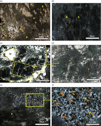
Dendritic thrombolites display well-defined clots containing numerous calcimicrobes, including Renalcis, Epiphyton bundles and subordinate Girvanella tubes visible in thin-sections (Fig. 4a). The Renalcis group is the most abundant dendritic microbial fossil recorded within the studied reefs, consisting of a growing-upward shrub-like array of connected chambers made up of aphanitic micrite (Fig. 4a, b). Micrites, microspar and rare scattered clotted peloids (50 to 100 µm thick) filled the voids separating the Renalcis microframes. The diameters of the chambers range from 0.2 to 1.5 mm, and they commonly occupy the void spaces between the archaeocyath skeletons (Fig. 2c). Calcified microbial thrombolites show a tabular morphology. The Epiphyton calcimicrobe occurs in archaeocyath dominated reefs, encrusting the archaeocyath skeleton (Fig. 4c, d). They are observed as radiating branches consisting of aphanitic micrites and are usually grouped in relatively large colonies (up to 5 mm in diameter) to form a chambered structure (Fig. 4c, d), similar to Renalcis. However, it is difficult to distinguish between both structures in some cases, especially when the filaments are densely connected. Spaced Girvanella tubes are rare and usually separated from the Renalcis and Epiphyton microframes. The light grey matrix surrounding the Renalcis and Epiphyton frameworks consists of micrite and microspar with randomly scattered dark grey micritic peloid-like aggregates with diffuse edges (Fig. 4e, f). Irregular cavities and geopetal structures are common and are filled by greyish microspar or fibrous calcite.
Microscopic features of thrombolites observed under plane-polarized light. (a, b) Micrographs showing the various forms of Renalcis chambers (R) and Girvanella tubes. (c) Photomicrograph showing Epiphyton chambers (E) attached to archaeocyaths and surrounded by sparitic matrix (Sp). (d) A close-up view of the boxed area in (c) showing Epiphyton chambers with radiating filaments. (e) Photomicrograph of peloidal fabrics in the cryptic space between Renalcis chambers. (f) A close-up view of the boxed area in (e) showing the peloid (P) microspar fabric with a rare cloud of iron oxides (IO) derived from pyrite oxidation.

4.a.3. The nature of the matrix
The matrices between the Renalcis and Epiphyton chambers consist predominantly of clotted micrite and microspar, occurring as scattered poorly sorted peloid ‘grainstone-packstone’ (Fig. 4e, f). Allochthonous fine silt particles, including recognizable quartz and feldspar, are locally incorporated into the thrombolite but are generally scarce and only visible in samples taken beneath the Amouslek Formation shales. The size of the peloids that dominate the matrix microfabric mostly range between 30 and 80 µm. The high-density and coarse-grained peloids are common within the empty spaces between the Renalcis chambers (Fig. 4a, e, f). Some peloids show well-defined boundaries with simple outlines, scattered within the microspar matrix. However, other peloids exhibit irregular forms and occur as clotted aggregates with no well-defined margins. In contrast, the fine-gained peloids are spaced relatively far from the Renalcis microframes (Fig. 4b, e). The Renalcis and Epiphyton chambers also host the fine-grained peloids but in a lower density. A little pyrite also occurs within the matrix under reflected light and is surrounded by a cloud of iron oxides (Fig. 4f).
4.b. Ultra-fabrics of thrombolites
SEM observations of the Renalcis and Epiphyton chambers revealed amorphous translucent sheet-like structures interpreted as extracellular polymeric substances (EPSs) (Arp et al. Reference Arp, Reimer and Reitner2001; Decho et al. Reference Decho, Visscher and Reid2005; Barrett et al. Reference Barrett, Spentzos and Works2009; Mishra et al. Reference Mishra, Fischer and Bäuerle2009; Jones, Reference Jones2011; Zatoń et al. Reference Zatoń, Kremer and Marynowskii2012; Zhang et al. Reference Zhang, Shi, Jiang, Tang and Wang2015; Mackey et al. Reference Mackey, Sumner, Hawes, Jungblut, Lawrence, Leidman and Allen2017). Interpreted bacterial remains, pyrite framboids and a closely associated organomineral complex (biologically induced and biologically influenced mineralization), including nanoglobules, polyhedrons and micropeloids, are scattered within the interpreted mineralized EPS matrix.
4.b.1. Extracellular polymeric substances (EPSs)
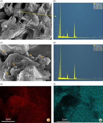
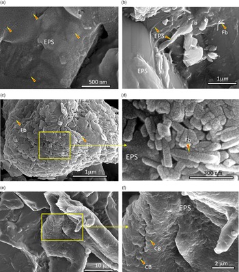
The EPSs show an amorphous flat and translucent mucus-like matrix (Fig. 5a, b, c) and form a meshwork of subpolygonal pits and walls (1–3 μm thick; Fig. 5a). EDS analyses show that the EPS matrix is composed of C, O, Ca, Si, Mg, Al, K, Cl and rare detectable Fe and S (Fig. 5b, d, e, f). The EPS surface in some areas shows nano-sized cracks (Fig. 6a). Rod-like forms and uniform spherical bodies are similar to those that have been reported in microbialites in many areas and interpreted as bacterial fossils (Perri & Tucker, Reference Perri and Tucker2007; Spadafora et al. Reference Spadafora, Perri, Mckenzie and Vasconcelos2010; Perri et al. Reference Perri, Tucker and Spadafora2012; Shen et al. Reference Shen, Qin, Tenger, Pan, Yang and Bian2017). Bacterial fossils are commonly attached or entombed within the EPS films in the well-preserved samples of Renalcis chambers (Figs 5c, 6b, c, d, e, f). These bacterial cell remains are mainly composed of filamentous and micro-spherical forms and are separated into filamentous tube-like fossils and filamentous rod-like to slightly ellipsoidal-shaped forms based on their different shapes and dimensions. The filamentous tube-like fossils have slightly curved tubes and are either isolated or grouped forming colonies (Figs 5c, 6b, c, d). The filamentous rod-like to slightly ellipsoidal-shaped cells (250–300 nm long and 50 nm wide) are usually found in associations to form colony-like clusters, up to 5 µm in diameter (Fig. 6c, d). Nanoscale SEM observations show the presence of fine granular textures on the surfaces of the possible bacteria fossils (Fig. 6c, d). Micro-spherical forms interpreted as coccoids, with uniform diameters of 600 nm, are grouped in colonies probably associated with irregular organic fragments (Fig. 6c, d).
Scanning electron microscopy photomicrographs and EDS of microbial structures in the micritic Renalcis chamber. (a) Close-up view of honeycomb-like structure consisting of translucent polygonal pits and walls interpreted as mineralized extracellular polymeric substance (EPS) matrix. (b) EDS spectrum and elemental quantitative data of the spot in (a). (c) EPS relics containing filamentous bacteria (Fb). (d) EDS spectrum and elemental quantitative data of the surface shown in (c) (Pt element resulted from platinum coating). (e, f) Elemental mappings of carbon (C) and calcium (Ca) of the surface in (c).

Scanning electron microscopy photomicrographs of EPSs and bacterial fossils. (a) EPSs showing nano-sized cracks that possibly result from dehydration of EPS films during early diagenesis. (b) EPS relics coating micrite crystals with filamentous bacterial fossils. (c) Filamentous bacteria (Fb) grouped in a colony. (d) Magnified view of the boxed area in (c) showing the fine granular texture of the surface of bacteria with nano-sized cracks. (e) Grainy surface on possible organic residue. (f) Magnified view of the boxed area in (e) showing the micro-spherical form of possible coccoidal bacteria (CB) covering the surface of organic remains and associated mineralized EPSs.

4.b.2. Organomineral complex
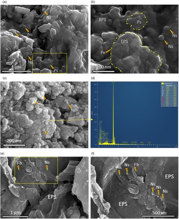
Higher magnification SEM observations of Epiphyton and Renalcis chambers and the clotted peloids show abundant nanoglobules that are 40–80 nm in size (Fig. 7a, b, c, d, e, f) and often fused into clusters to form anhedral polyhedrons of various sizes, depending on the number of the associated aggregates (Fig. 7a, b, c). The polyhedrons coalesce into irregular micrometre-scale micropeloids. The nanoglobules are closely related to EPS relics or clustered on their surfaces (Fig. 7e, f).
Scanning electron microscopy photomicrographs showing organominerals and detrital micrite in thrombolites. (a) Scattered nanoglobules (Ns) coalesced to form polyhedrons (Po) within EPS films. (b) Magnified view of the boxed area in (a) showing nanoglobules (Ns) and polyhedrons (Po) fused into micropeloids (Pe), closely associated with EPS relics. (c) Nanoglobules (Ns) coalesced to form polyhedrons (Po) and irregular micritic particles. (d) EDS spectrum and elemental quantitative data for nanoglobules (analysed spot (+) in (c)) (Pt element resulted from platinum coating). (e) Nanoglobules closely associated with EPSs and the probable filamentous bacteria (Fb). Nanoglobules (Ns) are visible on EPS flats. (f) Magnified view of the boxed area in (e) showing nanoglobules and nano-cracks resulting from dehydration of EPSs during early diagenesis.

4.b.3. Framboidal pyrite
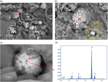
Pyrite framboids are common in the dark micrite forming the Renalcis and Epiphyton chambers. They are embedded in platy or amorphous EPSs (Fig. 8a, b, c). The diameter of the framboids varies between 4 µm and 40 µm, and they form euhedral (octahedral) microcrystals of 0.5–2 µm in size. The framboidal pyrite entombed in EPS relics contains Fe, S and Ca (Fig. 8d).
Scanning electron microscopy photomicrographs of pyrite framboids. (a, b) Abundant pyrite framboids (PF) consisting of equidimensional pyrite microcrystals associated with mucus-like EPS relics. (c) Close-up view of the boxed area in (b) showing pyrite framboid. (d) EDS spectrum of pyrite crystal in (c) (+ indicates the position of analysed spot). Fe, S and Ca elements are common in the pyrite crystals.

4.c. Mineralogical composition of thrombolites
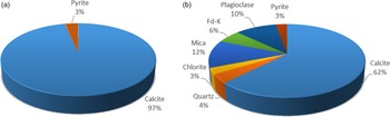
Mineralogical compositions of the thrombolite sediments show differences between the bottom and the top of the lower reef complex (Fig. 9a, b). Samples from the basal parts of the section contained mainly carbonate and a small amount of quartz and pyrite (Fig. 9a). However, the upper part of the complex, beneath the Amouslek calcareous shales, is dominated by carbonate and siliciclastic minerals represented by feldspar, mica, silica and a small amount of pyrite (Fig. 9b).
Bulk rock mineralogical composition of selected samples from (a) the bottom and (b) the top of the lower reef complex of the Igoudine Formation (see Fig. 1d).

5. Discussion
5.a. Components of thrombolite reefs
Early Cambrian microbial reefs containing archaeocyaths have been reported globally (Debrenne et al. Reference Debrenne, Gandin and Rowland1989; Rees et al. Reference Rees, Pratt and Rowell1989; James & Gravestock, Reference James and Gravestock1990; Gandin & Luchinina, Reference Gandin, Luchinina, Barattolo, De Castro and Parente1993; Gandin & Debrenne, Reference Gandin and Debrenne2010). In the Anti-Atlas, stromatolite-dominated microbial reefs remained unaffected until early Cambrian (Atdabanian) time, when thrombolite reefs with archaeocyaths became widespread (Álvaro & Debrenne, Reference Álvaro and Debrenne2010). These changes could be related to sea-level fluctuation, from shallow water restricted to deeper open sea conditions (Álvaro & Debrenne, Reference Álvaro and Debrenne2010).
The scattered small size of the archaeocyaths, their preservation in life position, microbial fabric attachments to Renalcis microframes, encrustation by Epiphyton chambers and a Girvanella crust, as well as their lower abundance (∼20 %) in thrombolite reefs ecosystems, suggest that they were reef dwellers rather than local framework builders. They usually required the presence of microbial elements to form frameworks (Debrenne, Reference Debrenne2007). Archaeocyath–Renalcis–Epiphyton reefs occur in low-energy subtidal conditions, mostly from lower subtidal to the outer ramps (Debrenne et al. Reference Debrenne, Gandin and Courjault-Radé2002; Gandin et al. Reference Gandin, Debrenne, Debrenne, Álvaro, Aretz, Boulvain, Munnecke, Vachard and Vennin2007), but they are also present on high-energy platform margins (Kruse et al. Reference Kruse, Zhuravlev and James1995; Riding & Zhuravlev, Reference Riding and Zhuravlev1995). The incorporation of fine-grained sediments into the thrombolite reefs suggests that they formed under low-energy conditions. The presence of erosion surfaces (Fig. 2a, c, d) indicates that reef growth was likely interrupted by episodic storm events. The remarkable change in diameter of archaeocyaths between both horizons L1 and L2 (Fig. 1d) could be related to variations in the chemical and/or hydrodynamic conditions of the environment.
The Igoudine thrombolite reefs are composed of calcimicrobes: Renalcis with subordinate Epiphyton and Girvanella (Fig. 4a, b, c, d). The taxonomic position of Renalcis is controversial, but is it interpreted as red algae, foraminifera and most commonly as cyanobacteria (Mamet, Reference Mamet and Riding1991). Pratt (Reference Pratt1984) interpreted Renalcis as a ‘diagenetic taxa’ resulting from the calcification of coccoid blue-green algae grouped in colonies. However, some authors suggested Renalcis constitutes fossilized biofilm clusters, resulting from the calcification process due to the activity of heterotrophic bacteria (Stephens & Sumner, Reference Stephens and Sumner2002; Turner et al. Reference Turner, James and Narbonne2000). Woo et al. (Reference Woo, Chough and Han2008) and Adachi et al. (Reference Adachi, Nakai, Ezaki and Liu2014) suggested that the Renalcis resulted from diagenetic alteration of Epiphyton chambers based on their similar structures. However, Luchinina (Reference Luchinina2009) suggested that the Renalcis and Epiphyton structures may represent two different steps in the lifecycle of cyanobacteria.
SEM observations of the dark micrite of the Renalcis chambers revealed an amorphous flat and translucent mucus-like matrix (Fig. 5a, b, c, d) interpreted by many authors as EPS biofilms (Arp et al. Reference Arp, Reimer and Reitner2001; Decho et al. Reference Decho, Visscher and Reid2005; Barrett et al. Reference Barrett, Spentzos and Works2009; Mishra et al. Reference Mishra, Fischer and Bäuerle2009; Jones, Reference Jones2011; Zatoń et al. Reference Zatoń, Kremer and Marynowskii2012; Zhang et al. Reference Zhang, Shi, Jiang, Tang and Wang2015; Mackey et al. Reference Mackey, Sumner, Hawes, Jungblut, Lawrence, Leidman and Allen2017). Some samples revealed honeycomb-like patterns similar to those observed in modern microbialites (Défarge et al. Reference Défarge, Trichet and Coute1994, Reference Défarge, Trichet, Jaunet, Robert, Tribble and Sansone1996; Dupraz et al. Reference Dupraz, Reid, Braissant, Decho, Norman and Visscher2009; Spadafora et al. Reference Spadafora, Perri, Mckenzie and Vasconcelos2010). Furthermore abundant bacterial fossils have been described including rod-like bacteria and uniform coccoidal bacteria similar to those reported in modern and ancient microbialites (Perri & Tucker, Reference Perri and Tucker2007; Spadafora et al. Reference Spadafora, Perri, Mckenzie and Vasconcelos2010; Perri et al. Reference Perri, Tucker and Spadafora2012; Shen et al. Reference Shen, Qin, Tenger, Pan, Yang and Bian2017) and usually associated with EPSs. However, in many cases, studied fossils of the probable bacteria in our thrombolite reefs are very small and close to the lowest size boundary for the prokaryotes (Crawford, Reference Crawford2007), but such small, usually coccoid, forms have previously been described from recent and fossil microbialites (Spadafora et al. Reference Spadafora, Perri, Mckenzie and Vasconcelos2010; Perri & Spadafora, Reference Perri, Spadafora, Tewari and Seckbach2011).
Our observations suggest a bacterial origin for the Renalcis chambers and support the explanation proposed by Stephens & Sumner (Reference Stephens and Sumner2002) and Turner et al. (Reference Turner, James and Narbonne2000) that suggested a bacterial origin for the Renalcis group, resulting from the calcification process of EPSs and heterotrophic bacteria. The particular forms of Renalcis and Epiphyton can be related to the significant difference in bacterial assemblages involved in their formation.
5.b. Origin of micrite
Micrite is widely used to refer to a rock composed of fine-grained calcite crystals (less than 4 µm) produced in situ or derived from physical transport and deposition of fine particles (Fluegel, Reference Fluegel2010). Microscopic study and higher magnification SEM observations of the micromorphological structures allowed us to recognize the autochthonous micrite deposited in situ through organomineralization of organic compounds and calcification of bacterial sheaths and EPSs.
5.b.1. Biomineralization
The formation of the Igoudine thrombolites is analogous to that recognized in modern microbialites (Dupraz et al. Reference Dupraz, Visscher, Baumgartner and Reid2004; Perri & Spadafora, Reference Perri, Spadafora, Tewari and Seckbach2011) and could involve two major biomineralization processes: (i) replacement of organic matter by biominerals and (ii) calcification or encrustation of bacterial sheaths and EPSs.
Nanoglobules observed in the Igoudine thrombolites are closely associated with EPS relics (Fig. 7a, b, c, e, f), implying their possible origin from anaerobic degradation of EPS biofilms (Aloisi et al. Reference Aloisi, Gloter, Kruger, Wallmann, Guyot and Zuddas2006; Sánchez-Román et al. Reference Sánchez-Román, Vasconcelos, Schmid, Dittrich, McKenzie, Zenobi and Rivadeneyra2008; Perri et al. Reference Perri, Tucker and Spadafora2012; Zhang et al. Reference Zhang, Shi, Jiang, Tang and Wang2015). Nanoglobules fuse into variably shaped polyhedrons and micropeloids; similar patterns have been reported in microbial oncoids (Zhang et al. Reference Zhang, Shi, Jiang, Tang and Wang2015). Polyhedrons formed by nanoglobules may have served as primary nuclei for subsequent carbonate crystal growth (Sánchez-Román et al. Reference Sánchez-Román, Vasconcelos, Schmid, Dittrich, McKenzie, Zenobi and Rivadeneyra2008; Spadafora et al. Reference Spadafora, Perri, Mckenzie and Vasconcelos2010; Perri & Spadafora, Reference Perri, Spadafora, Tewari and Seckbach2011; Tang et al. Reference Tang, Chen, Santosh, Zhong and Yang2013; Zhang et al. Reference Zhang, Shi, Jiang, Tang and Wang2015). These organominerals can replace the organic part when EPSs and bacterial sheaths have been largely degraded, after the microenvironment become anoxic or dysoxic (Dupraz et al. Reference Dupraz, Visscher, Baumgartner and Reid2004; Spadafora et al. Reference Spadafora, Perri, Mckenzie and Vasconcelos2010; Perri & Spadafora, Reference Perri, Spadafora, Tewari and Seckbach2011; Zhang et al. Reference Zhang, Shi, Jiang, Tang and Wang2015). During this process, precipitation of carbonate minerals happened after intensive decomposition of EPSs and bacterial sheaths so that microbial fossils are rarely preserved (Bartley, Reference Bartley1996; Zhang et al. Reference Zhang, Shi, Jiang, Tang and Wang2015).
Encrustation of bacterial sheaths and EPSs can occur by increasing alkalinity through photosynthetic removal of CO2 from surrounding waters via photosynthetic prokaryotes and eukaryotic microalgae (Arp et al. Reference Arp, Reimer and Reitner2001, Reference Arp, Reimer and Reitner2003; Obst et al. Reference Obst, Wehrli and Dittrich2009). Bacterial sheaths and EPSs previously secreted by bacteria absorb bivalent cations (Ca2+, Mg2+); the latter decreases the pH of the microenvironment and inhibits carbonate nucleation and precipitation (Dupraz et al. Reference Dupraz, Visscher, Baumgartner and Reid2004; Aloisi et al. Reference Aloisi, Gloter, Kruger, Wallmann, Guyot and Zuddas2006; Braissant et al. Reference Braissant, Decho, Dupraz, Glunk, Przekop and Visscher2007). Such a scenario can favour the preservation of EPSs because encrustation happens before advanced microbial organic matter degradation. For carbonate minerals, the primary environmental factor controlling the biomineralization is pH, and an increase in pH induces the carbonate biomineralization (Zhao et al. Reference Zhao, Yan, Tucker, Han, Zhao, Mao, Peng and Han2020). In the case of possible cyanobacteria, like ancient Girvanella, Epiphyton and Renalcis, photosynthetic uptake of CO2 and/or HCO3 could raise pH in ambient waters (Dupraz et al. Reference Dupraz, Visscher, Baumgartner and Reid2004; Zhao et al. Reference Zhao, Yan, Tucker, Han, Zhao, Mao, Peng and Han2020). On the other hand, heterotrophic bacteria, like sulfate-reducing bacteria at the lower levels of microbial mats, perform ammonification and carbonic anhydrase (CA) catalysis that also raises pH. Moreover, the presence of CA also greatly increases the concentration of bicarbonate in the medium, so that the carbonate minerals reach their saturation state faster (Pan et al. Reference Pan, Zhao, Tucker, Zhou, Jiang, Wang, Zhao, Sun, Han and Yan2019; Zhao et al. Reference Zhao, Yan, Tucker, Han, Zhao, Mao, Peng and Han2020). Increasing alkalinity due to active heterotrophic degradation of organic matter, especially by bacterial sulfate reduction (BSR) communities or other heterotrophic mechanisms, can facilitate mineralization of the bacterial sheaths and EPSs. BSR is active under anoxic to suboxic conditions (Sass et al. Reference Sass, Eschemann, Kuhl, Thar, Sass and Cypionka2002, Reference Sass, Cypionka and Babenzien2006); the bicarbonate (HCO3 −) produced via BSR can increase the alkalinity of the microenvironment (Dupraz et al. Reference Dupraz, Visscher, Baumgartner and Reid2004; Braissant et al. Reference Braissant, Decho, Dupraz, Glunk, Przekop and Visscher2007). The presence of pyrite framboids indicates suboxic to anoxic microenvironments where reduction of sulfate occurred in the water column or close to the sediment/seawater interface (Wilkin & Barnes, Reference Wilkin and Barnes1997; Ohfuji & Rickard, Reference Ohfuji and Rickard2005; Maclean et al. Reference Maclean, Tyliszczak, Gilbert, Zhou, Pray, Onstott and Southam2008; Zhang et al. Reference Zhang, Shi, Jiang, Tang and Wang2015).
5.b.2. Autochthonous peloidal micrite
Although clotted peloidal micrite is very common within the microbialites, numerous interpretations have been proposed to explain the occurrence of peloid clotted fabrics within the microspar. Cayeux (Reference Cayeux1935) suggested that clotted fabrics resulted from partial re-crystallization of peloid grains. However, many authors considered that clotted fabrics are bacterial in origin and formed from in situ precipitation of calcite during degradation of organic matter mediated by heterotrophic bacteria (Reid, Reference Reid1987; Sun & Wright, Reference Sun and Wright1989; Riding, Reference Riding2000). Chafetz (Reference Chafetz1986) and Riding (Reference Riding2002) noted that the clotted microfabric resembles bacterial colonies that form a microfabric of calcified bacterial biofilms. Similar clotted peloidal micrite has been reported from the Holocene reef microbialites in Tahiti, where lipid biomarkers used in laboratory experiments indicated that a sulfate-reducing bacteria-dominated microbial community degrades the organic matter (Heindel et al. Reference Heindel, Birgel, Brunner, Thiel, Westphal, Gischler, Ziegenbalg, Cabioch, Sjövall and Peckmann2012). In the thrombolites, a particular ‘grainstone-like’ peloidal fabric is well developed within the microsparitic matrix (Fig. 4e, f), and higher density and coarse-sized peloids are generally observed close to Renalcis chambers. Micro- and nanoscale SEM observations of the matrix show abundant probable irregular calcified bacterial microcolonies that contain clustered rod-like bacteria with associated calcified EPSs and nanoglobules. The size of the colonies is probably controlled by the number of bacteria involved in their formation and the exposure time to seawater before burial by detrital fractions. The higher density of peloids close to Renalcis microframes can be explained by a large number of bacteria that are regularly detached from these structures to form new biofilms and colonies. The bacteria associated with Renalcis may have consumed its metabolic products.
5.c. Genesis of thrombolites
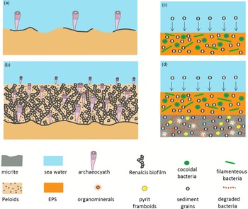
The Igoudine thrombolitic reefs containing archaeocyaths are interpreted to have developed under favourable conditions for microbial growth such as low siliciclastic input and low hydrodynamic energy with episodic high-energy events. These high-energy events generated numerous surfaces of reef-growth interruption (Fig. 2a, c, d). Under steady conditions, the resulting surfaces were colonized by various microbial populations. EPSs produced by bacteria were accumulated outside the bacterial cells and formed a protective gelatinous film that was used as substrate for attachment by bacterial colonies (Christensen & Characklis, Reference Christensen and Characklis1990). EPSs directly promoted the accretion of microbialites by favouring both mineral precipitation (i.e. biologically induced mineralization) and sediment trapping (Riding, Reference Riding2000). The trapping process can involve simple blockage of grain movement, their adhesion to EPSs and their incorporation (binding) into the mats (Riding, Reference Riding2000). The latter processes were facilitated where microbial mats had irregular surfaces. On the other hand, smooth films with little surface topography can only incorporate fine-grained particles if they are available (Riding, Reference Riding2000). In situ mineral precipitation (autochthonous micrite) is directly promoted through biogenic mineralization of organic compounds and calcification of bacterial sheaths and EPSs (Fig. 10c, d).
(a, b) Schematic model showing thrombolite growth and (c, d) EPS mineralization process.

Renalcis with subordinate Epiphyton and Girvanella dominate the archaeocyaths of the Igoudine Formation thrombolites. Renalcis boundstones are the main components of the studied thrombolites (Figs 2c, d, 10a, b). Their cryptic growth habitat is comparable to that of modern cryptic reef biofilms. The growth of Renalcis and archaeocyaths was probably synchronous with the influx of fine-grained allochthonous sediments, which are dominantly micrite and fine-grained microsparite. Archaeocyath skeletons are usually attached to microbial microframes (Fig. 2b, c, d), essentially Renalcis dendritic branches, which are in turn encrusted by minor Epiphyton chambers and Girvanella crusts. Small-sized archaeocyaths filled the remaining space between the Renalcis branches (Fig. 2c, d, f), indicating that the available space and the rate of encrustation by the microbial mats likely controlled their growth (Fig. 10a, b). In various examples of early Cambrian thrombolitic reefs containing archaeocyaths, archaeocyaths were considered reef dwellers rather than local framework builders (Debrenne, Reference Debrenne2007). However, they could play vital roles in filling the cavities, decreasing water velocity and accelerating the growth of thrombolite reefs, especially under a low rate of sediment supply.
The rate of multiplication and growth of bacteria probably controlled the size of isolated biofilms scattered within the sediments instead of their exposure time before burial by detrital fractions. Bacterial multiplication and the rate of EPS production was likely controlled by the growth of Renalcis microframes. Nutrients, oxidant supply and the length of surface exposure can play a primary limiting role for bacterial multiplication (Riding, Reference Riding2008). The siliciclastic supply increases upwards in the reef complex and reaches up to 35 % of the total rock volume (Fig. 9b), essentially above the Amouslek calcareous shales. Thereafter, the growth of thrombolites with archaeocyaths of the Igoudine Formation is stopped by the increase in siliciclastic input. The equilibrium between the EPS production and the sedimentation rate of detrital materials in the environment was likely interrupted when the supply of sediments exceeded a threshold value.
6. Conclusion
The first episode of the Atdabanian reef complex in the western Anti-Atlas consists of tabular and dendritic thrombolites. Small regular and irregular archaeocyaths occur in and around the reefs and are composed of three genera: Dictyocyathus, Erismacoscinus and Agastrocyathus. Abundance of archaeocyaths in this horizon does not exceed 20 % of the total rock volume. This suggests that archaeocyaths had a subordinate role in reef-building. The thrombolites containing archaeocyaths are dominated by Renalcis with subordinate Epiphyton and Girvanella. Petrographic study and higher resolution SEM observations of the dark micrite of the Renalcis chambers showed amorphous translucent sheet-like structures interpreted as EPSs, closely associated with organominerals including nanoglobules and polyhedrons. Exceptionally well-preserved Renalcis chambers contain possible bacterial fossils similar to those described in modern microbialites; micro-spherical coccoid fossils and filamentous bacteria coccoid fossils and filamentous tube-like bacteria are observed, isolated or in closely associated colonies, suggesting a bacterial origin for the Renalcis calcimicrobe. The growth and expansion of the Igoudine thrombolites took place under favourable conditions such as low hydrodynamic energy and low siliciclastic input.
Acknowledgements
The authors are grateful to Dr Stephen Kershaw and an anonymous reviewer for their constructive and encouraging comments. Financial support for this study was provided by Cadi Ayyad University Morocco; la Région Nouvelle Aquitaine and the University of Poitiers. The authors are grateful to Françoise Debrenne and Adeline Kerner for identifying the archaeocyaths. Lhoussain Ablouh (Centre des analyses chimique, UCA) and Emile Béré from Pole IMAGE UP, University of Poitiers are acknowledged for SEM and EDS analyses. Mouad Akboub, Idir Elhabib and Mouhssin Elhalim are thanked for their assistance during the fieldwork. We also appreciate Claude and Luis-Marie Bonneval for their warm reception and hospitality in Poitiers. OV was supported by an Estonian Research Council Grant (PRG836), the Sepkoski Grant and a grant by the Institute of Ecology and Earth Sciences, University of Tartu.
Conflict of interest
None.