Chylothorax is a rare condition in childhood, occurring most commonly in this age group following surgery for CHD.Reference Zuluaga 1 , Reference Haines, Walsh and Fletcher 2 Published estimates of the incidence of chylothorax following paediatric heart surgery vary widely, but the largest studies have found a rate of around 2.8–3.2%.Reference Haines, Walsh and Fletcher 2 , Reference Mery, Moffett and Khan 3 The mechanism for the development of postoperative chylothorax is not completely understood, but it is thought to result from mechanical damage either to the thoracic duct or to the proximal lymphatic tributaries, an increase in central venous pressure above the pressure in the intrathoracic lymphatic system, obstruction of lymphatic drainage caused by the development of postoperative thromboses of the central veins, or by a combination of all three of these mechanisms.Reference Zuluaga 1 , Reference Chan, Russell and Williams 4
Several studies have attempted to investigate potential preoperative and perioperative risk factors for the development of chylothorax following cardiac surgery in children.Reference Mery, Moffett and Khan 3 – Reference Doell, Bernet and Molinari 9 These studies vary widely in terms of study design and quality, and the results are not consistent. There is agreement among some of these studies that increasing surgical complexity,Reference Mery, Moffett and Khan 3 , Reference Borasino, Diaz and El Masri 5 , Reference White, Seckeler and McCulloch 8 increasing cardiopulmonary bypass times,Reference Borasino, Diaz and El Masri 5 , Reference Biewer, Zürn and Arnold 6 and the presence of genetic diseaseReference Mery, Moffett and Khan 3 , Reference Doell, Bernet and Molinari 9 are independently associated with an increased risk for chylothorax. There is, however, disagreement among the studies as to whether weight and age influence the risk for chylothorax.
Although relatively rare, postoperative chylothorax is of considerable interest to clinicians because of the substantial increase in mortality and morbidity associated with the condition. The flow of chylous fluid into the thoracic cavity results in immune compromise and nutritional impairment, as well as in respiratory compromise as the effusion increases in size.Reference Soto-Martinez and Massie 10 Compared with patients without chylothorax, those who develop the complication are more likely to die in the hospital.Reference Borasino, Diaz and El Masri 5 In addition, they have significantly longer ICU and total hospital stays, and have significantly increased total treatment costs.Reference Mery, Moffett and Khan 3 – Reference Borasino, Diaz and El Masri 5
Despite the clinical importance of this complication, there is little consensus or standardisation in management strategy across different centres.Reference Yeh, Brown and Kellogg 11 Initial management usually comprises drainage of the effusion using an indwelling chest drain.Reference Yeh, Brown and Kellogg 11 This is often accompanied by nutritional modification, with the aim of reducing chyle production. In the first instance, this usually takes the form of a feeding formula with a low proportion of long-chain triglycerides and a high proportion of medium-chain triglycerides.Reference Zuluaga 1 An alternative strategy is to withhold enteral feeding and administer total parenteral nutrition. The somatostatin analogue octreotide is used in some centres, often as a second-line therapy, should dietary modification be unsuccessful.Reference Caverly, Rausch and Da Cruz 12 Finally, surgical intervention can be considered if medical management has failed. This is usually in the form of either ligation of the thoracic duct or surgical pleurodesis.Reference Soto-Martinez and Massie 10 At the time of writing there were no randomised trials to support or refute the use, or timing, of any of these interventions in this patient group.
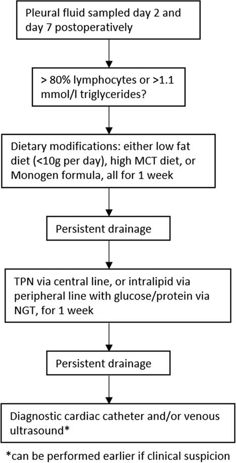
There is some evidence to suggest that the implementation of a standardised management protocol for the treatment of postoperative chylothorax can result in a significant decrease in time to diagnosis and length of hospital stay.Reference Yeh, Brown and Kellogg 11 Such a guideline was instituted in the ICU of our institution in January 2009 (Fig 1).
Management protocol for postoperative chylothorax, introduced to the Royal Children Hospital Melbourne in 2009. MCT = medium chain triglycerides; TPN = total parental nutrition; NGT = nasogastric tube.

The objectives of the present study were twofold. First, in view of the inconsistency of the existing literature regarding risk factors for the development of chylothorax, we aimed to investigate potential risk factors using a matched case–control study design. In addition, we aimed to investigate the impact of the management guideline on management strategies and outcome.
Methods
The study population included all patients diagnosed with chylothorax following cardiac surgery at the Royal Children’s Hospital, Melbourne, over a 48-month period beginning in January 2008. Cases were identified following a search of the hospital’s Cardiobase database, in which all postoperative complications are recorded. The diagnosis of chylothorax was confirmed by reviewing the hospital records, with the diagnostic criteria being a pleural fluid lymphocyte count of >80%, or a triglyceride level >1.1 mmol/L. To calculate the incidence of chylothorax, the total number of cardiac surgeries was obtained for the same period. A matched control group who also underwent cardiac surgery in the same period but did not develop chylothorax was generated from the Cardiobase database, matched for age at surgery – matched for age at surgery (within 3 months when the case was below 1 year of age, and within 1 year for older patients), date of surgery (within 1 year), and sex (exact match).
Demographic and clinical data were obtained by reviewing the medical records, either as a hard copy or in electronic format, as appropriate. Data were collected on the following parameters: preoperative diagnosis and surgery performed; type of surgery, namely open or closed, and arch or non-arch; weight and age at surgery; bypass time; incision type, namely sternotomy, thoracotomy, or already on central extracorporeal membrane oxygenation via limited sternotomy when taken to the operating theatre; virgin chest opening, namely that patient’s first ever sternotomy or thoracotomy, versus redo chest opening, namely that patient’s second or greater sternotomy or thoracotomy; management strategy used, namely dietary modification, use of parental nutrition and surgical intervention; and insertion and removal dates of chest drains. For each case and control, the risk adjustment for congenital heart surgery (RACHS-1) score of complexity was calculated as previously described.Reference Jenkins, Gauvreau and Newburger 13
Summary statistics were calculated, presented as means and standard deviation or as medians and range depending on the normality of the distribution, which was assessed by visual inspection of the relevant histogram. To examine risk factors for the development of chylothorax, univariable and multivariable logistic regression models were constructed with paired analysis. The multivariable model was constructed by including all variables that were significant at the 10% level on univariable analysis, and subsequently performing backwards elimination until all variables were significant at the 5% level. To examine the effect of a standardised management protocol on the duration of chyle drainage, and the use of both parenteral nutrition and dietary modification, data before and after the implementation of the guideline – that is, before and after 01 January, 2009 – were compared using either the χ2, Wilcoxon test for median, or Fisher’s exact test depending on the distribution and size of the data.
Results
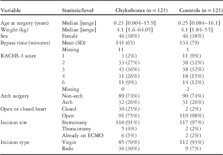
In total, 121 cases of chylothorax were identified following cardiac surgery in the defined time period, with 121 controls, matched for age at surgery, date of surgery, and sex. Over the same time period, 2273 paediatric cardiac surgeries were performed, resulting in a chylothorax incidence of 5.23%. Table 1 shows the summary statistics for the potential chylothorax risk factors, for both cases and controls. The median age at surgery was 0.23 years for the chylothorax cases, with a median weight at surgery of 4.1 kg. The majority (62%) of chylothorax cases were male.
Summary statistics.

ECMO = extra-corporeal membrane oxygenation
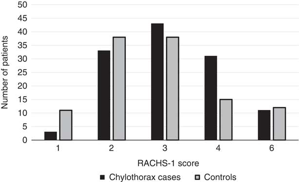
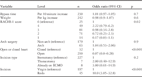
Table 2 shows the results of the univariable matched regression analysis, and Table 3 shows the results of multivariable regression analysis. Bypass time and patient weight were not associated with an increased risk for chylothorax; however, increasing surgical complexity as demonstrated by the RACHS-1 score is associated with an increasing risk for chylothorax. In the univariable model, a RACHS-1 score of 1 is associated with an odds ratio for the development of chylothorax of 0.17. RACHS-1 group 4 is associated with the highest risk, with odd ratios of 2.22 and 2.98. The distribution of the RACHS-1 score among cases and controls is also demonstrated in Figure 2.
RACHS-1 score for chylothorax cases and controls.

Univariable conditional logistic regression model.

ECMO = extra-corporeal membrane oxygenation
Multivariable conditional logistic regression model.

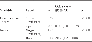
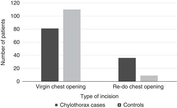
Whether the surgery was arch or non-arch, and the location of incision, namely sternotomy or thoracotomy, had no effect on chylothorax risk. Closed-heart surgeries, however, were significantly more likely to result in chylothorax compared with open-heart surgeries, and this association was seen in both univariable and multivariable models. Similarly, both models found an association between chylothorax and redo chest openings, with redo incisions associated with a significantly higher risk, demonstrated in Figure 3.
Type of chest opening for chylothorax cases and controls.

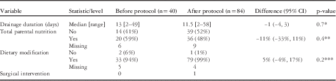
The cohort of chylothorax cases was split into two groups based on surgery date, to examine the effect of the implementation of a standardised management protocol. The results of this are shown in Table 4. No significant difference in drainage duration was seen between the two time periods. In addition, we could demonstrate no significant difference in the proportion of patients receiving either dietary modification or total parenteral nutrition. Only one surgical intervention for chylothorax was performed during the time period of this study after the introduction of the management protocol, a thoracic duct ligation.
The effect of a standardised management protocol: summary statistics and comparisons.

* Wilcoxon test for median as the distribution of drainage duration was skewed to the right
** χ2 Test
*** Fisher’s exact test was used because of small frequencies: that is, <5
Discussion
We identified 121 cases of chylothorax, making ours the second largest study in the literature examining postoperative chylothorax in children with CHD, and the largest single-centre study. Our incidence rate of 5.23% is in keeping with previously reported rates. The published incidence rates following paediatric congenital cardiac surgery vary from 0.85 to 15.8%,Reference Haines, Walsh and Fletcher 2 – Reference White, Seckeler and McCulloch 8 , Reference Milonakis, Chatzis and Giannopoulos 14 , Reference Nguyen, Shum-tim and Dobell 15 although this large variance in reported rates may represent differences in study design, particularly in case ascertainment and definition. A large-scale study in 2014 by Haines et alReference Haines, Walsh and Fletcher 2 and the British Paediatric Surveillance Unit found a postoperative incidence rate of 3.2% following paediatric congenital heart surgery. This is similar to the rate of 2.8% found by Mery et al,Reference Mery, Moffett and Khan 3 who performed a review of 77,777 children undergoing cardiac surgery in multiple centres across North America. Interestingly, Mery et al have also demonstrated a significant increase in chylothorax incidence rates over time from 2% in 2004 to 3.7% in 2011, even after accounting for other variables.
We found that the complexity of the surgical procedure was associated with the risk for postoperative chylothorax on univariable modelling, but this association did not remain when other factors were included in the multivariable model. Compared with RACHS-1 group 6, which includes the most complex surgical operations including surgeries such as the Norwood operation for hypoplastic left heart syndrome, patients in RACHS-1 group 1, which includes the least complex ones including surgeries such as atrial septal defect repair, had an odds ratio for chylothorax of only 0.17 in the univariable model. Our findings replicate those of several other studies in which increasing surgical complexity was associated with greater risk for chylothorax.Reference Mery, Moffett and Khan 3 , Reference Borasino, Diaz and El Masri 5 , Reference White, Seckeler and McCulloch 8
Our findings demonstrate that in our cohort the patients at the highest risk for chylothorax are those in RACHS-1 group 4, which includes surgeries such as repair of hypoplastic or interrupted aortic arch, complex arterial switch operations, and neonatal aortic valve operations. It is possible that there are anatomical reasons why the surgeries included in group 4 are more likely to result in mechanical disruption of the thoracic duct or lymphatic tributaries and, hence, make postoperative chylothorax more likely. Indeed, it has been postulated that surgery in the region of the aortic arch may predispose to chylothorax development by this mechanism,Reference Chen, Wang and Lue 16 and this is supported by our data showing that closed-heart surgeries, which will include some aortic arch operations as well as some systemic to pulmonary shunt operations, are significantly more likely to result in chylothorax than are open-heart surgeries. Conversely, this theory is not supported by our data examining aortic arch surgery directly, which do not demonstrate any increased risk for surgery specifically involving the aortic arch compared with surgery that does not. Our data, however, may suggest that surgery that does not specifically involve the aortic arch, but is in this region – such as the placement of a systemic to pulmonary shunt – may be associated with increased risk for chylothorax.
We did not find any association between the location of the chest opening, namely sternotomy versus thoracotomy, and risk for chylothorax. These findings, however, should be viewed with caution because of the small number of patients in our cohort in the thoracotomy group. We did find a large increase in risk among those patients who were undergoing a redo chest opening – that is, their second or greater sternotomy or thoracotomy – as part of their procedure. This is presumably as a result of the increased surgical difficulty in performing redo chest openings because of adhesions, making thoracic duct injury more likely. To our knowledge, this is the first time this association has been demonstrated in the literature.
Previous studies have found mixed results for weight as a potential risk factor.Reference Chan, Russell and Williams 4 – Reference Biewer, Zürn and Arnold 6 , Reference White, Seckeler and McCulloch 8 Clearly, weight is closely correlated with age, and the studies have varied in how they have controlled for this association. In our study, as the control group was matched for age, to a certain extent, they will be matched for weight by proxy. We, therefore, cannot interpret our data as showing that weight or age is not a risk factor for the development of chylothorax. Previous studies have shown a strong negative association between age and chylothorax risk, with the study by Mery et alReference Mery, Moffett and Khan 3 showing a multivariable odds ratio of 7.03 for neonates undergoing surgery compared with the reference group of teenagers. Although our data do not allow measurement of the effect of age because of our matching process, it is certainly possible that younger patients are at an increased risk in view of the high proportion of infants in our cohort; 69% of our chylothorax cases were under 12 months of age at the time of surgery.
The results of the impact of a standardised management protocol are disappointing. Yeh et alReference Yeh, Brown and Kellogg 11 found a significant reduction in the duration of chest-tube drainage after the introduction of their own management protocol, from an average of 20 days to 14 days. In contrast, we observed only a modest decrease in chest-drainage duration from a median of 13–11.5 days (Table 4) after the introduction of our protocol. The implementation of our protocol also did not result in a noticeable change in the proportion of patients receiving either dietary modification or TPN (Table 4). Over this time period, only one patient underwent ligation of the thoracic duct, and none underwent pleurodesis; thus, it is difficult to comment on these management techniques. Similarly, octreotide was not used in these patients over this time period.
Although the impact of our protocol was limited in terms of drainage duration and chylothorax management, it may be that the management strategy outlined in the protocol was already loosely used by clinicians at our centre. It is likely, therefore, that although the protocol is helpful in terms of formalising our approach and the timing of treatment, it would not necessarily be expected to significantly change either the outcome or overall management.
There are several limitations to the present study. Our study population is derived from an institutional database of cardiology patients, and is reliant on accurate recording of postoperative complications by the clinical team. As chylothorax is a serious and often prolonged source of postoperative morbidity, it is likely that this method will capture the majority of chylothorax cases, but it is possible that some cases were missed. There is, however, no reason to suspect that any missed cases should differ systematically from those included; hence, this should not introduce bias into our analysis of risk factors for chylothorax.
A further limitation is the lack of data on comorbidity. Two studies have demonstrated an increased risk for chylothorax among cardiac patients with genetic disease, particularly in those with Down’s Syndrome (trisomy 21)Reference Doell, Bernet and Molinari 9 and Turner’s Syndrome.Reference Mery, Moffett and Khan 3 We did not collect data on genetic conditions as part of this study and, thus, unable to comment on this possible association. In addition, our matched case–control study design made it impossible to examine the association of age or weight on the risk for chylothorax; however, the effect of age is well described in the literature, and we felt that the advantages of a matched design in controlling for cofounding variables outweighed this limitation.
Conclusion
This is the largest single-centre study in the literature examining the risk factors for chylothorax following paediatric cardiac surgery. We have replicated the previously reported association between surgical complexity and chylothorax risk, and have shown for the first time that redo chest openings are also associated with a significantly increased risk. The implementation of a standardised management protocol in our institution did not result in a significant change in either chylothorax drainage duration or in management strategy.
Acknowledgements
The Heart Research Group at the Murdoch Childrens Research Institute is supported by BigW and RCH 1000, RCH Foundation. The MCRI is supported by the Victorian Government’s Operational Infrastructure Support Program.
Financial Support
This research received no specific grant from any funding agency, commercial or not-for-profit sectors.
Conflicts of Interest
None.