INTRODUCTION
The whale shark, Rhincodon typus (Smith, Reference Smith1828), is designated as ‘Vulnerable' to extinction by the World Conservation Union (IUCN) Red List of Threatened Species and is listed under Appendix II of the Convention on International Trade in Endangered Species (CITES, 2002) and Appendix II of the Convention on Migratory Species (CMS, 2005). However, despite this status, there remain considerable gaps in our knowledge about R. typus (Bradshaw, Reference Bradshaw2007) due to the limited level of consistent, reliable and accurate data on whale shark biology and ecology, in particular with regard to population structure and growth rates.
Over the past decade technological advancements have led, or are leading, to substantial improvements in the development of whale shark databases, providing important information on aggregations found around the world. Such enhancements include the use of satellite tagging systems to track the horizontal and vertical movements of whale sharks, together with associated environmental data, including sea surface temperature (SST) and depth–temperature profiles, both measured by the tag and remotely sensed broadscale data such as SST, chlorophyll-a and geostrophic currents (Rowat & Gore, Reference Rowat and Gore2007; Sleeman et al., Reference Sleeman, Meekan, Stewart, Wilson, Polovina, Stevens, Boggs and Bradshaw2010). The genetic analysis of biopsy samples is beginning to identify global population structures to a molecular level (Bradshaw et al., Reference Bradshaw, Mollet and Meekan2007; Castro et al., Reference Castro, Stewart, Wilson, Hueter, Meekan, Motta, Bowen and Karl2007; Schmidt et al., Reference Schmidt, Schmidt, Ozer, Ernst, Feldheim, Ashley and Levine2009) and the application of automated spot-recognition programs to identify individual sharks through photo-analysis is a non-invasive tool enabling population estimates by capture–mark–recapture methods (Arzoumanian et al., Reference Arzoumanian, Holmberg and Norman2005; Speed et al., Reference Speed, Meekan and Bradshaw2007; Brooks et al., Reference Brooks, Rowat, Pierce, Jouannet and Vely2010). However, despite recent innovations for collecting valuable and accurate information on whale shark populations, the use of a robust, dependable and easily applied method for accurately and precisely measuring the length of the largest fish in the ocean remains limited.
Presently established methods used to record size data for free swimming R. typus include:
-
(a) Size estimates made to the nearest 0.5 m by experienced in water researchers or boat skippers, sometimes estimates are based on the length of a snorkeller or an object of a known size positioned alongside the shark (Meekan et al., Reference Meekan, Bradshaw, Press, McLean, Richards, Quasnichka and Taylor2006; Graham & Roberts, Reference Graham and Roberts2007; Norman & Stevens; 2007; Bradshaw et al., Reference Bradshaw, Fitzpatrick, Steinberge, Brook and Meekan2008).
-
(b) Measurements made using a tape measure, or a rope knotted at 1 m intervals, held underwater alongside the shark by two swimmers (Meekan et al., Reference Meekan, Bradshaw, Press, McLean, Richards, Quasnichka and Taylor2006; Bradshaw et al., Reference Bradshaw, Mollet and Meekan2007, Reference Bradshaw, Fitzpatrick, Steinberge, Brook and Meekan2008; Norman & Stevens, Reference Norman and Stevens2007; Riley et al., Reference Riley, Hale, Harman and Rees2010).
-
(c) Size estimations made by driving a boat alongside a shark swimming at the surface and aligning the tip of the tail with the stern of the boat, and estimating total length relative to the bow (Graham & Roberts, Reference Graham and Roberts2007).
-
(d) Size estimates made by spotter plane pilots by comparison to nearby vessels of a known length (Bradshaw et al., Reference Bradshaw, Fitzpatrick, Steinberge, Brook and Meekan2008).
-
(e) Through a combination of any of the methods (a) through to (d) (Bradshaw et al., Reference Bradshaw, Fitzpatrick, Steinberge, Brook and Meekan2008).
-
(f) Laser photogrammetry using projected total lengths derived from pre-caudal lengths of free-swimming and deceased shark specimens (Rohner et al., Reference Rohner, Richardson, Marshall, Weeks and Pierce2011).
Methods (a) through to (e) contain many variables and therefore the reliability of recorded sizes for free swimming whale sharks remains relatively low, with minimum standard errors (SEs) considered to be 0.5 m based on visual observations (Meekan et al., Reference Meekan, Bradshaw, Press, McLean, Richards, Quasnichka and Taylor2006; Graham & Roberts, Reference Graham and Roberts2007; Norman & Stevens, Reference Norman and Stevens2007; Bradshaw et al., Reference Bradshaw, Fitzpatrick, Steinberge, Brook and Meekan2008; Holmberg et al., Reference Holmberg, Norman and Arzoumanian2009). Rohner et al. (Reference Rohner, Richardson, Marshall, Weeks and Pierce2011) found an even greater margin of error (SE of +/− 68.77 cm) between visual estimates and their predicted total length (TL) computed from laser photo-grammetry measured pre-caudal length (PCL).
Precise and accurate size data become important when considering the biology and ecology of whale shark populations especially when establishing the maturity state of sharks based on length (Norman & Stevens, Reference Norman and Stevens2007; Stevens, Reference Stevens2007), population structures and size trends (Meekan et al., Reference Meekan, Bradshaw, Press, McLean, Richards, Quasnichka and Taylor2006; Bradshaw, Reference Bradshaw2007; Graham & Roberts, Reference Graham and Roberts2007; Bradshaw et al., Reference Bradshaw, Fitzpatrick, Steinberge, Brook and Meekan2008), and whale shark growth rates (Graham & Roberts, Reference Graham and Roberts2007; Norman & Stevens, Reference Norman and Stevens2007; Stevens, Reference Stevens2007).
There are some published data on the size and growth rates of R. typus, for sharks of total lengths between 0.6 m (neonatal) and 8 m (immature). However, these are mainly derived from measurements taken from captive individuals with consequently artificial environmental parameters (Chang et al., Reference Chang, Leu and Fang1997; Uchida et al., Reference Uchida, Toda, Kamei and Teruya2000) and are extremely small sample sizes, which may not be comparable to growth rates achieved in the wild. Neonatal specimens (N = 2) exhibited growth rates from 0.98 to 2.34 m y−1 while juveniles in excess of 3.5 m (N = 5) showed rates from 0.21 to 0.5 m y−1 with a mean of 0.29 m y−1 (Chang et al., Reference Chang, Leu and Fang1997; Uchida et al., Reference Uchida, Toda, Kamei and Teruya2000).
The only comparable study based on wild populations was taken from 15 deceased specimens in South Africa (sharks with a PCL of 4.18–7.7 m), where TL was shown to have a linear relationship with the number of annual growth rings contained within the shark vertebrae (Wintner, Reference Wintner2000). Predicted growth rates were back-calculated and found to be slightly slower than the captive rates observed by Uchida et al. (Reference Uchida, Toda, Kamei and Teruya2000), with a mean of 0.22 m y−1 for juveniles in excess of 3.5 m.
Precise and accurate methods of measurement for wild animals have been developed using various photographic based techniques, termed photogrammetry. Such systems have been used on a number of species in both terrestrial and marine environments (e.g. gorillas, Gorilla gorilla (Breuer et al., Reference Breuer, Robbins and Boesch2006); elephants, Loxondonta africana (Schrader et al., Reference Schrader, Ferreira and Aarde2006); bowhead whales, Balaena mysticetus (Koski et al., Reference Koski, Davis, Miller and Withrow1992); bluefin tuna, Thunnus thynnus thynnus (Costa et al., Reference Costa, Loy, Cataudella, Davis and Sardi2006); and killer whales, Orcinus orca (Durban & Parsons, Reference Durban and Parsons2006)). The method most commonly employed in marine environments is that of stereo-photogrammetry whereby two angled cameras are used simultaneously to create a three-dimensional image of the subject that can be accurately measured using specialized software (Shortis et al., 2009). Stereo photogrammetry, while very accurate is comparatively expensive to implement, due to the additional calibration materials and dedicated software necessary to accurately process measurements (Harvey et al., Reference Harvey, Cappo, Shortis, Robson, Buchanan and Speare2003), and the apparatus required can be cumbersome to use (Durban & Parsons, Reference Durban and Parsons2006).
Another much simpler, single camera system of laser photogrammetry or laser metrics has also been successfully employed to measure the physical traits of a number marine animals (e.g. Sebastes sp. (Gingras et al., Reference Gingras, Ventresca and McGonigal1998; Yoklavich et al., Reference Yoklavich, Greene, Sullivan, Lea and Love2000); Orcinus orca (Durban & Parsons, Reference Durban and Parsons2006); Cephalorhynchus hectori (Webster et al., Reference Webster, Dawson and Slooten2010); Manta alfredi (Deakos, Reference Deakos2010); and R. typus (Rohner et al., Reference Rohner, Richardson, Marshall, Weeks and Pierce2011)). This method is based on the principle that parallel lasers project light that is equidistant regardless of the distance from the origin (Rothman et al., Reference Rothman, Chapman, Twinomugisha, Wasserman, Lambert and Goldberg2008). Parallel laser beams a known distance apart are projected on to the side of the animal being observed creating a ‘scale-bar’ which is then photographed; provided the subject is perpendicular to the axis of the lasers (Rowe & Dawson, Reference Rowe and Dawson2009) measurements can be made from images taken at any distance from the subject at which the laser points are visible.
In terms of using laser photogrammetry on whale sharks in-water there are several potential advantages:
-
(a) significantly decreased average error values (Rothman et al., Reference Rothman, Chapman, Twinomugisha, Wasserman, Lambert and Goldberg2008; Rohner et al., Reference Rohner, Richardson, Marshall, Weeks and Pierce2011) when compared to established methods of measurement (Meekan et al., Reference Meekan, Bradshaw, Press, McLean, Richards, Quasnichka and Taylor2006; Graham & Roberts, Reference Graham and Roberts2007; Norman & Stevens, Reference Norman and Stevens2007; Bradshaw et al., Reference Bradshaw, Fitzpatrick, Steinberge, Brook and Meekan2008; Holmberg et al., Reference Holmberg, Norman and Arzoumanian2009);
-
(b) remotely and non-invasively obtained accurate measurements of whale sharks in situ;
-
(c) measurements, tests and calibrations can be performed accurately both in-water and on land (Muljowidodo et al., Reference Muljowidodo, Rasyid, SaptoAdi and Budiyono2009) as the image contains no effects of visual refraction;
-
(d) multiple measurements can be taken from a single image;
-
(e) ease of operation; and
-
(f) being relatively inexpensive and simple to construct.
The main disadvantage of the system is the requirement to ensure the camera and lasers are exactly perpendicular to the subject being photographed in order to obtain an accurate measurement.
Relative or morphometric measurements have been known to provide a suitable predictor for the size of the animal for some time (Huxley, Reference Huxley1932). This has been shown in some marine species (Strauss & Fuiman, Reference Strauss and Fuiman1985; Ross & Lima, Reference Ross and Lima1994; Hall et al., Reference Hall, Smith, de Lestang and Potter2006; Katsanevakis et al., Reference Katsanevakis, Thessalou-Legaki, Karlou-Riga, Lefkaditou, Dimitriou and Verriopoulos2007); in whale sharks the height of the first dorsal fin (Meekan et al., Reference Meekan, Bradshaw, Press, McLean, Richards, Quasnichka and Taylor2006) and the distance between the 5th gill slit and start of the 1st dorsal (Rohner et al., Reference Rohner, Richardson, Marshall, Weeks and Pierce2011) have been suggested as predictors for TL, while in Hector's dolphins (Cephalorhynchus hectori) the length of the base of the dorsal fin was found to be a better predictor than its height (Webster et al., Reference Webster, Dawson and Slooten2010).
The principle aims of this study were: (a) to build a precise, accurate, robust, reliable and relatively inexpensive in-water laser-photogrammetry system; (b) to determine accurate total length and morphometric measurements of R. typus; (c) to field test these on whale sharks observed during seasonal feeding aggregation in the Seychelles and Djibouti; and (d) to analyse sources of error to ascertain possible methods for increasing the precision, accuracy and versatility of the system. The apparatus and technique developed could then be employed to establish a database of measurements that, used in conjunction with the automated photo-identification programs, would allow accurate yearly size data from individual shark re-sightings, thereby providing vital information on growth rates within a free ranging whale shark population.
MATERIALS AND METHODS
Study area
The study was performed on populations of whale sharks that aggregate seasonally around the coastal waters of Mahe Island, Seychelles situated at the centre of a shallow continental plateau at 4°S and 55°E in the Western Indian Ocean, and off the coast of Djibouti, North Africa at 11°N and 43°E.
Whale shark photo-identification
All whale sharks measured in the study were photo-identified using the algorithmic I3S Interactive Individual Identification System (Van Tienhoven et al., Reference Van Tienhoven, Den Hartog, Reijns and Peddemors2007), which utilizes the unique spot pattern on the area posterior to the gill slits of each whale shark as a ‘finger print’ (Arzoumanian et al., Reference Arzoumanian, Holmberg and Norman2005); identified individuals were then catalogued on a master database with records for the Seychelles aggregation beginning in 2001 and in Djibouti in 2003. The use of the I3S system ensured that similarly marked individuals could be consistently identified and their measurements confidently compared.
Laser apparatus
The laser metric system used in the study employs the same principle described by Rowe & Dawson (Reference Rowe and Dawson2009), Deakos (Reference Deakos2010) and Rohner et al. (Reference Rohner, Richardson, Marshall, Weeks and Pierce2011) of horizontally mounting two parallel lasers a known distance apart to a fixed camera-base. The laser pointers used were commercially available ‘Underwater Green Laser Pointers’ (model DIVE-1, Z-Bolt®, Beam of Light Technologies, Inc. Oregon, USA; wavelength 532 nm, output power <5 mW). Green lasers were used because the beam wavelength of 532 nm dissipates at a slower rate in seawater compared to red lasers and can therefore be viewed over greater distances.
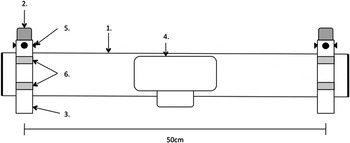
The development of the laser and camera mounting apparatus evolved through several stages to produce a robust, precise, accurate and fully adjustable set-up. In the final version used for this study, two lasers were fixed in aluminium tubes with centres 50 cm apart on an aluminium frame with a digital camera mounted centrally between them in a waterproof housing (Canon PowerShot G9 camera & WP-DC21 Waterproof Case Canon Inc., Tokyo, Japan) (Figure 1).
Schematic diagram of the final laser apparatus (top view) showing the aluminium frame (1), lasers (2) encased within an aluminium tube (3), underwater camera housing (4), calibration screws (5) and jubilee clips (6).

The Z-bolt lasers were not collimated, i.e. the axis of the laser beam was not parallel to the laser pointer's body, which needed to be mitigated by calibration to counteract the resultant error of refraction and non-parallel laser beams when the laser bodies were positioned 50 cm apart (see Supplementary Material Methods section Refraction Correction and Laser Calibration and Supplementary Figure 1 for details).
During testing, the unit was found to be easy to calibrate, robust and reliable in use. The cost to produce the final apparatus, including camera and underwater housing, was approximately £500.
Operation and calculation of measurements
Before and after each monitoring session, the lasers were calibrated and verified at 3, 5 and 10 m; if laser alignment was out by ≥ 5 mm at a distance of 5 m after a measurement session data from that session were rejected.
To operate the apparatus during a shark encounter, a swimmer positioned themselves both level and perpendicular to the head of the shark, and remaining motionless in the water, allowed the shark to move past while taking three overlapping photographs of the head, body and tail. A whale shark positioned perpendicular to the laser system is generally too big to fit onto one image, even with the use of a wide angled lens and wide-angle lens correction function (Deakos, Reference Deakos2010). The camera was kept level and perpendicular to the shark, with the body and tail straight and fully in shot as far as conditions allowed. This was commonly achieved by ensuring that photographs were taken during the glide phase of locomotion observed in R. typus during descents from the surface, when the shark's body was in the optimally straight position (Gleiss et al., Reference Gleiss, Norman and Wilson2011).
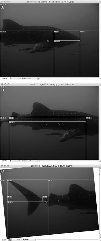
Images were then downloaded and analysed using Photoshop CS3 (v10.0.1 Adobe Systems Inc., Delaware, USA.) (Figure 2) (see Supplementary Material Methods section Calculation of Measurements for details). Prior to analysis, all images were screened for suitability and images were removed from the data set that were found to be out of focus, where the body axis of the whale shark was not perpendicular to the camera lens or where the tail was bent substantially from the vertical axis of the animal, thus increasing the robustness of the measurements (Durban & Parsons, Reference Durban and Parsons2006; Bergeron, Reference Bergeron2007; Rothman et al., Reference Rothman, Chapman, Twinomugisha, Wasserman, Lambert and Goldberg2008).
Screenshots from Photoshop of the head (A), body (B) and tail (C) photographs post-analysis, showing positions of the laser points (circled) and the Section Measurement Parameters defined using the ‘Splice tool’.

Precision and accuracy validation
Where multiple measurements of individual whale sharks were obtained in the same season, repeatability (r) of the laser metric system was calculated as an index of precision using the within-groups and among-groups mean square (MS) values obtained from a one-way analysis of variance. A coefficient is included in the calculation to account for the unbalanced distribution of multiple measurements and sample size. A detailed description for the calculation of r is provided by Lessels & Boag (Reference Lessels and Boag1987). Where multiple within-season measurements were taken of the same shark, the maximum period between sightings was 20 days with 95% of all measurements taken within 9 days of each other.
Due to the physical characteristics of the whale sharks in the study, and the nature of the marine environment, obtaining accurate, corresponding manual measurements of each subject was impractical. The lasers were therefore continually validated throughout the programme using measurements of known objects pictured both above and below the water as tests for accuracy and precision. Because each laser beam was calibrated for refraction and set perpendicular to the camera lens there is no light refraction apparent to the camera when submersed (Muljowidodo et al., Reference Muljowidodo, Rasyid, SaptoAdi and Budiyono2009), therefore, the camera distinguishes an object underwater as if it is in the air and consequently tests for accuracy can be completed in both mediums.
Validation tests were conducted capturing laser referenced images of objects of a fixed measurement (e.g. a whale shark ‘model’ or markings on a wall) at a variety of distances. The laser measurements for all validation tests were then compared using linear regression against manual lengths measured with a tape measure to the nearest millimetre.
Morphometric measurements
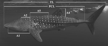
A number of defined areas were measured from the calibrated images to explore allometric relationships between the length of these measurements in proportion to directly measured TL and PCL. As a single image of the gill slit area can provide data needed for identification, a potential morphometric index in this area was preferred. The indices measured included the distance between the leading edge of the spiracle and the bottom of the 5th gill slit (Figure 3 A1), the distance between the top of the 1st gill slit and the top of the 5th gill slit (Figure 3 A2), the height of the 5th gill slit (Figure 3 A3) and the height of the 1st dorsal fin (Figure 3 A4) (Meekan et al., Reference Meekan, Bradshaw, Press, McLean, Richards, Quasnichka and Taylor2006). The indices also included the horizontal distance between the origin of the 1st dorsal fin and the 5th gill slit (Figure 3 A5) as described by Rohner et al. (Reference Rohner, Richardson, Marshall, Weeks and Pierce2011).
Laboratory tests: sources of parallax error
Parallax error caused by horizontal axis error (Webster et al., Reference Webster, Dawson and Slooten2010; Rohner et al., Reference Rohner, Richardson, Marshall, Weeks and Pierce2011), tail movement (Rohner et al., Reference Rohner, Richardson, Marshall, Weeks and Pierce2011) and the effects of a three-dimensional surface (Bergeron, Reference Bergeron2007) were identified as the principal sources of measurement error during the course of the study. Laboratory tests were therefore set up to quantify the three effects (see Supplementary Material Methods section Tests for Sources of Parallax Error and Supplementary Figures 2, 3 and 4).
Schematic of whale shark measurements taken for: TL, total length; PCL, pre-caudal length (after Wintner, Reference Wintner2000); A1, leading edge of spiracle to bottom of 5th gill slit; A2, top of 1st gill slit to top of 5th gill slit; A3, height of 5th gill slit; A4, height of 1st dorsal; and A5, the origin of the 1st dorsal to the 5th gill slit (photograph D. Robinson).

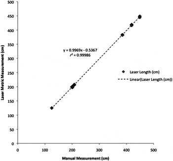
Regression of the measurement of 41 objects of a fixed size with a tape measure and lasers.

Analysis
Geometric mean (GM) or reduced major-axis linear regression was used to analyse size-on-size data (Laws & Archic, Reference Laws and Archic1981; McArdle, Reference McArdle1988, Reference McArdle2003; Mollet & Cailliet, Reference Mollet, Cailliet, Klimley and Ainley1996; Smith, Reference Smith2009) of morphometric and PCL measurements to TL. GM linear regression was employed due to the absence of an independent variable, the symmetry of results provided by GM and to reduce bias when interpreting allometric growth from morphometric measurements. GM regression parameters were calculated using b(GM) = b/r and a(GM) = mean y – b(GM) mean x (Mollet & Cailliet, Reference Mollet, Cailliet, Klimley and Ainley1996). Quality of fit for GM regression lines was assessed using standard error of estimate (SEE) and coefficient of determination r2; although r2 has been considered relatively ineffective as an indicator of best fit and reliability of estimation of data (Bass, Reference Bass1973; Mollet & Cailliet, Reference Mollet, Cailliet, Klimley and Ainley1996). In addition, because GM calculations do not provide the necessary data for the coefficient of determination and residual analyses, r2 values and confidence intervals were produced using the ordinary least squares (OLS) regression results.
The precision of the system was evaluated using the calculation for repeatability (Lessels & Boag, Reference Lessels and Boag1987) derived from the results of a one-way analysis of variance performed by CoStat 6.4 (CoHort Software, California, USA).
The Mann–Whitney U-test, performed by Statistica 6.0 (StatSoft Inc., Tulsa, USA), was used to verify possible variability in above and below water measurements by testing the null hypothesis that the distributions of the independent measurements from both groups were equal.
RESULTS
Precision and accuracy validation
COMPARING THE PRECISION AND ACCURACY OF LASER METRIC ESTIMATES AGAINST MANUALLY MEASURED OBJECTS
Comparing the lengths of 41 fixed objects measured above and below the water both manually and using the laser metric system resulted in a regression line that approached one. This indicated that the measurements obtained using the system were unbiased compared to the manual measurements (b = 0.997, SE = 0.002, r2 = 0.9999 P < 0.001). All 41 laser measured lengths fell within 1.78% of the corresponding manual length (Figure 4).
When terrestrial and in-water test results were analysed separately there was no significant difference (terrestrial: b = 0.997, r2 = 0.9999, P < 0.001; in water: b = 0.996, r2 = 0.9999, P <0.001) (Mann–Whitney U-test: Z = 0.243, N = 41, P = 0.808).
CALCULATION OF PRECISION (REPEATABILITY) WITH TOTAL LENGTHS OF R. TYPUS
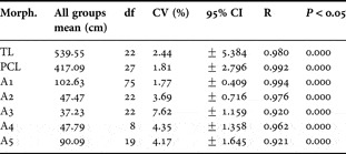
Twenty-three individual whale sharks, which were photographed on multiple occasions using the laser photogrammetric system, provided full photographic sets to measure TL (12 twice, 9 three times, 1 four times and 1 six times). Repeatability (r) for TL measurements was high (r = 0.9798), with a mean coefficient of variation (CV) of 2.44% (Table 1).
Group means, degrees of freedom (df), coefficients of variation (CV), 95% confidence intervals, repeatability (r) and P values calculated from multiple measurements of identified sharks for total, pre-caudal and morphometric lengths.

CALCULATION OF REPEATABILITY WITH PRE-CAUDAL LENGTHS OF R. TYPUS
Twenty-eight individual whale sharks, including all 23 sharks used in the calculation of r for TL, photographed on multiple occasions using the laser photogrammetric system, provided full photographic sets to measure PCL (12 twice, 10 three times, 3 four times, 2 five times and 1 on six occasions). Repeatability (r) for PCL measurements increased (r = 0.992) and CV of 1.81% (Table 1).
Morphometric measurements
A calculation of precision (repeatability) for the five candidate morphometric measurements of R. typus was carried out for the following indices: A1, the leading edge of spiracle to bottom of 5th gill slit; A2, the top of 1st gill slit to top of 5th gill slit; A3, the height of 5th gill slit; A4, the height of 1st dorsal fin; and A5, the origin of the 1st dorsal fin to the 5th gill slit (Table 1).
Predicting total length
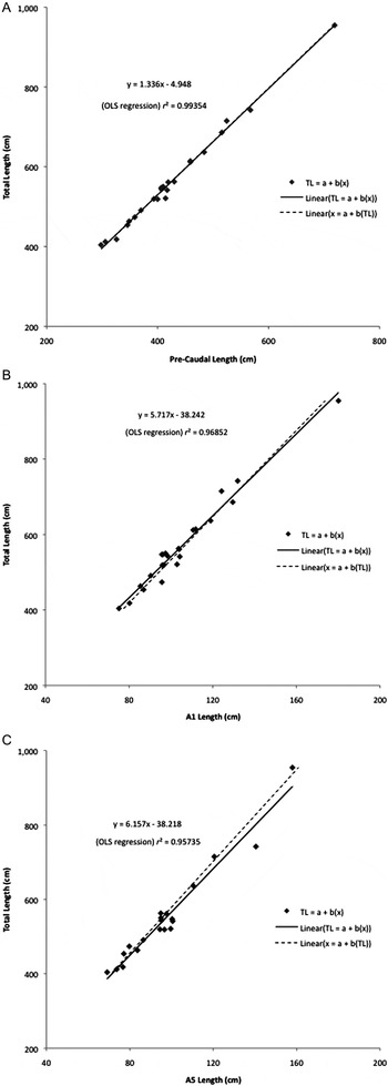
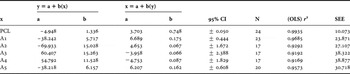
Twenty-four sharks, identified with straight head, body and tail positions throughout each laser-metric photoset, were further analysed using GM linear regressions to compare PCL and morphometric indices against the laser-measured TL measurements to produce predictor equations to estimate TL. The comparisons of all morphometric indices with TL exhibited strong linear relationships throughout (Table 2), in particular with regards to PCL, A1 and A5 (Figure 5) resulting in regression lines approaching one.
Reduced major axis of the slope of the regression y over x and the inverse of the slope x over y for the comparisons of pre-caudal length (A), leading edge of spiracle to bottom of 5th gill slit (B) and origin of the 1st dorsal to the 5th gill slit (C) with total length (TL) from 24 straight sample sharks. Shark TL ranged from 4 m to 9.5 m.

Reduced major axis of the slope of the regression y over x and the inverse of the slope x over y; morphometric indices (x), total length (y), estimate of intercept (a), estimate of slope (b), 95% confidence intervals, sample number (N), (ordinary least squares regression) coefficient of determination (r 2) and standard error of the estimate (SEE) calculated from sampled ‘straight’ sharks for total, pre-caudal and morphometric lengths.

The PCL to TL predictor equation of TL = –4.948 + 1.3318 × PCL (N = 24, range 298–719 cm PCL, SE = 0.024, SEE = 10.073 cm, 95% confidence interval on slope: 1.282 and 1.382, r2 = 0.994) is also in general agreement with the formula derived by Wintner (Reference Wintner2000): TL = 1.252PCL + 20.308 (N = 21, range 254–780 cm PCL, 95% confidence interval on slope: 1.18 and 1.325, r2 = 0.986). Using the Wintner (Reference Wintner2000) formula, for a 400 cm PCL, TL equals 521.11 cm, compared to a TL of 529.45 cm using the PCL formula described here. At 700 cm PCL, TL equals 896.71 cm compared to 930.25 cm respectively, a difference that equates to an 8.4 cm deviation for every 100 cm change in PCL.
From the morphometric data collected the A1 length (leading edge of the spiracle to the bottom of the 5th gill slit) was the most robust as a predictor of TL from a single image (95% CI =±0.444, r 2 = 0.9685, SEE = 23.871) (Table 2). A1 also recorded the most precise measurements, having the lowest CV (1.77%) and highest r value (0.994) overall when obtaining measurements from multiple images of the same shark (Table 1).
Operational analysis
The lasers were employed and analysed for operational analysis in the Seychelles during the 2009 and 2010 aggregations. In 2009 the focus of the operator was solely to obtain complete TL photosets, from which morphometric indices were defined and their suitability analysed. In 2010 the focus was changed to capture images of the suitable morphometric indices as a predictor of TL (A1), as defined by the analysis of the morphometrics obtained from the 2009 TL photosets.
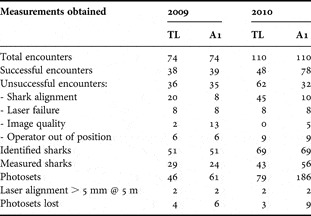
Over the course of the 2009 Seychelles season, 51% of in-water encounters (38/74) using the laser system produced useable TL measurements and 53% (39/74) produced usable A1 measurements.
Of the 36 failed attempts to capture TL measurements (A1 = 35) during in-water encounters in 2009; 56% (20/36) were due to shark alignment in photosets, in particular the position of the caudal keel and tail away from the central axis of the shark in the final of the three required photographs (A1 = 23%, 8/35).
In 2010, the percentage of TL measurements obtained reduced to 44% (48/110); while successful A1 measurements increased to 71% (78/110). Of the 62 failed attempts to obtain TL measurements from an encounter (A1 = 32); 72% (45/62) were due to incorrect shark alignment (A1 = 31%, 10/32).
In both 2009 and 2010 the lasers fell outside the calibration parameters for post-test analysis (≥ 5 mm) on two occasions, resulting in four and three TL photosets being omitted from the study (Table 3).
Operational results recorded from two years of measurements taken in the Seychelles seasonal feeding aggregation.

TL, total length; A1,the leading edge of spiracle to bottom of 5th gill slit.
Laboratory tests: sources of parallax error
The results of the test for horizontal axis error indicated that as the angle of the laser system from the perpendicular increased, the measured length of an object decreased, with length decreasing at a faster rate the more the angle moves through 0, 10, 20, 30 and 40 degrees. Conversely, the mean scale width of the laser points showed little variation with an increase to the object angle; these results were reflected almost identically at 2, 4 and 6 m (see Supplementary Material Results section Tests for Sources of Parallax Error and Supplementary Figure 5 for details).
The test for three-dimensional surface error revealed that when the vertical plane of a perpendicular object was closer to the camera, the pixel measurement of the scale length increased progressively as the angle increased; with the percentage change to the scale measurement diminishing with distance from the camera (see Supplentary Material Results section Tests for Sources of Parallax Error and Supplementary Figure 6 for details).
The test for the effects of tail bend showed that when the ‘tail’ was bent away from the camera it resulted in a decreased tail measurement that followed a straight linear regression. Conversely, when the ‘tail’ length was bent towards the camera there was an increase in recorded lengths (see Supplementary Material Results section Tests for Sources of Parallax Error and Supplementary Figure 7 for details).
DISCUSSION
The laser photogrammetry system trialled here was an adaptation of that used by several cetacean research groups (Durban & Parsons, Reference Durban and Parsons2006; Rowe & Dawson, Reference Rowe and Dawson2009; Webster et al., Reference Webster, Dawson and Slooten2010) but differs in that it is used in-water (Deakos, Reference Deakos2010; Rohner et al., Reference Rohner, Richardson, Marshall, Weeks and Pierce2011) and allows measurement of the total body length without the need for morphometric projections. This was important because there is a dearth of manually measured whale shark specimens with morphometric indices in whale shark literature (Wintner, Reference Wintner2000).
Precision and accuracy validation
Results of the experimental testing procedure from the laser photogrammetry set-up produced non-biased, highly accurate and precise measurements that were close to the manually measured length of all objects measured both in and out of the water. Our results (r2= 0.999, b = 0.9969, SE = 0.0019) equate favourably to similar laser metric tests carried out by Bergeron (Reference Bergeron2007) (r2 = 0.999, b = 1.003, SE = 0.003) and Rothman et al. (Reference Rothman, Chapman, Twinomugisha, Wasserman, Lambert and Goldberg2008) (r2 = 0.999, b = 1.002, SE = 0.005). Furthermore, when the terrestrial and in-water tests are separated there is no significant difference between manual and laser measurements in either medium (Mann–Whitney U-test, P = 0.808) confirming light refraction does not affect the performance of the laser system (Muljowidodo et al., Reference Muljowidodo, Rasyid, SaptoAdi and Budiyono2009).
In terms of the in situ measurements, the variance recorded in repeated measurements of the total length (TL) of individual whale sharks can be mainly attributed to the effects of shark body angle and movement and not to discrepancies in the laser system itself. This is supported by the results of the calculations of pre-caudal length (PCL), where percentage coefficient of variation (CV) was reduced from 2.44% for TL to 1.81% for PCL, and the measure of precision increased from r = 0.980 to r = 0.992 when the influence of caudal fin movement was removed from the final measurement. This was enhanced further to CV = 1.77% and r = 0.994 using the A1 morphometric measurement, which essentially removed variability resulting from lateral undulations of the body in motion. A result which equates favourably to that of the repeatability value attained for Capra ibex (Bergeron, Reference Bergeron2007) of r = 0.992.
Variability caused by lateral flexion was highlighted by our experimental measurements showing the effect of a ‘tail’ bending towards and away from the laser apparatus, which resulted in both an increase and decrease to measurements of TL from the central axis (Supplementary Figure 7). This is in apparent contradiction to previous laser photogrammetric studies where it was assumed that any deviation away from the perpendicular would always decrease the apparent length of a morphological feature (Bergeron, Reference Bergeron2007; Rothman et al., Reference Rothman, Chapman, Twinomugisha, Wasserman, Lambert and Goldberg2008); consequently, we did not assume that the longest calculated length for repeated TL measurements on individual sharks would be the most accurate estimate when analysing the results.
Morphometric measurements
The accuracy (Figure 4) and precision of the system (Table 1) promotes its use for in situ measurements of additional morphological relationships between the dimensions of specific body parts or defined areas on a shark with the TL or PCL, as previously indicated in the linear relationship between dorsal fin height and TL (A4) (Meekan et al., Reference Meekan, Bradshaw, Press, McLean, Richards, Quasnichka and Taylor2006) and the horizontal distance between the 5th gill slit and the origin of the 1st dorsal and TL (A5) (Rohner et al., Reference Rohner, Richardson, Marshall, Weeks and Pierce2011). The morphometric measurements A1 through to A4 compared in this study were chosen due to their uniformly defined positions helping to reduce the level of measurement error caused by observer interpretation, an error magnified by their use as a predictor equation for TL. For example, the origin of the anterior edge of the 1st dorsal fin as defined in A5 exhibits a smooth transition from the body to the fin, so does not provide a clear mark for measurement by an observer without the need to draw inferences from other identified areas of the dorsal, leading to possible measurement error. In contrast, the transition from the upper caudal keel to the anterior edge of the upper caudal lode is clearly defined by the pre-caudal notch and is therefore easily and precisely identified in most photographs incorporating the tail. In an attempt to reduce measurement error, this study defined the origin of the 1st dorsal as the lowest point of the dorsal surface where the dorsal ridge creates a small concave shape leading in to the 1st dorsal.
Morphological indices were largely chosen due to their relative position from the origin of the 1st dorsal fin; features forward of this point have reduced levels of variance caused by the lateral sub-carangiform undulation of the body posterior to the pectoral fins during swimming, a motion observed in both adolescent and adult R. typus (Martin, Reference Martin2007). The effect that body or tail undulation may have on TL measurements reinforces the benefit of these morphometric predictors to calculate TL.
In addition to reducing the effects of body movement, the use of the head area when defining morphometric measurements was supported to some extent by the results of the I3S Interactive Individual Identification System (Van Tienhoven et al., Reference Van Tienhoven, Den Hartog, Reijns and Peddemors2007) on R. typus (Speed et al., Reference Speed, Meekan and Bradshaw2007). This system uses three reference points (top of the 5th gill slit, the point on the flank corresponding to the posterior edge of the pectoral fin, and the bottom of the 5th gill slit), to define a reference frame within which a spot pattern ‘finger print’ is produced. These are matched using a two-dimensional linear algorithm to other similarly ‘fingerprinted’ images, and despite increases in the size of animals over time (from 9– 12 years), the product of the algorithm still successfully matches each shark (Speed et al., Reference Speed, Meekan and Bradshaw2007; Rowat et al., Reference Rowat, Speed, Meekan, Gore and Bradshaw2009; Brooks et al., Reference Brooks, Rowat, Pierce, Jouannet and Vely2010). This therefore suggests that these morphological reference points grow uniformly with TL. Additionally, the integration of the laser ‘scale’ into the requisite identification images produces both a ‘finger print’ and the necessary data to calculate TL measurements from a single image.
Predicting total length
The close linear relationship recorded between PCL, morphometric measurements and TL (P < 0.001) implied that measurements of TL could be obtained from the proportional dimensions of R. typus recorded in this study.
The use of GM linear regression to calculate predictor equations also allowed the interpretation of allometric growth for each morphometric index in relation to TL. When comparing the two PCL to TL formulas, from Wintner (Reference Wintner2000) and this study, Wintner (Reference Wintner2000) describes a positive b value in the size on size equation for the specified proportional dimensions of R. typus, which implies positive allometry for PCL (Mollet & Cailliet, Reference Mollet, Cailliet, Klimley and Ainley1996) suggesting an accelerated growth of PCL proportional to TL. In comparison, the negative b value from this study describes negative allometry. The result of negative allometry for PCL is unpredicted when considering previous reports regarding changes in the proportional dimensions of many disparate shark species (not including R. typus) that indicate positive allometry in the trunk area when compared to the head and tail (Garrick, Reference Garrick1960; Bass, Reference Bass1973; Mollet & Cailliet, Reference Mollet, Cailliet, Klimley and Ainley1996). However, when interpreting the results of fork length to TL from 13 shark species over four families, Kohler et al. (Reference Kohler, Casey and Turner1995) reported only six species from three different families with positive allometry and seven species with negative allometry.
The divergence of the two formulae and the result of negative allometry for PCL may, in part, be due to differences in methods, small sample sizes, the tail angle of the deceased and live specimens and measurements taken ‘as accurately as conditions allowed’ (Wintner, Reference Wintner2000). The associated problems of measuring shark carcasses are described in more detail by Ecopacifico (2010) and are incrementally magnified with large species the size of whale sharks.
This study expands on previously published literature (Rohner et al., Reference Rohner, Richardson, Marshall, Weeks and Pierce2011) by presenting total laser metric length measurements, which include caudal lobe lengths, taken directly from live, free-swimming whale sharks during the straight glide phase of locomotion. As such differences between Wintner (Reference Wintner2000) and this study could have resulted from the ‘natural’ position of the whale shark, and specifically the caudal fin, when suspended in water compared to the prone position of a whale shark on land and the differing measurement errors associated with both methods (Deakos, Reference Deakos2010; Ecopacifico, 2010; Rohner et al., Reference Rohner, Richardson, Marshall, Weeks and Pierce2011). Consequently, the five morphometric indices tested in this study (A1 to A5) are the first comparison of direct total length measurements of R. typus taken using laser photogrammetry.
Analysis of the five morphometric indices inferred that the A1 morphometric, with the lowest CV and highest r value, had the least variation caused by body movement and camera perspective; although lateral flexion of the body while swimming is minimal in the head area, there was an element of dorso-ventral flexion of the head observed during feeding when the mouth is open. To reduce the levels of error caused by dorso-ventral flexion, images with the mouth open were rejected when analysing A1 to A3.
When comparing the analysis of A1 against PCL, it is apparent that despite the increased precision of results when obtaining A1 measurements compared to PCL (CV = 1.77%, r = 0.994 compared to CV = 1.81%, r = 0.992), as an accurate predictor of TL, it is still preferential to obtain full PCL images when possible (95% CI =± 0.050, r 2 = 0.9935, SEE = 10.073) as indicated in the increased deviation of the slope of the regression lines y over x and the inverse of the slope x over y for the GM regression scatter plots for PCL, A1 and A5 (Figure 5a–c).
The greater level of variation and reduced precision for morphometrics A2 and A3 is likely due to the flexible nature of the gill slit area and so has not been considered as a suitable measurement for predicting TL. The height of the dorsal fin (A4) was also rejected as a TL predictor (r 2 = 0.9169, SEE = 38.877); however, this lower precision may be due to how measurements were taken using the laser system. The scale measurements were taken from the side of the body at varying vertical angles thus affecting perspective when measurements of the fin were taken. Therefore, the suitability of the dorsal fin height as a predictor of TL may be improved by projecting the laser ‘scale bar’ directly on to the dorsal fin itself.
The horizontal length from the origin of the 1st dorsal fin to the 5th gill slit (A5) is similarly affected by perspective due to the difference in the three-dimensional planes of the two morphometric parameters. When perpendicular to the shark, the origin of the 1st dorsal fin is further from the camera lens than the 5th gill slit, and so when the three-dimensional object is converted in to a two-dimensional image the measurements of length are affected in the same manner as the laboratory test for three-dimensional error (see Supplementary Material). Therefore, as a shark increases in width as it grows so does the distance between the two A5 measurement parameters in relation to the camera, and consequently the ratio of A5 to TL will naturally decrease over time. Further validation would therefore be required to verify to what level three-dimensional error affects A5 lengths and its suitability as a measure of TL growth in whale sharks. The effects of the three-dimensional surface will also influence measurements of A5 depending on the proximity of the camera to the subject and the vertical angle of the photographer to the horizontal mid-line of the shark; again further testing is required to quantify the influence of these possible sources of error.
Such effects may be the reason why the A5 linear regression r 2 value obtained in this study (r 2 = 0.96) was very close to that of A1 (r 2 = 0.97) and exceeded that of the comparative value for A5 obtained by Rohner et al. (Reference Rohner, Richardson, Marshall, Weeks and Pierce2011) (r 2 = 0.93) when taken from ‘optimal’ photosets of straight sharks, although the SEE was still comparatively low at 30.718. While the percentage coefficient of variation and repeatability were markedly lower in comparison to A1 and PCL (CV = 4.17% and r = 0.92) when analysed against multiple photosets of the same sharks.
However, notwithstanding the issue of perspective when measuring A5, and by implementing a standard method of precisely locating the origin of the 1st dorsal, use of an A5 predictor equation should help achieve a good indication of TL, particularly when a shark is feeding and the operator is unable to obtain suitable images of the A1 morphometric.
Operational analysis
In addition to accuracy and precision, the robustness and reliability of the laser apparatus developed to acquire complete TL measurements was evident from the operational analysis of two complete seasons from the Seychelles feeding aggregation in 2009 and 2010 (Table 3). Although only 51% of in-water encounters during the 2009 season and 44% in 2010 resulted in usable TL photosets for analysis (N = 38 and N = 48 respectively), this increased to 53%, (N = 61) and 71% (N = 78) when incorporating A1 morphometric measurements for 2009 and 2010. The percentage of useable photosets obtained per session also increased through each season with an increased level of operator experience and training.
In addition, the increase to 73% (45/62) of failed encounters due to improper shark alignment when obtaining TL measurements in 2010 can mainly be attributed to change of focus of the in-water operator to principally obtain multiple images appropriate for morphometric measurements. Effectively, in 2010 a greater amount of time was spent photographing the head of each shark with less time per encounter spent obtaining a suitable image incorporating a ‘straight’ tail, leading to negatively biased results.
Notwithstanding the apparent negative bias and the use of morphometric projections, the success rate of the system when obtaining full TL measurements due to improper shark alignment is still relatively low, which could be improved with the use of Stereo Diver Operated Video Systems (Stereo-DOV) (Shortis et al., Reference Shortis, Harvey and Abdo2009). However, the additional frame width required for a Stereo-DOV (~1 m) compared to the laser system, and consequently its bulk and drag in the water, would likely increase the percentage of failed encounters due to the operator being out of position.
In each year the laser system only failed calibration parameters (≥ 5 mm at 5 m) on two occasions, at the cost of four and three TL photosets respectively, indicating a high level of robustness and reliability of the apparatus. The high level of precision and reliability, coupled with the ability of a single operator to easily acquire the necessary underwater digital images to obtain morphometric, PCL or TL measurements, confirms the system to be a suitable and accurate method of remote measurement, for repeated and continued use in the marine environment with R. typus and other large marine species.
Sources of error
The apparatus did have some operational limitations, including the relatively short range that the lasers remain visible underwater (judged to be approximately <6 m depending on horizontal in-water visibility) and parallax error. The effects of such limitations are reduced by the versatility of the system allowing the operator to take multiple images of the same subject from different positions, and screening photographs for suitability post-encounter. However, by analysing the sources of error and understanding how measurements are obtained and altered in the two-dimensional image, it should also be possible to develop the system further to increase precision and accuracy, and diminish measurement error.
The test for horizontal axis error showed that when a straight object containing the scale-bar is moved from the perpendicular, measured lengths decrease at the same rate with increasing angle, notwithstanding the distance between the camera lens and the object. Indicating that whale shark images taken within the parameters of 0–10 degrees horizontal axis error should still provide reliable size estimates (Webster et al., Reference Webster, Dawson and Slooten2010).
However, at an angle of 10 degrees between vertical planes of a three-dimensional object, percentage change at 2, 4 and 6 m was 1.955, 0.995 and 0.667% respectively (see Supplementary Material). Therefore, assuming a 1% maximum margin of error, only at a distance of > 4 m would a 10 degree difference in vertical planes provide reliable estimate of size. As such, a measurement of distance may be useful in increasing the predicted accuracy of the system over an uneven surface.
It should also be noted, that although there is no documented evidence of lasers causing a negative reaction when they are exposed to whale sharks or other marine species, further study is required to monitor to what extent, if any, shark behaviour could be altered through the use of underwater lasers, even though risks associated with brief exposures are minimal (Durban & Parsons, Reference Durban and Parsons2006; Bergeron, Reference Bergeron2007; Rothman et al., Reference Rothman, Chapman, Twinomugisha, Wasserman, Lambert and Goldberg2008). As defined by the safety regulations for lasers administered by the US Food and Drug Administration, the units employed in this study were designated as Class IIIa lasers with a power output <5 mw, which ‘depending on power and beam area, can be momentarily hazardous when directly viewed or when staring directly at the beam with an unaided eye’.
After a few sharks were observed eliciting avoidance behaviour when the laser system was initially employed, in an effort to reduce exposure to the lasers and any potential to promote a failed encounter, a standard operating procedure (SOP) was also implemented. For the SOP the operator would approach and dive down to a shark behind the pectoral fins, and remaining between 3 m to 5 m from the flank, move forward towards the head with lasers pointed away from the shark at all times. Only when photographs were being taken, as the shark swam past the operator, were the lasers directed at a shark.
Conclusion
Our study was able to expand on previous work (Rohner et al., 2011) by validating the accuracy and precision of the method, and by refining suitable morphometric projections to improve the estimation of TL in whale sharks. The results also substantiate that the apparatus used in this study can be confidently employed to obtain accurate and precise in-water morphometric, PCL and TL measurements for R. typus. Consequently, due to the high inter-annual re-sighting of many individual whale sharks in the Seychelles and Djibouti feeding aggregations (Brooks et al., Reference Brooks, Rowat, Pierce, Jouannet and Vely2010), continued implementation of this system to record precise multiple measurements of total lengths for sharks over a number of years, used in conjunction with I3S as an effective identification system (Speed et al., Reference Speed, Meekan and Bradshaw2007; Van Tienhoven et al., Reference Van Tienhoven, Den Hartog, Reijns and Peddemors2007) should go some way to obtaining definitive growth rates for a wild whale shark population, where even small variations in measurements may bias results with current estimates of growth being only 0.29 m y−1 (Chang et al., Reference Chang, Leu and Fang1997; Uchida et al., Reference Uchida, Toda, Kamei and Teruya2000) and 0.22 m y−1 (Wintner, Reference Wintner2000) for juveniles in excess of 3.5 m. This could then be incorporated into population dynamics and maturation status models (Cailliet et al., Reference Cailliet, Smith, Mollet and Goldman2006; Bradshaw, Reference Bradshaw2007) when combined with the relevant environmental and behavioural data from each season. This should prove valuable as a starting point to understand how life-history traits may act in predicting the responses of populations to various perturbations (Musick, Reference Musick1999). Such data would subsequently have a positive impact on our overall knowledge of the biology of R. typus and assist in the conservation of the species.
The small size and simple calibration allow the system to be easily transported both locally and internationally for use on remote aggregation sites, and at a cost of approximately £500, the implications and applications of this comparatively inexpensive set-up could therefore be wide-ranging in the study of R. typus, and other large marine fauna.
ACKNOWLEDGEMENTS
Studies in Seychelles and Djibouti were supported with grant aid from the Save Our Seas Foundation. We thank all 2009, 2010 and 2011 Marine Conservation Society, Seychelles staff and volunteers who assisted in the development and implementation of the study.
Supplementary materials and methods
The Supplementary material referred to in this article can be found online at journals.cambridge.org/mbi.