INTRODUCTION
Studies on the deep-water nudibranch fauna of the North Atlantic (below the continental shelf) are scarce. A handful of species were described in early expedition reports such as the Challenger (Bergh, Reference Bergh, Thompson and Murray1885), Ingolf (Bergh, Reference Bergh1899) and Travailleur and Talisman (Vayssière, Reference Vayssière and Milne-Edwards1902). The first modern approach to the systematics of North Atlantic deep-sea nudibranchs was the work of Bouchet (Reference Bouchet1977), which was followed by a handful of papers describing new species (Valdés & Ortea, Reference Valdés and Ortea1996; Valdés & Bouchet, Reference Valdés and Bouchet1998a, Reference Valdés and Bouchetb). Most of these studies deal mainly with tropical or temperate species, and the deep-water nudibranch fauna of polar and subpolar regions remains poorly understood. Additionally, all these papers are based solely on morphological examination of specimens. There are virtually no available molecular data available for deep-water Atlantic nudibranchs, making classification and identification more challenging.
In this paper we examine a collection of nudibranchs from the North Atlantic waters off the coast of Newfoundland, including some rare and poorly known species. We provide new bathymetric and geographic records as well as anatomical descriptions of rare and uncommon species. We also provide sequence data of the mitochondrial barcoding gene COI for species for which this was possible.
MATERIALS AND METHODS
Source of specimens
Material examined for this study came from two research programmes. Most of the material was obtained during groundfish bottom trawl surveys carried out by the Spanish Institute of Oceanography (Instituto Español de Oceanografía, IEO) together with the Instituto de Investigaciones Marinas (IIM-CSIC) and the Instituto Português do Mar e da Atmosfera (IPMA) on board the RV ‘Vizconde de Eza’. The remainder came from rock dredge and scallop gear samples taken by the RV ‘Miguel Oliver’, operated by the Spanish General Secretariat of the Sea (Secretaría General del Mar) under the NEREIDA project. Groundfish bottom trawl surveys are carried out annually between spring and summer using a random stratified sampling design with standardized 30 min tows and vessel speed around 3 knots. They cover the Tail and Nose of the Grand Bank of Newfoundland, the Flemish Cap, and the Flemish Pass (north-west Atlantic Ocean) to 1500 m depth. A Campelen 1800 bottom-trawl gear was used in the Flemish Pass and the Grand Bank of Newfoundland, whereas Lofoten bottom-trawl gear was used at Flemish Cap. NEREIDA surveys were undertaken in the Flemish Pass, Flemish Cap and slope of the Grand Bank of Newfoundland at depths of 700–2000 m. These surveys were carried out during spring and summer using a rock dredge and scallop gear. Tows of 15 min were made at a vessel speed of about 1.5 knots. Specimens were preserved onboard in 70% ethanol.
All specimens are deposited at the Invertebrate Collection of the California State Polytechnic University (CPIC) and the research collections of the Instituto Español de Oceanografía (IEO). Type material is deposited at the Natural History Museum of Los Angeles County (LACM).
Abbreviations: PLA, Platuxa, common name of the groundfish survey along the Tail of the Grand Bank; FC, Flemish Cap; FN3L, Fletan Negro 3L, common name of the groundfish survey along the Nose of the Grand Bank and Flemish Pass; L, Lance (tow set); NEREIDA, the project (Nafo potEntial vulneRable marine Ecosystems. Impacts of Deep-seA fisheries); RD, Rock dredge; SG, Scallop gear.
Morphological examination
At least two specimens (in best preserved condition) of each species were dissected. Specimens were dissected by dorsal incision. The reproductive system was drawn with the aid of a camera lucida. The buccal mass was dissected to extract the radula and jaws by dissolving the surrounding tissue in sodium hydroxide 10%. Once the tissue was dissolved the radula and jaws were rinsed in distilled water and mounted on a stub, and sputter coated for examination under a Hitachi S-3000N scanning electron microscope (SEM).
Genetic barcoding
The universal barcoding gene, mitochondrial cytochrome c oxidase 1 (CO1), was amplified from each species (when possible) to provide additional information for identification. DNA extractions were performed using ~1–3 mg of tissue taken from the foot of the animals. Three extraction methods were used, the Qiagen DNeasy extraction kit, the Omega Mollusk EZNA extraction kit, or the hot Chelex® extraction protocol. The manufacturers' protocols with minor modifications were followed.
Polymerase chain reaction (PCR) was used to amplify portions of the CO1 gene using Folmer et al. (Reference Folmer, Black, Hoeh, Lutz and Vrijenhoek1994) universal primers (LCO1490 5′-GGTCAACAAATCATAAAGATATTGG-3′, HCO2198 5′TAAACTTCAGGGTGACCAAAAAATCA-3′). In cases in which this CO1 fragment did not amplify, the internal primer EIC 5′-ACATCTTGCTGGTATGTCTTCTATTTT-3′ was used with the HCO2198 primer (Ornelas-Gatdula et al., Reference Ornelas-Gatdula, DuPont and Valdés2011). PCRs were performed in a 50 µl reaction volume containing 0.25 µl 5 U µl−1 Taq polymerase, 5.00 µl 10× buffer, 5.00 µl 25 mm MgCl2, 1.00 µl 40 mm dNTPs, 1.00 µl each 10 mm primer, 34.75 µl H2O, and 2.00 µl extracted DNA. Reaction conditions for COI involved an initial denaturation of 95°C for 3 min, 35 cycles of 94°C for 45 s (denaturation), 45°C for 45 s (annealing), and 72°C for 2 min (extension), followed by a final elongation step of 72°C for 10 min. Confirmation of amplification was done using agarose gel electrophoresis with ethidium bromide to detect the presence of DNA. PCR products were sent to Source Bioscience (Santa Fe Springs, California) for Sanger sequencing.
Sequences were assembled and edited using Geneious Pro 4.8.3 (Drummond et al., Reference Drummond, Ashton, Cheung, Heled, Kearse, Moir, Stones-Havas, Thierer and Wilson2010). Assembled sequences were identified using the BLAST-n program on the GenBank website (http://www.ncbi.nlm.nih.gov).
SYSTEMATICS
CLADOBRANCHIA
Family AEOLIDIIDAE Gray, 1827
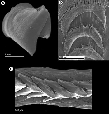
Aeolidiella sp. (Figures 1A, B & 2A).
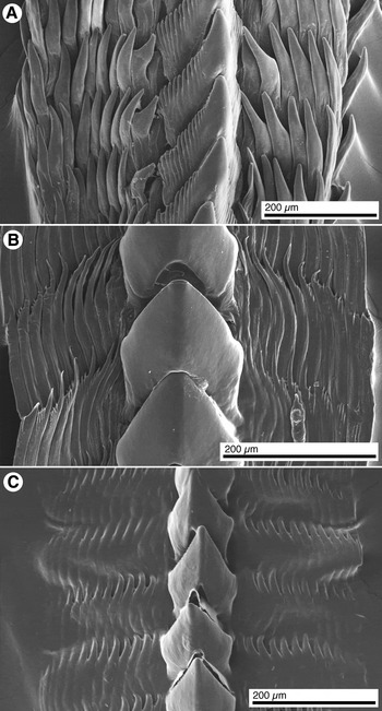
SEM of the radula and jaws of aeolids. (A) Aeolidiella sp., jaw. (B) Aeolidiella sp., radular teeth. (C) Flabellina salmonacea (Couthouy, Reference Couthouy1838), radular teeth.

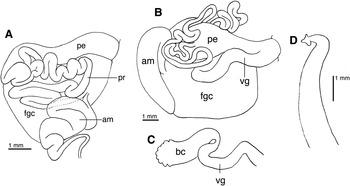
Reproductive systems of Aeolidiella sp. and Tritonia newfoundlandica n. sp. (A) Aeolidiella sp., dorsal view of the reproductive organs. (B) Tritonia newfoundlandica n. sp., dorsal view of the reproductive organs. (C) Tritonia newfoundlandica n. sp., bursa copulatrix. (D) Tritonia newfoundlandica n. sp., penis. am, ampulla; bc, bursa copulatrix; fgc, female gland complex; pe, penis; pr, prostate; vg, vagina.

MATERIAL EXAMINED
FC07 L23 (CPIC 01333), 46°21′54″–46°21′4.2″N 45°59′48.6″–45°57′40.8″W, depth 527–542 m, 26 June 2007, 1 specimen 12 mm preserved length; FC07 L108 (CPIC 01332), 47°38′4.2″–47°39′41.4″N 46°2′44.4″–46°1′54″W, depth 707–683 m, 7 July 2007, 1 specimen 11 mm preserved length; FC07 L138 (CPIC 01532), 48°42′18.6″–48°43′20.4″N 44°42′43.8″–44°44′24″W, depth 1362–1364 m, 12 July 2007, 1 specimen 10 mm preserved length; FC07 L178 (CPIC 01331), 47°6′10.8″–47°7′45.6″N 46°37′7.8″–46°36′39.6″W, depth 866–878 m, 19 July 2007, 1 specimen 9 mm preserved length.
EXTERNAL MORPHOLOGY
Live animals orange with deep red rhinophores. Photographs of live animals (not illustrated) lack resolution for a detailed description, thus the following description is based on preserved specimens. Body elongate, tapering posteriorly into the posterior end. The anterior end of the foot has two conspicuous, triangular foot corners. Oral tentacles elongate. Rhinophores smooth. Cerata arranged in 16 rows with 7–15 cerata per row. Anus located on the upper right hand side of the body between ceratal rows 4 and 5.
ANATOMY
The radula consists of a single row of 15 wide teeth (formula 15 × 0.1.0). Each tooth has a characteristic horseshoe shape with numerous elongate denticles and no distinct cup. The denticles vary in number between 26–42. Jaws with a distinct but smooth masticatory border.
Reproductive system with a large and convoluted ampulla, connecting directly into the female gland complex (Figure 2A). Penis wide and elongate, narrowing into a long, tightly folded prostate that connects into the female gland complex. No bursa copulatrix or vagina was observed.
GENETIC BARCODE
The DNA from the three specimens collected could not be amplified.
BIOLOGY
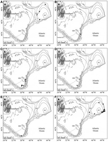
This species was collected in the eastern side of Flemish Cap between 527 and 1364 m depth (Figure 3A), mainly in sandy-silty bottoms with temperatures and salinities between 3.5 and 3.9°C and 34.87 and 34.90‰, respectively. Other species commonly found were the sea pen Anthoptilum grandiflorum, the cup coral Flabellum alabastrum, the black coral Stauropathes arctica, the sponge Asconema foliata or the sea urchin Phormosoma placenta.
Maps of collection localities. (A) Aeolidiella sp. (B) Flabellina salmonacea (Couthouy, Reference Couthouy1838). (C) Dendronotus niveus Ekimova et al., Reference Ekimova, Korshunova, Schepetov, Neretina, Sanamyan and Martynov2015. (D) Dendronotus frondosus (Ascanius, Reference Ascanius1774). (E) Dendronotus robustus A. E. Verrill, Reference Verrill1870. (F) Tritonia newfoundlandica n. sp. GB, Grand Bank; FC, Flemish Cap; FP, Flemish Pass.

REMARKS
Species level identification of this species was not possible. We were unable to amplify DNA and the anatomical information obtained from the damaged preserved specimens was inconclusive.
Family FLABELLINIDAE Bergh, 1889
Flabellina cf. salmonacea (Couthouy, Reference Couthouy1838) (Figure 1C).
Eolis salmonacea Couthouy, Reference Couthouy1838, pp. 68–69, pl. 1, Figure 2.
See Kuzirian (Reference Kuzirian1979) for a full list of synonyms.
MATERIAL EXAMINED
PLA07 L79 (CPIC 01329), 43°55′10.2″–43°57′42″N 48°57′42″–48°58′38.4″W, depth 666–635 m, 14 June 2007, 1 specimen 25 mm preserved length.
EXTERNAL MORPHOLOGY
Live animal was orange with deep red rhinophores. The specimen was severely damaged beyond recognition. The cerata were detached.
ANATOMY
Radular formula n × 1.1.1. Rachidian teeth with a large, robust, triangular cusp and 5–9 small denticles on each side (Figure 1C). Lateral teeth elongate, with a narrow base and a long sharp cusp. Each tooth has 5–7 small denticles on the inner side.
GENETIC BARCODE
DNA from the specimen collected could not be amplified.
BIOLOGY
This species was collected in one locality of the continental slope of the Tail of the Grand Bank (Figure 3B), in silty-sandy bottom with temperature and salinity of 3.9°C and 34.86‰, respectively. Thirty-two other benthic species were found in this locality, between them, some characteristics of the slope were the sponges Tentorium semisuberites and Polymastia uberrima.
REMARKS
The single specimen here examined was damaged beyond recognition. It was tentatively identified as Flabellina salmonacea (Couthouy, Reference Couthouy1838) based on the radular morphology. The radula of the specimen here examined has a large rachidian tooth with several denticles on each side of the central cusp, and relatively large lateral teeth with a sharp cusp and denticles on its inner side. This is very similar to the description of Flabellina salmonacea by Kuzirian (Reference Kuzirian1979, figure 11).
Flabellina salmonacea was originally described from Charles River, Massachusetts (Couthouy, Reference Couthouy1838). Kuzirian (Reference Kuzirian1979) redescribed this species and compiled numerous records from the North Atlantic, all of them from relatively shallow waters (above 160 m depth). Wakeling (Reference Wakeling2002) reported F. salmonacea from the North Pacific. Here we report this species for the first time from 600 m depth.
Family DENDRONOTIDAE Allman, 1845
Dendronotus frondosus (Ascanius, Reference Ascanius1774) (Figures 4A & 5C)
Amphitrite frondosa Ascanius, Reference Ascanius1774, pp. 155–158, pl. 5, Figure 2A, B.
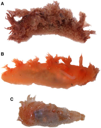
Photographs of live Dendronotus. (A) Dendronotus frondosus (Ascanius, Reference Ascanius1774). (B) Dendronotus niveus Ekimova et al., Reference Ekimova, Korshunova, Schepetov, Neretina, Sanamyan and Martynov2015. (C) Dendronotus robustus A. E. Verrill, Reference Verrill1870.

SEM of the radula and jaws of Dendronotus. (A) Dendronotus robustus A. E. Verrill, Reference Verrill1870. (B) Dendronotus niveus Ekimova et al., Reference Ekimova, Korshunova, Schepetov, Neretina, Sanamyan and Martynov2015. (C) Dendronotus frondosus (Ascanius, Reference Ascanius1774).

See Robilliard (Reference Robilliard1970) and Ekimova et al. (Reference Ekimova, Korshunova, Schepetov, Neretina, Sanamyan and Martynov2015) for a full list of synonyms.
MATERIAL EXAMINED
PLA07 L21 (CPIC 01490), 44°20′9″–44°18′33.6″N 49°12′30″–49°12′37.8″W, depth 59 m, 5 June 2007, 1 specimen 25 mm preserved length; PLA07 L63, 43°17′7.2″–43°18′25.2″N 49°48′39″–49°49′46.8″W, depth 61–62 m, 11 June 2007, 1 specimen 27 mm preserved length.
EXTERNAL MORPHOLOGY
Live animals reddish brown with numerous dark brown patches scattered all over the body, including the rhinophores and the cerata (Figure 4A). Body narrow, elongate, with 9 highly ramified cerata on each side, one pair located next to the rhinophores. Velum composed of 8 highly ramified appendages. Rhinophores with an elongate sheath, having three highly ramified papillae on its outer, distal edge. Rhinophoral club perfoliate, with about 16 lamellae.
ANATOMY
Radular formula n × 10.1.10. Rachidian tooth rhomboid in shape, short and wide, with numerous tiny denticles on each side (Figure 5C). Lateral teeth long and narrow, with a curved, elongate cusp and 3–4 sharp denticles on their outer side.
The reproductive anatomy was not examined. It will be described in future review of the genus Dendronotus in progress.
GENETIC BARCODE
DNA from the two specimens collected could not be amplified.
BIOLOGY
This species was collected on the continental shelf of the Tail of the Grand Bank between 52 and 62 m depth (Figure 3D), in sandy bottoms with temperatures and salinities between −0.5 and −0.2°C and 33.14 and 33.15‰, respectively. Other species commonly found in these localities were the sea cucumber Cucumaria frondosa, the bryozoan Eucratea loricata, the crabs Hyas coarctatus and Hyas araneus, the soft coral Gersemia rubiformis or the hydroid Halecium muricatum.
REMARKS
The specimens here examined were identified as Dendronotus frondosus based on the external morphology and radula. Robilliard (Reference Robilliard1970) described the radula and anatomy of D. frondosus, which differ from those of our specimens, but his drawings were probably based on specimens collected in the Pacific Ocean, which belong to a different species, Dendronotus venustus MacFarland, 1966 (see Stout et al., Reference Stout, Pola and Valdés2010). Pola & Stout (Reference Pola and Stout2008) illustrated the radula of specimens from Scotland, which are nearly identical to that of the material here examined.
Dendronotus robustus A. E. Verrill, Reference Verrill1870 (Figures 4C & 5A).
Dendronotus robustus Verrill, Reference Verrill1870, pp. 405–406, Figure 1.
See Robilliard (Reference Robilliard1970) and Ekimova et al. (Reference Ekimova, Korshunova, Schepetov, Neretina, Sanamyan and Martynov2015) for a full list of synonyms.
MATERIAL EXAMINED
PLA07 L53 (CPIC 01524), 42°52′00″–42°52′28.8″N 50°14′1.8″–50°11′51.6″W, depth 231–212 m, 10 June 2007, 1 specimen, fragmented into small pieces, size unknown.
EXTERNAL MORPHOLOGY
Live animal reddish-grey, with numerous opaque white spots all over the body (Figure 4C). The single specimen was fragmented into small pieces and the live animal photographed was too contracted to allow a detailed description.
ANATOMY
Radular formula n × 12.1.12. Rachidian tooth triangular, elongate, with 13–14 large denticles on each side of the cusp (Figure 5A). Lateral teeth long and narrow, smooth, lacking denticles.
The single specimen was fragmented thus examination of the reproductive anatomy was not possible.
GENETIC BARCODE
GenBank accession number (KU695599). A BLAST-n of this sequence resulted 99% identical to a sequence of Dendronotus robustus (KC660038).
BIOLOGY
This species was collected in one locality on the upper continental slope of the Tail of the Grand Bank (Figure 3E), in silty-sandy bottom with temperature and salinity of 1.5°C and 34.01‰, respectively. Forty-seven other benthic species were found in this locality, between them, some characteristics to the end of the continental shelf and beginning of the slope as the sea star Crossaster papposus or the sea urchin Strongylocentrotus droebachiensis.
REMARKS
The single specimen was positively identified as Dendornotus robustus based on DNA sequence data and radular morphology, based on the data provided by Ekimova et al. (Reference Ekimova, Korshunova, Schepetov, Neretina, Sanamyan and Martynov2015). This species is circumpolar, including records from Norway, Spitzbergen, Greenland, Siberia, Cape Cod, Nova Scotia, the White Sea, the Barents Sea, the Kara Sea and the Bering Strait (Robilliard, Reference Robilliard1970; Ekimova et al., Reference Ekimova, Korshunova, Schepetov, Neretina, Sanamyan and Martynov2015).
Dendronotus niveus Ekimova, Korshunova, Schepetov, Neretina, Sanamyan and Martynov, Reference Ekimova, Korshunova, Schepetov, Neretina, Sanamyan and Martynov2015 (Figures 4B & 5B)
Dendronotus niveus Ekimova et al., Reference Ekimova, Korshunova, Schepetov, Neretina, Sanamyan and Martynov2015, pp. 864–869, figures 6D, 8F, 13B, 14 & 15.
MATERIAL EXAMINED
PLA07 L21 (CPIC 01519), 44°20′9″–44°18′33.6″N 49°12′30″–49°12′37.8″W, depth 59 m, 5 June 2007, 1 specimen 23 mm preserved length; PLA07 L23 (CPIC 01520), 44°12′15.6″–44°10′41.4″N 49°21′00″- 49°21′2.4″W, depth 54–52 m, 6 June 2007, 1 specimen 30 mm preserved length; PLA07 L24 (CPIC 01518), 44°1′1.8″–43°59′39.6″N 49°17′30.6″–49°18′41.4″W, depth 46–47 m, 6 June 2007, 1 specimen 35 mm preserved length; PLA07 L51 (CPIC 01528), 43°12′26.4″–43°11′00″N 50°9′44.4″–50° 9′44.4″W, depth 68–69 m, 9 June 2007, 10 specimens, between 29 and 45 mm preserved length; PLA07 L63, 43°17′7.2″–43°18′25.2″N 49°48′39″–49°49′46.8″W, depth 61–62 m, 11 June 2007, 3 specimens, between 30 and 65 mm length; PLA07 L64 (CPIC 01517), 43°20′48″–43°21′57.6″N 50°4′54.6″–50°3′21″W, depth 66–63 m, 11 June 2007, 1 specimen fragmented into small pieces, size unknown.
EXTERNAL MORPHOLOGY
Live animals uniform deep orange-red in colour (Figure 4B). Body narrow, elongate, with 5 highly ramified cerata on each side. Velum composed of 8 highly ramified appendages. Rhinophores with an elongate sheath, having one anterior and three posterior simple papillae. Rhinophoral club perfoliate, with about 12 irregular lamellae.
ANATOMY
Radular formula 39 × 9.1.9. Rachidian tooth robust, short and wide, lacking denticles (Figure 5B). Lateral teeth long and narrow, with a curved, elongate cusp and 3–5 sharp denticles on their outer side.
The reproductive anatomy was not examined. It will be described in future review of the genus Dendronotus in progress.
GENETIC BARCODE
GenBank accession number (KU695598). A BLAST-n of this sequence resulted 99% identical to two sequences of Dendronotus niveus (KC660036, KC660037).
BIOLOGY
This species was collected on the continental shelf of the Tail of the Grand Bank between 46 and 69 m depth (Figure 3C), in sandy bottoms with temperatures and salinities between −0.2 and 1.2°C and 33.15 and 33.32‰, respectively. Other species commonly found in these localities were the sea cucumber Cucumaria frondosa, the bryozoan Eucratea loricata, and the crabs Hyas coarctatus and Hyas araneus.
REMARKS
Ekimova, Korshunova, Schepetov, Neretina, Sanamyan and Martynov (Reference Ekimova, Korshunova, Schepetov, Neretina, Sanamyan and Martynov2015) has been recently described from the White and Barents Seas. In this paper we report this species for the first time in the Atlantic Ocean. The radular morphology of the specimens here examined is very similar to that in the illustrations by Ekimova et al. (Reference Ekimova, Korshunova, Schepetov, Neretina, Sanamyan and Martynov2015). Also the Atlantic specimens are genetically nearly identical to the specimens of Dendronotus niveus from northern Russia; therefore they are confidently assigned to this species.
Family TRITONIIDAE Lamarck, 1809
Tritonia newfoundlandica n. sp. (Figures 6 & 7).
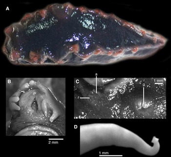
External morphology and anatomy of Tritonia newfoundlandica n. sp. (A) Dorsal view of the live animal. (B) Ventral view of the mouth region and velum. (C) Lateral view of the right side of the body. (D) Penis. a, anus; g, genital opening; r, renal opening.

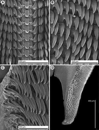
SEM of the radula and jaws of Tritonia newfoundlandica n. sp. (A) Rachidian and inner lateral teeth. (B) Lateral teeth. (C) Outer teeth. (D) Masticatory border of the jaw.

TYPE MATERIAL
Holotype (LACM 3433): FC07 L26 (CPIC 01489), 46°26′55.2″–46°28′28.2″N 45°45′14.4″–45°46′20.4″W, depth 538–492 m, 26 June 2007, 38 mm preserved length.
MATERIAL EXAMINED
FC07 L26 (CPIC 01489), 46°26′55.2″–46°28′28.2″N 45°45′14.4″–45°46′20.4″W, depth 538–492 m, 26 June 2007, 1 specimen, 40 preserved length; FC07 L44, 46°44′48.6″–46°46′13.8″N 44°7′1.8″–44°5′31.2″W, depth 480–471 m, 29 June 2007, 2 specimens, between 45 and 60 mm length; FC07 L74, 48°13′36″–48°14′45.6″N 44°43′51″–44°45′46.2″W, depth 596–599 m, 3 July 2007, 1 specimen 51 mm length; FC07 L164, 47°2′46.8″–47°1′1.8″N 43°39′25.2″–43°39′46.8″W, depth 759–764 m, 17 July 2007, 1 specimen 47 mm length; FC07 L166, 46°53′13.2″–46°52′1.8″N 43°23′28.8″–43°25′6.6″W, depth 1242–1245 m, 17 July 2007, 3 specimens, between 30 and 49 mm length; FC07 L168, 46°53′14.4″–46°52′0.6″N 43°29′17.4″–43°30′59.4″W, depth 1094–1083 m, 17 July 2007, 6 specimens, between 26 and 39 mm length; FN3L08 L69, 46°42′54″–46°41′25.2″N 46°55′55.2″–46°56′7.8″W, depth 1232 m, 5 August 2008, 2 specimens, between 26 and 27 mm preserved length; NEREIDA0509 RD10, 48°0′1.88″–48°0′32.54″N 43°45′38.65″–43°45′39.27″W, depth 1554–1607 m, 5 June 2009, 1 specimen 50 mm preserved length; NEREIDA0509 SG19, 47°9′51.52″–47°9′57.11″N 43°28′34.78″–43°28′41.27″W, depth 1137–1132 m, 14 June 2009, 4 specimens, between 34 and 42 mm preserved length; NEREIDA0509 SG20, 47°4′20.09″–47°4′15.27″N 43°26′56.91″–43°27′5.91″W, depth 1122–1113 m, 15 June 2009, 11 specimens, between 30 and 37 mm preserved length; NEREIDA0509 SG21, 46°50′45.84″–46°50′44.23″N 43°43′3.51″–43°43′18.88″W, depth 870–871 m, 16 June 2009, 2 specimens, between 26 and 35 mm preserved length; NEREIDA0509 SG22, 46°50′24.04″–46°50′31.74″N 43°38′25.41″–43°38′32.13″W, depth 956–943 m, 17 June 2009, 2 specimens, between 34 and 40 mm preserved length; NEREIDA0509 SG23, 46°46′29.47″–46°46′25.82″N 43°51′54.37″–43°52′10.76″W, depth 1127–1108 m, 18 June 2009, 18 specimens, between 23 and 38 mm preserved length; NEREIDA0509 SG24, 46°41′39.2″–46°41′31.92″N 43°58′6.95″–43°58′15.55″W, depth 1104–1112 m, 19 June 2009, 3 specimens, between 30 and 35 mm preserved length.
EXTERNAL MORPHOLOGY
Live animals are black, with the rhinophores, rhinophoral sheaths, velar appendages and secondary gills brownish-red (Figure 6A). Body elongate, with the anterior end rounded, tapering posteriorly into a pointy, triangular end. Oral veil undivided, with only one differentiated portion and bearing 10 simple, tentacular processes (Figure 6B). Dorsum smooth, lacking tubercles. Edge of the mantle with 10 short, ramified gills on each side. Rhinophores perfoliated with 6 branched vertical lamellae. Rhinophoral sheaths with two posterior extensions bearing 2 papillae each.
The reproductive opening is located on the anterior right hand side of the body. The anus and renal opening are located more posteriorly, also on the right hand side (Figure 6C).
ANATOMY
The radula of a 40 mm preserved length specimen was examined. The radular formula is 46 × 52.1.52. The rachidian teeth are broad and short, with an apical depression in the centre of each tooth (Figure 7A). The cusp is short with 3–4 denticles on each side. There is a large denticle on each side of the cusp. The innermost lateral teeth are hook-shaped but slightly shorter and wider that the rest of the inner laterals (Figure 7A). Mid lateral teeth hook-shaped, lacking denticles (Figure 7B). Outer lateral teeth with proportionally longer cusps, also lacking denticles, becoming smaller towards the outer edge of the radula (Figure 7C). Jaws with a masticatory border composed of irregular cell-like structures (Figure 7D).
The reproductive system has a relatively large ampulla with 2 distinct folds, connecting directly into the female gland complex (Figure 2B). The penial sac is large and narrows sharply into a long and convoluted prostate that connects directly into the female gland complex. The penis, located inside the penial sac is elongate, having a characteristic mushroom-like structure at the tip that could function as a scoop (Figure 2D, 6D). The vagina is long and curved, opening into an oval bursa copulatrix. The bursa has several characteristic small and rounded vesicles on its distal end (Figure 2C).
GENETIC BARCODE
DNA from the specimens collected could not be amplified.
BIOLOGY
This species was collected in the Flemish Cap, mostly in the south-east corner, between 471 and 1607 m depth (Figure 3F), mainly in silty-sandy bottoms with temperatures and salinities between 3.5 and 3.9°C and 34.86 and 34.91‰, respectively. Other species found in these localities were the sponge Asconema foliata, the soft corals Duva florida and Drifa flavescens, the bamboo coral Acanella arbuscula, or the sea stars Henricia perforata and Henricia spongiosa.
REMARKS
Tritonia newfoundlandica n. sp. is morphologically distinct from other species of Tritonia described to date. Tritonia ingolfiana is a very rare species, originally described by Bergh (Reference Bergh1899) from the mid-Atlantic ridge based on a single specimen collected at 887 m depth. Bergh (Reference Bergh1899) described his preserved specimen as greyish blue with yellow rhinophores. This is different from T. newfoundlandica n. sp., which has a black body and the rhinophores, gills and velum are much lighter even in the preserved specimens (orange in the live animals). Other differences include the shape of the penis, which is very elongate and tightly packed in the penial sheath of T. ingolfiana and lacks a scoop at the tip (Bergh, Reference Bergh1899, pl. 3; Figure 9). The penis of T. newfoundlandica n. sp. is much shorter, straight and has a characteristic scoop.
The only other Atlantic species with an apical scoop in the penis is Tritonia episcopalis Bouchet, Reference Bouchet1977 described from deep waters (1035–2170 m depth). This species is clearly different from T. newfoundlandica n. sp. in several aspects. First, T. episcopalis has the body covered by small tubercles (Bouchet, Reference Bouchet1977), whereas T. newfoundlandica n. sp. has a smooth body. The rachidian teeth of T. episcopalis lack denticles on the cusp, present in T. newfoundlandica n. sp., and the inner lateral teeth of T. episcopalis have small denticles, absent in T. newfoundlandica n. sp. The external colouration of T. episcopalis is bright violet with the gills of the same colour and the rhinophores red, whereas in T. newfoundlandica n. sp. the body is black and both the rhinophores and gills are brownish-red.
Family DORIDOXIDAE Bergh, Reference Bergh1899
Doridoxa ingolfiana Bergh, Reference Bergh1899 (Figures 8, 9 & 11A–C).
Doridoxa ingolfiana Bergh, Reference Bergh1899, pp. 15–17, pl. 2, Figures 3–15, pl. 3, Figures 1–3.
See Schrödl et al. (Reference Schrödl, Wägele and Willan2001) for a full list of synonyms.
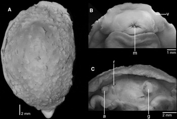
Photographs of a preserved specimen of Doridoxa ingolfiana Bergh, Reference Bergh1899. (A) Dorsal view of the animal. (B) Ventral view of the mouth region. (C) Lateral view of the right side of the body. a, anus; g, genital opening; m, mouth opening; r, renal opening; v, velum.

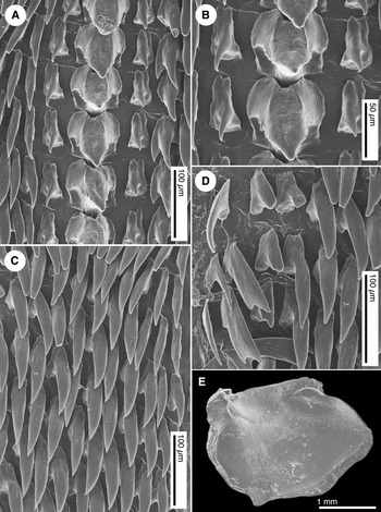
SEM of the radula and jaws of Doridoxa ingolfiana Bergh, Reference Bergh1899. (A) Rachidian and inner lateral teeth. (B) Detail of the rachidian teeth. (C) Lateral teeth. (D) Outer teeth. (E). Jaw.

MATERIAL EXAMINED
PLA06 L35 (CPIC 01531), 43°3′50.4″–43°4′42.6″N 51°0′4.2″–51°1′41.4″W, depth 290–300 m, 12 June 2006, 1 specimen 26 mm preserved length; PLA07 L110 (CPIC 01052), 45°48′49.2″–45°47′40.8″N 48°15′25.8″–48°17′1.2″W, depth 120–119 m, 19 June 2007, 1 specimen 23 mm preserved length; FC07 L44 (CPIC 01514), 46°44′48.6″–46°46′13.8″N 44°7′1.8″–44°5′31.2″W, depth 480–471 m, 29 June 2007, 2 specimens, between 25 and 27 mm preserved length; FC07 L168 (CPIC 01513), 46°53′14.4″–46°52′0.6″N 43°29′17.4″–43°30′59.4″W, depth 1094–1083 m, 17 July 2007, 2 specimens, between 15 and 27 mm preserved length; FN3L07 L92 (CPIC 01515), 46°4′8.4″–46°5′40.8″N 46°59′57″–47°0′2.4″W, depth 1358–1357 m, 9 August 2007, 2 specimens, between 28 and 30 mm preserved length; FN3L08 L96 (CPIC 01516), 46°9′31.8″–46°9′1.8″N 48°5′9.6″–48°3′12.6″W, depth 110–114 m, 10 August 2008, 1 specimen 18 mm preserved length; NEREIDA0509 SG23, 46°46′29.47″–46°46′25.82″N 43°51′54.37″–43°52′10.76″W, depth 1127–1108 m, 18 June 2009, 2 specimens, between 11 and 23 mm preserved length.
EXTERNAL MORPHOLOGY
Live animals are deep-reddish brown. Body elongate, with the anterior end rounded, tapering posteriorly into a pointy, triangular end (Figure 8A). The posterior end of the foot is visible on a dorsal view. The notum does not cover the head, but is fused anteriorly with the oral veil as indicated by Schrödl et al. (Reference Schrödl, Wägele and Willan2001). Oral veil wide, devoid of papillae (Figure 8B). Dorsum covered with irregular papillae, varying in density between specimens; in some specimens they are completely lacking, suggesting they could be autotomizable cerata. Rhinophores perfoliated with 25 narrow lamellae. There is no evidence of the presence of specialized gills.
The reproductive opening is located on the anterior right hand side of the body. The anus and renal opening are located more posteriorly, also on the right hand side (Figure 8C).
ANATOMY
The radular formula is 28 × 14.1.1.1.14 in a 27 mm preserved length specimen. The rachidian teeth are robust, short and wide, with a large ventral cusp bearing several small denticles on each side (Figure 9A, B). The innermost lateral teeth are narrow and elongate, having a sharp and elongate cusp (Figure 9A). The rest of the lateral teeth are hook-shaped, with a long and narrow cusp devoid of denticles (Figure 9C). The only exceptions are some inner teeth on the left half-row that can have a secondary cusp (Figure 9A). The lateral teeth diminish in size towards the outer end of the radula (Figure 9D). The jaws are featureless plates, lacking a masticatory border (Figure 9E).
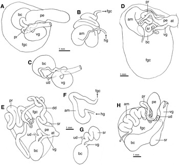
The reconstruction of the reproductive system here conducted (Figure 11A–C) is similar to the drawing by Schrödl et al. (Reference Schrödl, Wägele and Willan2001, Figure 6) with some differences. The ampulla is large, long and convoluted and appears to connect directly into the female gland complex (Figure 11B). The penis is large, wide, smooth, devoid of armature, connected to a short and convoluted deferent duct that expands into a short prostate (Figure 11A). The prostate opens into the female gland complex near the connection of the ampulla. The vagina is elongate, convoluted, connecting into a large, muscular, oval bursa copulatrix (Figure 11C). A narrow uterine duct connects the bursa copulatrix with the female gland complex opening near the connection of the ampulla and the prostate.
GENETIC BARCODE
GenBank accession number (KP871640). A molecular phylogenetic analysis including sequence data from the specimens here examined have been recently published (Mahguib & Valdés, Reference Mahguib and Valdés2015).
BIOLOGY
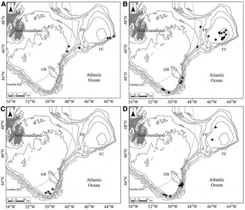
This species was collected in the south-east of Flemish Cap, south of Flemish Pass and Nose of the Grand Bank, between 110 and 1358 m depth (Figure 10A), in silty-sandy, sandy and clayed silty bottoms with temperatures and salinities between −0.1 and 3.8°C and 33.44 and 34.89‰, respectively. Other species commonly found in these localities were the sea stars Henricia perforata and Henricia spongiosa and the soft coral Drifa flavescens.
Maps of collection localities. (A) Doridoxa ingolfiana Bergh, Reference Bergh1899. (B) Aldisa zetlandica (Alder & Hancock, 1854). (C) Onchidoris bilamellata (Linnaeus, Reference Linnaeus1767). (D) Colga villosa (Odhner, Reference Odhner1907). GB, Grand Bank; FC, Flemish Cap; FP, Flemish Pass.

REMARKS
Doridoxa ingolfiana is an extremely rare species, until recently known from a handful of preserved specimens (Schrödl et al., Reference Schrödl, Wägele and Willan2001). Despite the lack of data on the anatomy of D. ingolfiana, this species has been considered essential to understand the early evolution of nudibranchs and has been the centre of vigorous debate (Odhner, Reference Odhner1934; Tardy, Reference Tardy1970; Wägele, Reference Wägele1989; Salvini-Plawen, Reference Salvini-Plawen1990; Schrödl et al., Reference Schrödl, Wägele and Willan2001). More recently Mahguib & Valdés (Reference Mahguib and Valdés2015) using molecular data from one of the specimens here examined concluded that D. ingolfiana is a cladobranch, rather than a basal nudibranch.
Doridoxa ingolfiana was originally described based on specimens collected during the Ingolf expedition, off the coast of Greenland at 103 m depth and subsequently reported from the Faroe Islands (Sneli et al., Reference Sneli, Schiøtte, Jensen, Wikander and Stokland2005) at 603 m depth, and Norway (Evertsen & Bakken, Reference Evertsen and Bakken2013) at 881–907 m depth. Schrödl et al. (Reference Schrödl, Wägele and Willan2001) re-examined the holotype in great detail, as well as two partially dissected or damaged specimens collected during the Ingolf expedition and by Lemche respectively. Schrödl et al. (Reference Schrödl, Wägele and Willan2001) provided numerous anatomical details, however, because all the specimens available were either dissected or damaged, the radula and the reproductive system could not be illustrated. In this paper, we provide for the first time SEM images of the radula and jaw as well as an interpretation of the reproductive anatomy. The characteristics of the specimens here examined closely match the re-description by Schrödl et al. (Reference Schrödl, Wägele and Willan2001) of the holotype of D. ingolfiana and therefore are confidently assigned to this species.
Until now D. ingolfiana was known only from Greenland, the Faroe Islands and Norway from 103–907 m depth. This paper expands both the geographic and bathymetric range of the species to the western Atlantic down to 1358 m depth.
DORIDACEA
Family CADLINIDAE Bergh, 1891
Aldisa zetlandica (Alder & Hancock, 1854) (Figures 11D & 12A, B).
Doris zetlandica Alder & Hancock, Reference Alder and Hancock1845–1855 [1854]: 102.
Reproductive systems of some species examined. (A–C) Doridoxa ingolfiana Bergh, Reference Bergh1899, dorsal view of the entire reproductive system (A); dissected ampulla, not visible in a dorsal view (B); dissected female organs, partially covered by the penis (C). (D) Aldisa zetlandica (Alder & Hancock, 1854), dorsal view of the reproductive system. (E–G) Onchidoris bilamellata (Linnaeus, Reference Linnaeus1767), dorsal view of the entire reproductive system (E); dissected ampulla, not visible in a dorsal view (F); dissected female organs, partially covered by the penis (G). (H) Colga villosa (Odhner, Reference Odhner1907), dorsal view of the reproductive system. am, ampulla; at, genital atrium; bc, bursa copulatrix; dd, deferent duct; fgc, female gland complex; hg, hermaphroditic gland; pe, penis; pr, prostate; sr, seminal receptacle; ud, uterine duct; vg, vagina.

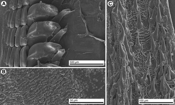
SEM of the radulae of two dorids. (A) Lateral teeth of Aldisa zetlandica (Alder & Hancock, 1854). (B) Detail of the cusp and denticles of the lateral teeth of A. zetlandica. (C) Radular teeth of Onchidoris bilamellata (Linnaeus, Reference Linnaeus1767).

MATERIAL EXAMINED
FN3L06 L34, 47°32′7.8″–47°30′40.2″N 47°9′41.4″–47°10′34.8″W, depth 529–506 m, 6 August 2006, 1 specimen 18 mm preserved length; PLA07 L44 (CPIC 01496), 43°3′52.8″–43°2′4.2″N 51°1′15.6″–50°59′30″W, depth 450–458 m, 9 June 2007, 1 specimen 18 mm preserved length; PLA07 L45 (CPIC 01500), 42°59′21.6″–42°58′24″N 50°53′16.2″–50°51′30″W, depth 336–332 m, 9 June 2007, 2 specimens, between 18 and 25 mm preserved length; PLA07 L52 (CPIC 01494), 42°48′1.8″–42°47′30.6″N 50°23′36″–50°21′30.6″W, depth 370–390 m, 10 June 2007, 2 specimens, between 18 and 22 mm preserved length; PLA07 L72 (CPIC 01498), 43°30′22.2″–43°30′24″N 49°12′44.4″–49°14′52.8″W, depth 390–418 m, 13 June 2007, 3 specimens, between 19 and 23 mm preserved length; FC07 L17 (CPIC 01510), 46°53′25.8″–46°54′38.4″N 46°21′25.8″–46°23′10.8″W, depth 335–330 m, 25 June 2007, 1 specimen 16 mm preserved length; FC07 L29 (CPIC 01511), 46°38′53.4″–46°37′35.4″N 45°36′57.6″–45°35′12.6″W, depth 257–252 m, 27 June 2007, 1 specimen 23 mm preserved length; FC07 L34 (CPIC 01495), 46°40′34.8″–46°42′2.4″N 44°57′28.8″–44°58′48″W, depth 178–180 m, 27 June 2007, 1 specimen 30 mm preserved length; FC07 L35 (CPIC 01493), 46°45′49.2″–46°47′30.6″N 44°56′46.2″–44°57′21.6″W, depth 169–170 m, 27 June 2007, 1 specimen 23 mm preserved length; FC07 L36 (CPIC 01497), 46°49′49.8″–46°51′24″N 44°44′30.6″–44°45′23.4″W, depth 144–145 m, 27 June 2007, 1 specimen 21 mm preserved length; FC07 L39 (CPIC 01509), 46°46′51.6″–46°46′51.6″N 44°48′6.6″–44°50′38.4″W, depth 141–147 m, 28 June 2007, 4 specimens, between 22 and 27 mm preserved length; FC07 L48 (CPIC 01492), 47°0′3″–47°1′45.6″N 44°34′14.4″–44°34′34.2″W, depth 138–148 m, 30 June 2007, 3 specimens, between 14 and 22 mm preserved length; FC07 L90 (CPIC 01501), 47°17′57.6″–47°16′22.8″N 44°35′12.6″–44°34′8.4″W, depth 181–178 m, 5 July 2007, 1 specimen 22 mm preserved length; FC07 L93 (CPIC 01530), 47°10′9.6″–47°10′6.6″N 44°46′5.4″–44°43′30.6″W, depth 159–162 m, 5 July 2007, 2 specimens, between 19 and 29 mm preserved length; FC07 L96 (CPIC 01512), 47°5′43.8″–47°4′29.4″N 45°1′45.6″–45°3′37.2″W, depth 163–160 m, 6 July 2007, 1 specimen 20 mm preserved length; FC07 L97 (CPIC 01499), 47°5′57″–47°7′39.6″N 45°13′21.6″–45°13′34.8″W, depth 181–183 m, 6 July 2007, 1 specimen 26 mm preserved length; FC07 L98 (CPIC 01491), 47°8′58.2″–47°9′11.4″N 45°15′5.4″–45°17′42″W, depth 193–198 m, 6 July 2007, 1 specimen 22 mm preserved length; PLA08 L2, 43°38′16.2″–43°39′41.4″N 49°6′22.2″–49°6′6.6″W, depth 332–306 m, 27 May 2008, 1 specimen 24 mm preserved length; PLA08 L82, 43°54′1.8″–43°55′22.8″N 48°59′47.4″–49°0′35.4″W, depth 450–414 m, 9 June 2008, 1 specimen 18 mm preserved length; FN3L09 L91, 46°8′16.8″–46°7′23.4″N 47°29′10.2″–47°30′55.8″W, depth 508–498 m, 10 August 2009, 2 specimens, between 16 and 17 mm preserved length.
EXTERNAL MORPHOLOGY
Live animals white, semi-translucent, with the pale-cream to reddish-brown viscera visible through the dorsum. Dorsum covered with a number of large, conical tubercles, varying in size, typically larger in the centre of the dorsum and smaller on the mantle edge. Gill composed of 7 bipinnate branchial leaves, arranged in a circle around the anus. Rhinophores lamellate, with 11–12 lamellae in the specimens examined (18–30 mm preserved length). Anterior border of the foot grooved but not notched. Mouth area large, with two blunt lateral expansions, lacking oral tentacles.
ANATOMY
The internal anatomy of this species was described in detail by Millen & Gosliner (Reference Millen and Gosliner1985) and Valdés (Reference Valdés2002), the specimens here examined match these descriptions. The radula of a 30 mm preserved length specimen was examined, it contains a number of hair-like, elongate teeth as in other species of Aldisa (Figure 12A). The teeth have a wide triangular base and a rounded upper edge as well as a series of 19–22 elongated denticles on their outer and upper edges (Figure 12B). The radular formula is impossible to determine due to the large number of overlapping teeth.
The reproductive system has a large, wide, folded ampulla, which merges with the proximal end of the prostate before entering the female gland complex (Figure 11D). The vagina is short and straight, connecting directly into the spherical bursa copulatrix. A short but convoluted duct connects the bursa to the muscular, pear-shaped seminal receptacle. Mid-length from this latter duct a short uterine duct emerges, connecting to the female gland complex. The deferent duct is wide and short, it narrows into an elongate duct that expands into the short but convoluted prostate.
GENETIC BARCODE
GenBank accession number (KU695603). A BLAST-n of this sequence resulted in a 91% identical to a sequence of Aldisa albatrossae (KP871632.1). This confirms that the specimens were correctly placed in the genus Aldisa and that A. zetlandica and A. albatrossae are genetically distinct.
BIOLOGY
This species was collected in the top of Flemish Cap, and on the edge of the continental shelf and on the continental slope of the Grand Bank, between 138 and 1005 m depth (Figure 10B), in sandy and silty-sandy bottoms with temperatures and salinities between 2.5 and 4.0°C and 34.18 and 34.86‰, respectively. Other species commonly found in these localities were the brittle-star Ophiopholis aculeata, the sea stars Henricia perforata and Leptychaster arcticus, or the soft coral Duva florida.
REMARKS
The specimens here assigned to Aldisa zetlandica are confidently identified based on their external, reproductive and radular morphology and based on Millen & Gosliner's (Reference Millen and Gosliner1985) redescription of this species.
Aldisa zetlandica was studied in detail by Millen & Gosliner (Reference Millen and Gosliner1985) based on specimens from Norway and Iceland. According to these authors the known range of the species includes several localities in the North-eastern Atlantic (Finland, Norway, Shetland Islands and Iceland). Therefore this is the first record from the North-western Atlantic. Additionally, Millen & Gosliner (Reference Millen and Gosliner1985) reported A. zetlandica as usually occurring between 70 and 320 m depth with one specimen found at 1900 m. The present study corroborates the abundance of this species between 138 and 529 m depth.
Family ONCHIDORIDIDAE Gray, 1827
Onchidoris bilamellata (Linnaeus, Reference Linnaeus1767) (Figures 11E–G & 12C).
Doris bilamellata Linnaeus, Reference Linnaeus1767, p. 1083.
Doris fusca O. F. Müller, Reference Müller1776, p. 229.
Onchidoris leachii de Blainville, Reference de Blainville1816, pp. 95–97.
MATERIAL EXAMINED
PLA07 L48 (CPIC 01334), 43°7′36″–43°8′37.2″N 50°24′52.8″–50°26′30″W, depth 80–79 m, 9 June 2007, 1 specimen 21 mm preserved length; PLA07 L51 (CPIC 01330), 43°12′26.4″–43°11′N 50° 9′44.4″–50° 9′44.4″W, depth 68–69 m, 9 June 2007, 5 specimens, between 15 and 25 mm preserved length; PLA07 L59, 43°4′44.4″–43°5′34.8″N 49°58′43.2″–49°56′53.4″W, depth 70 m, 10 June 2007, 1 specimen 27 mm length; PLA08 L12, 43°24′13.2″–43°22′48.6″N 49°42′49.8″–49°43′33.6″W, depth 55–57 m, 28 May 2008, 7 specimens, between 16 and 21 mm preserved length.
EXTERNAL MORPHOLOGY
Live animals light brown with a darker irregular median band. Mantle margin with dark brown mottling against the light brown background. Photographs of the live animal (not illustrated) lack resolution for a detailed description, thus the following description is based on preserved specimens. Body elongate, oval, with round tubercles covering the dorsum. Larger tubercles surrounded by a ring of smaller ones; other small tubercles not surrounding larger tubercles. Rhinophores with 14 lamellae. Gill with 17–20 unipinnate brachial leaves arranged in a horseshoe pattern, surrounding the anus. The foot is smooth with no foot corners. The mouth lacks oral tentacles and has irregular lips under the velum.
ANATOMY
The radular formula is 28 × 1.1.1.1.1. The rachidian teeth are narrow and elongate lacking a distinct cusp, but having a central, longitudinal ridge (Figure 12C). The inner lateral teeth are broad, with an apical, elongate cusp lacking denticles. The outer lateral teeth are simple plates.
The reproductive system has an elongate and curved prostate that connects directly into the female gland complex (Figure 11F). The genital atrium is wide, from it emerges the long, muscular and convoluted deferent duct that narrows into a thin tube (Figure 11E). This tube expands again into the very long and convoluted prostate that also connects into the female gland complex. The vagina is short and straight, it opens into the bursa copulatrix (Figure 11G). Another duct connects the bursa copulatrix with the muscular seminal receptacle. The uterine duct is short, it emerges from the duct connecting the bursa copulatrix with the muscular seminal receptacle and opens into the female gland complex.
GENETIC BARCODE
GenBank accession number (KU695604). A BLAST-n of this sequence resulted 99% identical to a sequence of Onchidoris bilamellata (KF643873), from New Brunswick, Canada, a locality close to where CPIC 01330 was collected.
BIOLOGY
This species was collected on the continental shelf of the Grand Bank, between 55 and 80 m depth (Figure 10C), in sandy bottoms with temperatures and salinities between 0.1 and 1.8°C and 32.91 and 33.53‰, respectively. This species was commonly found between the branches of the bryozoan Eucratea loricata.
REMARKS
The external morphology and anatomy of Onchidoris bilamellata have been described by Thompson & Brown (Reference Thompson and Brown1984) and Marcus (Reference Marcus1961) based on specimens from the British Isles and California respectively, and both match the characteristics of the specimens examined here.
Onchidoris bilamellata has a broad geographic range including the North Pacific and North Atlantic but has been reported from the shallow subtidal waters. In this paper we provide records from deeper water, down to 80 m depth.
As mentioned above, BLAST-n of the partial COI sequence here obtained from specimen CPIC 01330 was 99% identical to several specimens of Onchidoris bilamellata from New Brunswick, Canada (KF643873, KF643475, KF644026, KF643245), sequenced by Layton et al. (Reference Layton, Martel and Hebert2014), suggesting all belong to the same species. Hallas & Gosliner (Reference Hallas and Gosliner2015) also sequenced COI from a specimen identified as O. bilamellata collected in California, but this sequence (KP340408) has a pairwise identity of only 78.5% with CPIC 01330. Subsequent blasting of KP340408 revealed it has a 99% pairwise identity with specimens of Doris montereyensis (Family Dorididae) (KF643914, KF643446) also sequenced by Layton et al. (Reference Layton, Martel and Hebert2014). This suggests that Hallas & Gosliner (Reference Hallas and Gosliner2015) specimen is misidentified or the sequence is the result of contamination. There are no COI sequences available for Pacific specimens of O. bilamellata to verify whether they are the same species as the Atlantic specimens.
Family POLYCERIDAE Alder & Hancock, 1845
Colga villosa (Odhner, Reference Odhner1907) (Figures 11H & 13)
Issa villosa Odhner, Reference Odhner1907, p. 100, pl. 1, Figure 21. For a full list of synonyms see Martynov & Baranets (Reference Martynov and Baranets2002).
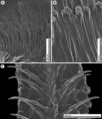
SEM of the radula, jaws and penis of Colga villosa (Odhner, Reference Odhner1907). (A) Half-row of the radula. (B) Jaw elements. (C) Penial spines.

MATERIAL EXAMINED
PLA07 L1 (CPIC 01488), 43°42′28.2″–43°43′41.4″N 49°9′28.8″–49°10′33″W, depth 250–234 m, 29 May 2007, 2 specimens, between 19 and 20 mm preserved length; PLA07 L45 (CPIC 01526), 42°59′21.6″–42°58′24″N 50°53′16.2″–50°51′30″W, depth 336–332 m, 9 June 2007, 2 specimens 28 mm preserved length; PLA07 L46 (CPIC 01486), 42°55′7.8″–42°54′30″N 50°40′24.6″–50°38′26.4″W, depth 228–238 m, 9 June 2007, 1 specimen 26 mm preserved length; PLA07 L53 (CPIC 01529), 42°52′00″–42°52′28.8″N 50°14′1.8″–50°11′51.6″W, depth 231–212 m, 10 June 2007, 1 specimen 36 mm preserved length; PLA07 L75 (CPIC 01525), 43°38′40.2″–43°39′34.2″N 49°18′45″–49°19′55.2″W, depth 450–368 m, 13 June 2007, 1 specimen 20 mm preserved length; PLA07 L77 (CPIC 01522), 43°43′52.8″–43°45′29.4″N 49°0′3.6″–48°59′3″W, depth 620–575 m, 14 June 2007, 1 specimen 24 mm preserved length; PLA07 L78 (CPIC 01487), 43°50′21.6″–43°51′56.4″N 49°2′13.8″–49°2′12.6″W, depth 320–306 m, 14 June 2007, 1 specimen 19 mm preserved length; FC07 L11 (CPIC 01523), 46°58′7.2″–46°56′22.2″N 45°58′24″–45°58′20.4″W, depth 306–299 m, 24 June 2007, 1 specimen 18 mm preserved length; FC07 L113 (CPIC 01521), 47°49′51.6″–47°48′34.8″N 45°37′37.2″–45°39′28.2″W, depth 337–342 m, 8 July 2007, 1 specimen 29 mm preserved length; NEREIDA0810 RD99, 43°36′52.75″–43°37′19.59″N 49°4′41.17″–49°4′49.96″W, depth 548–520 m, 3 August 2010, 1 specimen 22 mm preserved length. FN3L08 L58 (CPIC 01485), 46°56′20.4″–46°54′47.4″N 47°39′38.4″–47°39′54.6″W, depth 179–175 m, 3 August 2008, 1 specimen 29 mm preserved length.
EXTERNAL MORPHOLOGY
Live animals uniform off-white, with the rhinophores and the gill pale yellow. Body elongate, narrow, tapering into the posterior end of the foot. Rhinophores perfoliate, with 20 lamellae. Gill non-retractable, composed of 6 bipinnate branches, forming a circle surrounding the anus and open posteriorly. Dorsum covered by numerous long and narrow papillae. In some specimens the papillae located along the central mid-line are close to each other forming a longitudinal crest. Papillae located around the mantle margin also close to each other forming a rim around the mantle margin.
ANATOMY
Radular formula 15 × 5.1.1.1.1.1.5. Innermost lateral teeth narrow and elongate with an inconspicuous apical triangular cusp and a secondary cusp near the base of each tooth (Figure 13A). Second lateral teeth much wider, also having an apical triangular cusp and a secondary cusp mid-length. Outer teeth are simple plates, with no cusps or denticles, decreasing in size towards the radular margin. Rachidian teeth are also simple plates, nearly square, with an inconspicuous thickened crest in the middle. Jaws with a number of irregular elements (Figure 13B).
Reproductive system with a long and convoluted ampulla, connecting directly into the female gland complex (Figure 11H). The deferent duct is wide and muscular and narrows into a tubular, indistinctive prostate that connects directly into the female gland complex near the ampulla connection. The penis contains a number of translucent spines (Figure 13C). The vagina is long and wide and connects into the irregular bursa copulatrix. The seminal receptacle connects into the vagina mid-length, close to the distal opening of the uterine duct, which connects to the female gland complex proximally.
GENETIC BARCODE
GenBank accession numbers (KU695600–KU695602). A BLAST-n of these three (99.8% pairwise identity) sequences resulted to be 82% identical to Hypselodoris cf. nigrolineata (JQ727899), the closest match found. However, there are no other Colga sequences in GenBank, which may account for this result. Further investigation is needed to determine the phylogenetic position of Colga considering the closest match is a chromodorid nudibranch and not a member of Polyceridae.
BIOLOGY
This species was collected on the continental slope of the Grand Bank and in the Flemish Cap, between 175 and 620 m depth (Figure 10D), mainly in silty-sandy bottoms with temperatures and salinities between 1.4 and 4.2°C and 33.14 and 34.86‰, respectively. Other species commonly found in these localities were the sea anemone Actinauge cristata, the brittle-stars Ophiopholis aculeata and Ophiura sarsii, or the sea star Ceramaster granularis.
REMARKS
Martinov & Baranets (Reference Martynov and Baranets2002) reviewed the genus Colga and recognized three species: Colga villosa (Odhner, Reference Odhner1907), Colga pacifica Bergh, 1894, and Colga minichevi Martinov & Baranets, Reference Martynov and Baranets2002, the former found in the North Atlantic, and the two latter in the North Pacific. The three species are distinguished based on external and reproductive anatomy as well as radular morphology. One of the most distinctive features of Colga villosa is the presence of colourless penial spines. The specimens here examined match the description of C. villosa by Martinov & Baranets (Reference Martynov and Baranets2002) and they are confidently assigned to this species. Colga villosa has been reported from Cape Cod in the North-west Atlantic to the Barents Sea (Martinov & Baranets, Reference Martynov and Baranets2002).
The BLAST-n of the Colga villosa sequences here obtained returned unexpected results, as the closest species in the GenBank database was Hypselodoris cf. nigrolineata, a member of the family Chromodorididae. Martinov & Baranets (Reference Martynov and Baranets2002) classified Colga in the family Polyceridae based on morphological evidence. Because of these unexpected results, additional specimens of Colga were sequenced to discard contamination problems. All Colga villosa sequences obtained from different specimens were 99.8% identical, which suggests contamination was not a problem. There are two likely explanations for these results: (1) Because COI is a highly variable gene, saturation may result in overall similarities with phylogenetically distant taxa; (2) The absence of a comprehensive coverage of the family Polyceridae in GenBank may have resulted in the erroneous results. A phylogenetic analysis for this group, using a combination of nuclear and mitochondrial genes needs to be conducted to resolve the systematic position of this group.
ACKNOWLEDGEMENTS
This paper was made possible with support of the Spanish Institute of Oceanography (IEO) and the Spanish Government (Secretaría General del Mar). The authors would like to acknowledge the scientific staff involved in NAFO groundfish bottom trawl and NEREIDA surveys, and the heads of these surveys, especially Esther Roman, for facilitating the data collection. NEREIDA is a multidisciplinary research project involving scientists from Instituto Español de Oceanografía (IEO), Fisheries and Oceans Canada (DFO), Natural Resources Canada (NRCAN), Centre for the Environment, Fisheries and Aquaculture Science (Cefas), Instituto de Investigaciones Marinas (IIM-CSIC), Secretaría General del Mar (SGM), Polar Research Institute of Marine Fisheries and Oceanography (PINRO), and the P.P. Shirshov Institute of Oceanology (RAS). Thanks are also due to the crews of the research vessels RV ‘Vizconde de Eza’ and RV ‘Miguel Oliver’ for assistance at sea. NAFO groundfish surveys are co-funded by the EU and the Spanish Government. SEM work was conducted at the Cal Poly Pomona SEM laboratory supported by the US National Science Foundation (NSF) grant DMR-1429674, and the Natural History Museum of Los Angeles County SEM laboratory supported by the NSF grant DBI-0216506. Ellen Kenchington and an anonymous reviewer made constructive comments on the manuscript. Lindsey Groves helped with the curation of specimens.