Introduction
Carcinoma of the tongue can be locally advanced and has a strong tendency to metastasise to regional lymph nodes.Reference Shah, Patel, Singh and Shah 1 , 2 Initial assessment of local tumour invasion via a physical examination can be inaccurate owing to ulceration or submucosal spread. Identification and treatment of cervical lymph node metastasis and possible extracapsular spread are important for disease prognosis and treatment.Reference Ding, Liang, Shen, Xie, Huang and Zhang 3 Cervical metastasis with extracapsular spread represents one of the most significant adverse prognostic indicators in oral squamous cell carcinoma (SCC).Reference Shaw, Lowe, Woolgar, Brown, Vaughan and Evans 4
Magnetic resonance imaging (MRI) can show clear delineation of normal tongue architecture and its interface with cancer, and has been useful for predicting the depth of invasion of oral tongue cancer.Reference Lwin, Hanlon, Lowe, Brown, Woolgar and Triantafyllou 5 – Reference Lam, Au-Yeung, Cheng, Wei, Yuen and Trendell-Smith 10 Tumour thickness is related to cervical nodal metastasis, and numerous studies have attempted to define this relationship. To this end, 1.5 Tesla MRI has been widely used to evaluate cervical lymph node status.Reference Ding, Liang, Shen, Xie, Huang and Zhang 3 According to Okura et al., 1.5 Tesla MRI has a 53 per cent sensitivity, 89 per cent specificity and 77 per cent overall accuracy for detecting cervical lymph node metastasis.Reference Okura, Iida, Aikawa, Adachi, Yoshimura and Yamada 8
However, there are a number of technical benefits and challenges when imaging at a field strength greater than 1.5 Tesla.Reference Frayne, Goodyear, Dickhoff, Lauzon and Sevick 11 The most appealing features of 3 Tesla are the increased signal-to-noise ratio (proportional to the magnetic field strength), increased T1 relaxation time, decreased T2 relaxation time, increased magnetic susceptibility contrast and increased spectral resolution.Reference Lu, Nagae-Poetscher, Golay, Lin, Pomper and van Zijl 12 The increased signal-to-noise ratio and larger susceptibility result in higher spatial resolution, leading to improved imaging and diagnostic strength.
Advantages of 3 Tesla MRI for clinical radiology have been demonstrated in many clinical studies.Reference Lu, Nagae-Poetscher, Golay, Lin, Pomper and van Zijl 12 Most 3 Tesla MRI studies have been designed to evaluate brain tumoursReference Kim, Lekovic, White and Karis 13 – Reference Watanabe, Yamasaki, Kajiwara, Takayasu, Nosaka and Akiyama 17 and prostate cancerReference Patel, Dasgupta, Challacombe, Cahill, Brown and Patel 18 – Reference Ferda, Kastner, Hora, Hes, Finek and Topolcan 21 ; only a few are aimed at clinically evaluating tongue cancer. Previous publications using 3 Tesla MRI in head and neck oncology have tried to determine the apparent diffusion coefficient values for carcinoma and tumour-involved lymph nodes, and to find associated reference values.Reference Srinivasan, Dvorak, Rohrer and Mukherji 22 , Reference Lee, Tsai, Chuang, Liu and Chen 23 This study defined the role of 3 Tesla MRI, which has a higher signal-to-noise ratio than 1.5 Tesla MRI, in the initial evaluation of oral tongue cancer. The primary objective was to evaluate the efficacy of pre-operative 3 Tesla MRI in predicting the thickness of the oral cancer compared with histological measures. Secondary objectives were to compare radiographic and pathological (i.e. histological) nodal staging, assess the relationship between the thickness of oral tongue cancer invasion and the presence of cervical lymph node metastasis, and assess the capacity of 3 Tesla MRI to predict extracapsular extension.
Materials and methods
Study design
This prospective study evaluated 29 patients with histologically proven SCC of the tongue who underwent evaluation with 3 Tesla MRI before surgery (range 1–40 days) between 2009 and 2012. Of these, 25 patients were included and 4 were excluded because of technical difficulties (cancelled MRI and/or surgery, imaging artefacts caused by dental hardware or extreme motion). One patient who did not undergo neck dissection but had no clinical or radiographic evidence of metastasis after three years of follow up was considered to have a clinically staged tumour–node–metastasis (TNM) node-negative (N0) neck and was included in one analysis (i.e. correlation analysis of MRI depth of tumour invasion and cervical lymph node metastasis).
The study protocol was approved by the Institutional Review Board at the University of Cincinnati College of Medicine (protocol 08-10-23-07-EE). Medical records (including radiology and pathology reports) of all patients were reviewed for clinical information and surgical details.
Based on pre-operative 3 Tesla MRI of the head and neck (with and without gadolinium contrast), tumour thickness was (1) measured directly and (2) reconstructed based on tumour characteristics (exophytic or endophytic), and cervical lymph node metastasis was evaluated. Radiological findings were compared with the corresponding histological findings.
Radiological measurements
For the 25 patients who underwent pre-operative MRI on a 3-Tesla superconducting magnet (GE Healthcare, Milwaukee, Wisconsin USA), the scanning protocol included a T2 sagittal localising sequence, axial fast spin echo T1 imaging, axial and coronal fast spin echo T2 fat saturation at 3 mm, post-contrast axial and coronal fast spin echo T1 fat saturation at 3-mm slice thickness, and post-contrast T1 coronal fast spoiled gradient echo three-dimensional 1.3–1.5 mm slices. Axial images were parallel to the hard palate. The scanned area covered the middle cranial fossa to the thoracic inlet. The neurovascular coil was used to image the soft tissue of the neck according to the protocol. In patients with a contraindication to gadolinium administration, contrast sequences were not performed. Detailed parameters for individual MRI sequences are listed in Table I. There were occasional minor variations in technical factors based on the body habitus of patients and their ability to cooperate. All measures were performed on axial images.
Parameters of individual magnetic resonance imaging sequences

NEX = number of excitations; FLIP = flip angle; TR = repetition time; TE = echo time; TI = inversion time; Fat sat = Fat saturation; 3D = three-dimensional
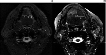
On MRI, tumour thickness was directly measured in the axial plane as the distance from the lateral tumour surface to the deepest point of tumour invasion (direct thickness; Figure 1, distance T), whereas the reconstructed measure of tumour thickness (reconstructed thickness) was determined using methodologies previously described by Iwai et al.Reference Iwai, Kyomoto, Ha-Kawa, Lee and Yamashita 24 and Preda et al.Reference Preda, Chiesa, Calabrese, Latronico, Bruschini and Leon 9 (shown in detail in Figure 1.) Briefly, measure A was the distance from the surface to the lingual septum on the unaffected side, and measure B was the distance from the septum to the point of deepest tumour invasion. For cancer that did not extend beyond the midline, the MRI reconstructed thickness was the difference between measures A and B. For cancer that had invaded beyond the midline, the reconstructed thickness was the sum of measures A and B (Figure 1). Tumour thickness was measured on MRI scans in duplicate at the same time by a single radiologist (RSC) for all available axial sequences for each patient. If there were discrepancies in thickness measurements between sequences, the largest measurement was recorded.
Radiological 3 Tesla magnetic resonance imaging (MRI) scans showing measures of (a) superficial (i.e. tumour does not extend beyond the midline) and (b) deeply invasive (i.e. tumour extends beyond the midline) tongue cancer. T = Direct MRI thickness, the distance from the tumour surface to the deepest point of tumour invasion; A = the distance from the surface to the lingual septum on the unaffected side; B = the distance from the septum to the point of deepest tumour invasion. Reconstructed MRI Thickness is A-B (a) and A + B (b).

The MRI scans were also used to identify the presence and number of lymph nodes involved by metastatic disease, and the possibility of extracapsular spread. Lymph nodes were considered malignant if the largest diameter of a node exceeded 8 mm for retropharyngeal nodes or 10 mm for all other nodal levels. In addition, any node with frank necrosis, heterogeneous enhancement or a round (not oval) shape was considered malignant. Imaging evidence of extracapsular spread included loss of the adjacent fat plane, a poorly defined nodal border and inability to separate the node from adjacent structures.
Histological measurements
Tissue specimens were fixed overnight in 10 per cent buffered formalin, processed with a standard protocol for surgical pathology specimens, embedded in paraffin, and stained with hematoxylin and eosin. Histological tumour thickness was measured using direct and reconstructed methods.
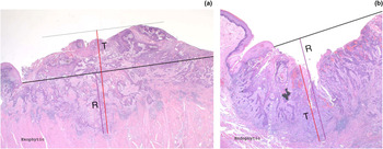
Preda et al. previously defined direct measurement as the distance from the tumour surface to the deepest point of tumour invasion, T.Reference Preda, Chiesa, Calabrese, Latronico, Bruschini and Leon 9 A reconstructed measurement, R, was used for ulcerated and exophytic lesions: this entailed drawing a straight line linking the non-neoplastic mucosal surfaces on each side of the lesion.Reference Preda, Chiesa, Calabrese, Latronico, Bruschini and Leon 9 The reconstructed thickness was defined as the distance from this line to the deepest point of tumour invasion (Figure 2), measured using both an ocular micrometer and a millimetre ruler.
Histological measures of (a) exophytic and (b) endophytic tongue cancer (H&E; ×10). The red line indicates the direct measure of tumour thickness (i.e. the distance from the tumour surface to the deepest point of tumour invasion); the purple line indicates the reconstructed measure of thickness (i.e. the distance from the line linking the mucosal surfaces on each side of the lesion (heavy black line) to the point of maximum tumour invasion).

Statistical analysis
Measures were classified into four groups for statistical analysis: direct MRI measures, reconstructed MRI measures, direct histological measures and reconstructed histological measures. The Kolmogorov–Smirnov test was used to test for normal distribution of the variables. Correlation between MRI and histological tumour thickness was tested using Pearson correlation coefficient for both direct and reconstructed thickness measures. Statistical significance was defined as a p value of 0.05 or under. A paired sample t-test was performed to compare MRI measured vs reconstructed thickness. Variables included tongue tumour thickness based on MRI direct measurements, histological direct measurements, MRI reconstructed measurements and histological reconstructed measurements. Magnetic resonance imaging scans were evaluated for the presence of pathological cervical lymph nodes and for the relationship between depth of oral tongue cancer invasion and presence of cervical nodal metastasis. The sensitivity, specificity, positive and negative predictive values, and accuracy of 3 Tesla MRI for evaluating cervical lymph node metastasis and extracapsular spread were also determined.
Results
In total, 25 patients (age range 29–80 years, mean 58 years) underwent 3 Tesla MRI, surgery and histological testing (of the tongue and/or neck), including 17 men (68 per cent) and 8 women (32 per cent); 24 were Caucasian and 1 was African-American.
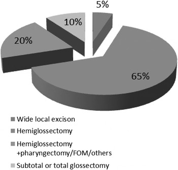
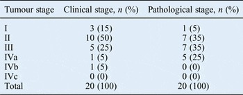
After excluding 5 patients with dental artefacts on MRI, radiological and histological measures of the tongue were available for 20 patients (14 men, 6 women). A summary of tumour stages is provided in Table II. Lesion location was lateralised to the left or right hemitongue in an equal number of patients. Surgical reports showed that several procedures had been performed to treat these malignancies, including wide local excision (5 per cent), hemiglossectomy (65 per cent), hemiglossectomy with pharyngectomy (20 per cent), and subtotal or total glossectomy (10 per cent; Figure 3). In all, 16 (80 per cent) patients underwent microvascular free flap reconstruction.
Pie chart showing the proportions of the different types of surgical procedures performed. FOM = floor of mouth

Clinical and pathological tumour stage

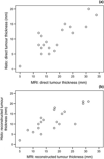
The Pearson correlation coefficient for direct measures of tumour thickness by MRI and histopathology was 0.84 (p < 0.0001). The correlation coefficient for reconstructed tumour thickness was 0.81 (p < 0.0001). The coefficient of determination (r2 ) was 0.712 for direct MRI and histological measures and 0.656 for MRI and histological reconstructed measures. In a paired sample t-test, mean (± standard deviation) values for direct (18.2 ± 7.3 mm) and reconstructed (17.9 ± 7.2 mm) MRI tumour thickness did not differ significantly (r = 0.879; Figure 4).
Scatter plots showing the correlation between (a) direct and (b) reconstructed measures of 3 Tesla magnetic resonance imaging (MRI) and histological tumour thickness. Hist = histological

In comparisons of MRI and histological tumour thickness for each patient, MRI tumour thickness was always significantly greater than histological tumour thickness. The mean direct MRI tumour thickness was 8.3 mm greater than the mean direct histological tumour thickness (95 per cent confidence interval (CI), 6.29 to 10.30; p < 0.001), whereas the mean reconstructed MRI tumour thickness was 5.51 mm greater than the mean reconstructed histological tumour thickness (95 per cent CI, 3.5 to 7.51; p < 0.001).
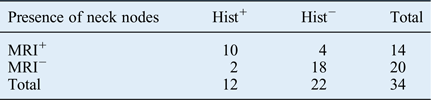
Of the 25 patients included in the study, 13 (52 per cent) underwent ipsilateral neck dissection, 4 (16 per cent) underwent ipsilateral radical neck dissection with contralateral selective neck dissection, 6 (24 per cent) underwent bilateral selective neck dissection and 1 (4 per cent) underwent ipsilateral radical neck dissection. For a total of 34 neck specimens, both radiographic and histological measures were available for comparison. In this sample group, 3 Tesla MRI had a sensitivity of 83.3 per cent (95 per cent CI, 55 per cent to 95 per cent), a specificity of 81.8 per cent (95 per cent CI, 61 per cent to 92 per cent) and an accuracy of 82.4 per cent for detecting cervical lymph node metastasis. The positive predictive value was 71.4 per cent and the negative predictive value was 90.0 per cent. Nodal stage was correctly predicted by 3 Tesla MRI in 8 out of 14 patients (57 per cent); of these, 3 had TNM nodal stage N1 and 5 had stage N2b. If N0 necks were also considered, 3 Tesla MRI predicted the cervical nodal stage in 26 out of 34 necks (76 per cent; Table III).
Node detection: radiological vs histological findings

Hist+ = positive histology findings; Hist− = negative histology findings; MRI+ = positive 3 Tesla MRI findings; MRI− = negative 3 Tesla MRI findings
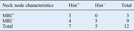
For evaluating extracapsular spread, only histologically positive neck dissection specimens were considered (n = 12). In this sample group, 3 Tesla MRI had a sensitivity of 42.8 per cent (95 per cent CI, 15 per cent to 75 per cent), a specificity of 100 per cent (95 per cent CI, 56 per cent to 100 per cent), a positive predictive value of 100 per cent, a negative predictive value of 55 per cent and an accuracy of 66 per cent for detecting extracapsular spread. (Table IV)
Nodal extracapsular extension: radiological vs histological findings

Hist+ = positive histology findings; Hist− = negative histology findings; MRI+ = positive 3 Tesla MRI findings; MRI− = negative 3 Tesla MRI findings
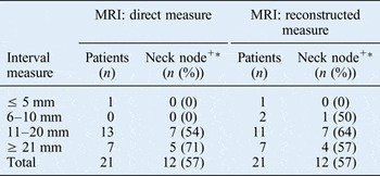
The relationship between MRI-determined tumour invasion depth and cervical metastasis was assessed in 21 patients with 3 Tesla MRI tongue measurements and corresponding neck histological findings (20 patients had undergone neck dissection and 1 had a clinically staged TNM N0 neck). No patient with an MRI tumour thickness of less than 5 mm had cervical metastasis. However, 12 out of 21 patients (57 per cent) with an MRI tumour thickness exceeding 5 mm had cervical metastatic disease at the time of surgery. There were no significant differences between direct and reconstructed MRI measures (Table V).
Radiological thickness predicts cervical lymph node metastasis

MRI = magnetic resonance imaging. *Determined histologically.
Discussion
The oral cavity is 1 of the 10 commonest cancer sites worldwide, with three-quarters of tumours affecting people in developing countries. Squamous cell carcinoma is the most frequent primary malignancy of epithelial origin and the most common cancer of the oral cavity. In the Western world, the tongue and floor of the mouth are the most common subsites for SCCs within the oral cavity.Reference Shah, Patel, Singh and Shah 1 An estimated 450 000 new cases of oral cancer are diagnosed annually worldwide, 25 and the American Cancer Society estimated 48 330 new cases of oral cancer in the USA for 2016 and 9570 deaths per year due to this disease. The five-year relative survival rate for tongue cancer patients is 78 per cent for those with local disease, 63 per cent for those with regional disease and 36 per cent for those with distant metastasis. 2 Accurate imaging of these cancers can help direct treatment.
Tumour thickness, nodal metastasis and extracapsular spread are all factors affecting patient treatment and survival. Studies have demonstrated that MRI tumour thickness directly correlates with histological tumour thickness, and can help guide glossectomy and lymphadenectomy.Reference Park, Jung, Joo, Jung, Cho and Kim 6 , Reference Preda, Chiesa, Calabrese, Latronico, Bruschini and Leon 9 , Reference Lam, Au-Yeung, Cheng, Wei, Yuen and Trendell-Smith 10 Since tumours can be ulcerated or exophytic, tumour thickness was calculated using both direct and reconstructed measures, as previously described.Reference Preda, Chiesa, Calabrese, Latronico, Bruschini and Leon 9 , Reference Iwai, Kyomoto, Ha-Kawa, Lee and Yamashita 24
This is the first prospective study to examine the utility of 3 Tesla MRI for evaluating oral tongue cancer. The signal-to-noise ratio of 3 Tesla MRI is approximately double that of 1.5 Tesla MRI,Reference Stehling, Vieth, Bachmann, Nassenstein, Kugel and Kooijman 26 thus enabling increased in-plane resolution and decreased slice thickness, as well as improving temporal resolution and decreasing scan time.Reference Lu, Nagae-Poetscher, Golay, Lin, Pomper and van Zijl 12 In a cohort of patients with previously untreated T1–3 tongue SCC, there was a high correlation between 3 Tesla MRI direct tumour thickness and histological tumour thickness (r = 0.844, p < 0.001). Reconstructed tumour thickness also correlated with histological tumour thickness (r = 0.81, p < 0.001). Comparison of predictive reliability between direct and reconstructed measures showed that direct MRI tumour thickness was predictive of direct histological tumour thickness in 71.2 per cent of patients; this predictive rate fell to 65.6 per cent for reconstructed measures. Correlation coefficients in the present study were higher than those reported by Preda et al. for both direct and reconstructed tumour thickness (r = 0.77 and r = 0.80, respectively),Reference Preda, Chiesa, Calabrese, Latronico, Bruschini and Leon 9 by Iwai et al.Reference Iwai, Kyomoto, Ha-Kawa, Lee and Yamashita 24 for direct tumour thickness (r = 0.60) and by Yesuratman et al.Reference Yesuratnam, Wiesenfeld, Tsui, Iseli, Hoorn and Ang 27 (average tumour thickness, r = 0.66). Other studies reported even higher correlation coefficients of 0.839 (reconstructed tumour thickness; Iwai et al.Reference Iwai, Kyomoto, Ha-Kawa, Lee and Yamashita 24 ), 0.94 (average tumour thickness; Lam et al.Reference Lam, Au-Yeung, Cheng, Wei, Yuen and Trendell-Smith 10 ), 0.88 (Lwin et al.Reference Lwin, Hanlon, Lowe, Brown, Woolgar and Triantafyllou 5 ), 0.876 (Bashir et al.Reference Bashir, Manzoor, Majeed, Khan, Hassan and Murtaza 7 ) and 0.95 (Park et al.Reference Park, Jung, Joo, Jung, Cho and Kim 6 ). The three latter reports used classic 1.5 Tesla MRI. The only two studies to use both direct and reconstructed measures (Preda et al. and Iwai et al.) showed higher correlations when using reconstructed tumour thickness.Reference Preda, Chiesa, Calabrese, Latronico, Bruschini and Leon 9 , Reference Iwai, Kyomoto, Ha-Kawa, Lee and Yamashita 24 In contrast, in the present study, 3 Tesla MRI direct tumour thickness correlated more closely with histological direct tumour thickness than with MRI reconstructed tumour thickness. Moreover, there was no significant difference in mean direct and mean reconstructed tumour thickness for MRI scans. Therefore, in the current cohort, reconstructed measures were unnecessary.
In the present study, 3 Tesla MRI had high sensitivity (83.3 per cent), specificity (81.8 per cent) and accuracy (82.3 per cent) for predicting cervical nodal metastasis. In fact, these values were higher than those previously reported by Okura et al. (53 per cent sensitivity, 77 per cent accuracy),Reference Okura, Iida, Aikawa, Adachi, Yoshimura and Yamada 8 Zeng et al. (60.8 per cent sensitivity and 75 per cent specificity),Reference Zeng, Liang, Zhou, Zheng and Zeng 28 Ding et al. (79.5 per cent sensitivity),Reference Ding, Liang, Shen, Xie, Huang and Zhang 3 and Lwin et al. (51 per cent sensitivity)Reference Lwin, Hanlon, Lowe, Brown, Woolgar and Triantafyllou 5 using a 1.5 Tesla scanner. The negative predictive value was 90.9 per cent for the current cohort. Most importantly, 3 Tesla MRI correctly predicted neck nodal stage in 76.5 per cent of patients.
In contrast to Lwin et al., who previously reported 0 per cent sensitivity when working with a 1.5 Tesla scanner, the present study found 43 per cent sensitivity, very high specificity (100 per cent) and a very high positive predictive value (100 per cent) for 3 Tesla MRI in detecting adenopathy with extracapsular spread. Extracapsular extension often requires more extensive dissection combined with post-operative radiotherapy or chemoradiotherapy. As the ability of 3 Tesla MRI to evaluate the presence of extracapsular extension in cervical lymph node metastases may be clinically very useful, additional studies are needed.
-
• Magnetic resonance imaging is useful to predict the depth of oral tongue cancer invasion and is essential for pre-operative planning
-
• The increased signal-to-noise ratio and larger susceptibility of 3 Tesla magnetic resonance imaging result in higher spatial resolution, improving diagnostic strength
-
• Correlation between radiological and histological tongue tumour thickness was determined for direct and reconstructed measures
-
• The ability of 3 Tesla magnetic resonance imaging to detect positive cervical node and extracapsular spread was evaluated
-
• This is the first prospective study to examine the utility of 3 Tesla MRI for evaluating oral tongue cancer
Some studies suggest that neck dissection should be considered for patients with a histological tumour thickness of 1.5–6 mm.Reference Preda, Chiesa, Calabrese, Latronico, Bruschini and Leon 9 Radiographically measured tumour thickness is also predictive of cervical metastasis. When evaluating cervical lymph nodes with 1.5 Tesla MRI, numerous authors have found a wide range of cut-offs for depth of tumour invasion: 3–10 mm for Lwin et al.Reference Lwin, Hanlon, Lowe, Brown, Woolgar and Triantafyllou 5 ; 3–9 mm for Lam et al.Reference Lam, Au-Yeung, Cheng, Wei, Yuen and Trendell-Smith 10 ; 9 mm for Zeng et al.Reference Zeng, Liang, Zhou, Zheng and Zeng 28 , 9.7 mm for Okura et al.Reference Okura, Iida, Aikawa, Adachi, Yoshimura and Yamada 8 ; 6 mm for Iwai et al.Reference Iwai, Kyomoto, Ha-Kawa, Lee and Yamashita 24 ; and 9.5 mm for Park et al.Reference Park, Jung, Joo, Jung, Cho and Kim 6 Recently, Kwon et al. retrospectively evaluated tongue cancer thickness using 3 Tesla MRI and concluded that a radiographic thickness of at least 6.7 mm detects occult lymph nodes metastasis in the axial plane.Reference Kwon, Moon, Nam, Lee, Kim and Lee 29 In the present study, MRI measures of tumour thickness were classified as (1) direct or (2) reconstructed and then were stratified as a thickness of less than 5, 6–10, 11–20 and 21 mm. For a cut-off point of 11 mm or greater, more than half of patients in the direct and reconstructed groups had cervical lymph node metastasis.
Study limitations
This study, which has a level of evidence 4, aimed to define the role of 3 Tesla MRI in the pre-operative evaluation of oral cancer; however, there were several limitations. A disadvantage of 3 Tesla imaging is its high susceptibility to artefacts generated by ferromagnetic implants compared with 1.5 Tesla imaging. Although most dental amalgam does not cause a significant artefact upon MRI, other metallic dental hardware (e.g. dental implants, metallic dental crowns, metallic orthodontic braces, bonded retainers) can produce pronounced artefacts. In such instances, 3 Tesla MRI of the oral cavity may prove to be non-diagnostic. In addition, radiological measures used in the present study were performed in the axial plane, and other dimensions were not included for comparison. This limited the determination of how the depth of radiological tumour invasion relates to cervical metastasis. Finally, this study does not directly compare 1.5 and 3 Tesla MRI data. Further studies should aim to compare these modalities.
Conclusion
This prospective study examined the utility of 3 Tesla MRI in the initial evaluation of oral cancer. There was a statistically significant correlation between radiological and histological tumour thickness. The findings provide evidence for the predictive value of 3 Tesla MRI in evaluating disease extent in SCC of the tongue by direct measurement of tumour thickness. Furthermore, there was a strong relationship between 3 Tesla MRI thickness and cervical lymph node metastasis. In the study sample, 3 Tesla MRI had a higher sensitivity, specificity, accuracy and negative predictive value compared with published reports for lower magnetic field 1.5 Tesla MRI in detecting cervical lymph node metastasis, and correctly predicted the nodal stage in three-quarters of patients. Lastly, this imaging modality was useful for evaluating malignant adenopathy with extracapsular spread. This study opens the way for future prospective investigations into the utility of 3 Tesla MRI.
Acknowledgements
The authors thank Dr K Wilson, Dr K Casper and Dr E Manders for their assistance with patient enrolment, and C Talbott for her collaboration with the Pathology Department.